
Abstract
The current lifestyle contributes to the increase in the incidence of life-threatening illness like cancer. Methods for early detection of the same can help better patient outcomes. Effective, inexpensive and non-invasive diagnostic methods are often sensitive as well as specific compared to conventional mechanisms. Advances in biosensor technologies have paved the way for low-cost, non-invasive diagnostic techniques that report greater levels of sensitivity and specificity. Organic Light-Emitting Diodes (OLEDs) have emerged as a proficient biosensor for clinical diagnostics. This research rigorously investigates the application of OLED-based biosensors for early-stage cancer detection. In addition, a comprehensive analysis of OLED characteristics pertinent to the detection of various cancer types, such as cutaneous and ovarian malignancies, is also reported. We also explore the integration of OLEDs into portable and adaptable diagnostic devices, with the aim of improving their screening capabilities. A parameterized evaluation framework is used to assess the design attributes and operational efficacy of OLED biosensors, emphasizing their suitability for oncological diagnostic applications.
Introduction
Cancer consists of a collection of disorders characterized by excessive growth, spread of malignant cell. 1 Abnormal cells can generate tumors that can infiltrate and obliterate adjacent healthy tissue. 1 Numerous non-invasive techniques exist for cancer detection, although they are prohibitively costly. OLEDs may be used to identify and forecast cancer probabilities depending on characteristics such as blood, serum, saliva, urine, and pH value. 2 Expensive cancer detection tests may be unavoidable for many individuals. The influence of early identification of cancer biomarkers at the treatment in intial stage has attracted the attention of many studies throughout years of research.
Therefore, a non-invasive portable device using an OLED based biosensor is suggested for the detection of cancer. The primary reason for conducting this research is the fact that 80% of patients diagnosed cancer at later stages. 3 Its asymptomatic nature and limitations of current diagnostic techniques, the lack of public knowledge, contribute to delayed detection and poor survival rates. Therefore, early identification of cancer is essential to improve clinical results. The higher mortality rate and worse prognoses result from this. The investigation of new technologies is necessary due to the pressing need for highly accurate, non-invasive and reasonably priced diagnostic instruments. Consequently, the focus is on creating accurate, reliable and affordable diagnostic-tool. This article analyses the operational mechanism of OLEDs and presents the potential for developing a mobile, versatile device for cancer screening and detection.
Originally developed for lighting and display applications, OLEDs are rapidly emerging as a viable biosensor design technique. 4 They are ideal for developing highly sensitive and specific cancer detection devices due to their special qualities. 4 The American Cancer Society (ACS) indicates that 5-year survival rates for different types of cancer increased from 50% to 66% between 1996 and 2004.1,5,6 Technological advances that facilitate early identification and improved treatment are responsible for this increase in survival rates. 7
Advanced biosensors can significantly improve patient quality of life by facilitating early detection, potentially decreasing cancer mortality rates. 8 This research emphasizes the assessment of meticulously designed biosensors with the demonstrations of innovative, quick and fast response. These biosensors have potential to transform early diagnostic and therapy options for many malignancies. 8 Employing advanced biosensor technologies might substantially enhance early cancer diagnosis. 4 This is particularly vital as several cancer forms advance gradually and have inadequate responses to treatment.
Organic light emitting diodes: Advancing cancer treatment and detection with innovative properties
An Organic Light Emitting Dioses (OLEDs) generates light using electroluminescence, with several thin organic layer interposed between a transparent-anode with a metallic cathode. Upon the application of voltage, the anode injects hole and the cathode injects electrons; these charges move through the organic layers until they meet at the emissive layer, recombining to form an exciton. This exciton-decays to its ground-state, releasing energy manifested as a photon. Hue of photon is determined by qualities of the substance in emissive-layer. The emitted light spreads in all directions, and structures are frequently employed to increase light extraction efficiency. OLED technology can improve cancer diagnosis by increasing sensitivity, multiplexing capability, mobility, cost-effectiveness, and real-time monitoring.
The OLED has many advantages like broad emission angles, high color purity, rapid speed of response, increased efficiencies, diverse spectrum of potential emission wavelengths, exhibits improved tolerances for bending and twisting, allowing fabrication on a diverse array of substrates, enhanced sensitivity, quick reaction times, cost efficiency, and novel uses in point-of-care diagnostics, promoting more precise and prompt cancer treatment etc. Although OLEDs have potential in cancer treatment applications such as Photodynamic Therapy (PDT), they possess some drawbacks. This includes reduced optical power output relative to alternative PDT light sources, 9 perhaps requiring extended treatment durations.
Figure 1 illustrates the various merits and features of OLEDs. The OLEDs necessitate certain device characteristics for biomedical applications, including mechanical flexibility and robust encapsulation for worn or implanted devices, which might be difficult to provide. The challenges of employing OLED as biosensor are as follows:
Certain demographic groups, including neonates, older one may exhibit heightened vulnerability pertaining biological-effects induced by blue enriched LEDs.
13
Concerns over Age-Related Macular Degeneration (AMD) due to continuous light exposure have been expressed, while epidemiological research on the relationship between sun exposure and AMD yield contradictory results.
13
Safety in light-based technologies for sensitive tissues, especially concerning eye toxicity and blue-light risks, is guaranteed by meticulous assessment of light sources, compliance with safety regulations i.e., international standards (such as International-Electrotechnical-Commission (IEC-60601, 60825series), American-National-Standards-Institute (ANSI-Z136)), which establish maximum permissible exposure limits based on wavelength, power, duration of exposure to avert harm.
15
Protective procedures encompass the use of specialist eyeglasses and corneal shields that filter deleterious wavelengths, namely the blue-violet spectrum (415–455 nm) linked to retinal photochemical injury, while permitting advantageous light to pass.
16
The advancement of these coatings has consistently enhanced performance while streamlining the production flow, minimizing time, and lowering costs.
20
The enhanced coating exhibited superior antifouling efficacy against diverse biological-fluids.
20
The concepts of antifouling coatings are significantly applicable to optical biosensors, such as OLED-based systems, to sustain performance in biological settings, despite breakthroughs being mostly in electrochemical sensors.
20
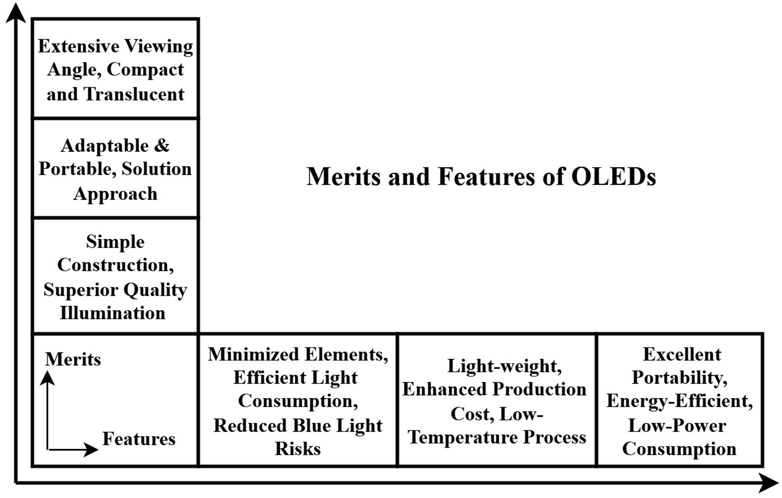
Various Merits and Features of Organic Light Emitting Diodes.
Challenges in manufacturing of large-scale OLED-based biosensor and their impact
Large-scale production of OLED-based biosensors faces challenges in scaling from laboratory to industry, affecting cost and consistency. The challenges in manufacturing large-scale OLED-based biosensors are as follows:
Impact on reproducibility and affordability:
Producing consistent OLED-based biosensors on a large scale is difficult due to manufacturing variations. Table 1 illustrates the impact of large-scale OLED-based biosensor manufacturing on reproducibility and affordability. This inconsistencies affect film quality and lead to non-uniform performance of device. The development of innovative technologies and materials, such as solution-processed all-organic sensors, is crucial to making OLED biosensors more accessible for widespread use, though these solutions are still in early development.
Impact of challenges in large-scale OLED-based biosensor manufacturing on reproducibility and affordability.

Addressing environmental and physiological variability in OLED biosensor
OLED biosensors address environmental and physiological fluctuations, including pH, temperature, and biofouling, by smart sensor design with innovative anti-biofouling coatings.26–28 Unregulated chemical interactions may result in experimental artifacts in electronics designed for signal processing. 29 These devices provide prospects for chemical modification, 30 doping, mixing at the interface, facilitating the development of innovative designs, functional transistors, bio-detection instruments, and flexible electronics.
Temperature fluctuations can affect enzyme processes, molecular transport and the stability of organic materials. 31 Increased temperatures 32 may accelerate the deterioration of organic layers, resulting in decreased device longevity, performance variability. Furthermore, variations in temperature might influence the binding-affinity, hence modifying sensor’s selectivity and sensitivity. 33 Katchman et al. 19 investigated the impact of temperature on general biosensor performance, such as a portable temperature-humidity control device for lateral flow assays. 19 Recent progress in pH sensing devices include tiny pH biosensors utilizing electrochemically modified materials, designed to enhance sensitivity and reliability.
An electrochemical biosensor intended for customized treatment has a mechanism to monitor and regulate pH and temperature fluctuations in patient samples. 34 The detection of pharmaceuticals using cytochrome P450-based electrochemical sensors is critical, as pH variations can influence both the potential, which indicates drug kind, and the current, which signifies drug concentration. 34 In 2011, De Venuto et al. 34 presented a novel architecture for multiplexing molecular biosensing with temperature and pH measurements. Molecular sensing is utilized for drug detection. The biosensor prototype for drug detection employs P450 3A4, facilitating extensive application for a range of pharmaceuticals. 34 It incorporates pH and temperature monitoring for accurate electrochemical detection in various biological fluids. This technology offers a viable approach for multi-drug monitoring in customized therapy. Bio-fouling severely reduces the lifespan and functioning of biosensors, especially those that are implanted in the body or function in complicated biofluids. 35 This process may result in the progressive passivation of the transducer surface, obstructing direct interaction with the target analyte and significantly compromising sensitivity, repeatability, stability, and overall dependability. 35
The remaining sections of the paper are arranged in following sections. The Section “Introduction” Introduction itself, having three subsections i.e., “Organic light emitting diodes: Advancing cancer treatment and detection with innovative properties”, discusses the advancing cancer treatment and detection with innovative properties, “Challenges in manufacturing of large-scale OLED-based biosensor and their impact” illustrates the challenges in manufacturing of large-scale OLED-based biosensor and their impact, “Addressing environmental and physiological variability in OLED biosensor” addresses the environmental and physiological variability in the OLED-based biosensor. Section “Biosensor architecture and key functional elements” describes the architecture of the biosensor. Further, Section “Cancer detection using organic semiconductors” contains cancer detection techniques using organic semiconductors, Furthermore, Section “OLEDs Potential in cancer diagnostics” provides a detailed exploration of various cancer detection techniques using OLED, and subsection “Ovarian-cancer detection utilizing oled technology” illustrates ovarian cancer detection using OLED and subsection “Detection of skin cancer using OLED” identifies skin cancer using OLED and subsection “Detection of various cancers using OLED”, discusses different techniques using OLED for cancer detection.
Subsequently, Section “Assessment using photo-dynamics theraphy” reveals the different assessment using Photo-Dynamics Theraphy (PDT), referring to Section “Limitations of light penetration in human tissues during PDT” which describes the limitations of light penetration in human tissues during PDT and overcoming those limitations in subsection “Overcoming light penetration limitations with OLED-based PDT devices”. Additionally, Section “OLED as Biosensor for disease identification” discusses the OLEDs as Biomarker for identification of various human diseases having subsection “Multiplexed detection techniques of OLED-based biosensor” describes the multiplexed detection techniques used in OLEDs Biosensor and subsection “Comparative analysis of OLED-based detection technique with gold standard techniques” illustrates the comparative analysis of OLED-based detection technique with gold standard techniques. Finally, Section “Conclusion” concludes the OLED techniques used for cancer detection and leads the researcher to a comprehensive understanding of the importance of the study and leading prospective areas for future research on cancer detection.
Biosensor architecture and key functional elements
A biosensor is a technology that detects biological-analytes, which may come from environment or human-body. 8 Details include whether or whether analyte levels and presence are turned into a quantifiable electrical signal that could measured, studied. Proteins and nucleic acids, and other bio-logical substances are examples of analytes. Biosensors can be used in medicine to diagnose and track cancer, identify infections, and check blood glucose levels in diabetics. Biosensors can be utilized in environment settings for identify dangerous germs, in food, water or air. The creation of biosensors to combat bioterrorism is of great interest to the military. Biosensors created with three main parts: a signal transducer, a recognition device, signal-processor showing data. The identification of molecules When an analyte is detected by the component as a ”signal” from the environment, the transducer translates biological signal into an electrical-output. The functional architecture of the biosensor (which may be mechanical, optical, electrochemical, electrical, or optoelectronic) is illustrated in Figure 2. A biosensor is an analytical instrument that identifies biological substances and transforms their presence or concentration into a quantifiable signal. It generally comprises three primary components organized sequentially: Bioreceptor, Transducer, and Signal Processor. The bioreceptor, which may be an enzyme, antibody, or DNA strand, precisely interacts with the target analyte, such as glucose, toxins, or infections, and initiates a biological reaction. The recognition element, crucial for selectivity, can be naturally occurring (e.g., purified proteins, antibodies, enzymes, nucleic acids) or synthetic (designed for improved stability and reproducibility).
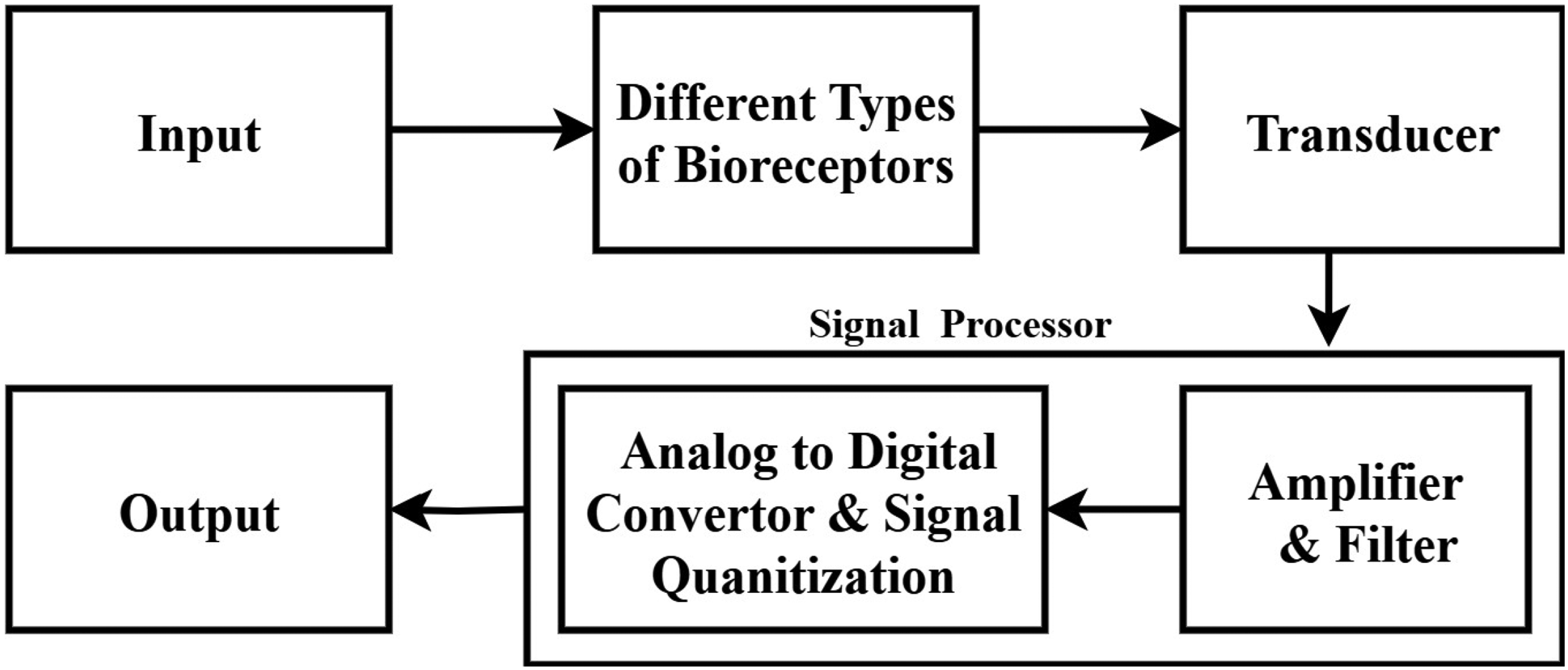
Basic Components and Functional Architecture of A Biosensor.
Examples include antibodies (like anti-PSA for prostate cancer detection), cell-surface receptors (though their use is complex due to membrane requirements), and aptamers (oligonucleotides selected for high target affinity via SELEX technology, enabling discovery of cancer biomarkers. The reaction is transmitted to the transducer, that transforms bio-logical contact into quantifiable signal—such as electrical, optical, thermal, or mass-based signals transformation of a molecule’s recognition process into a quantifiable signal (amperometric, potentio-metric), mass based, and temperature-based i.e., calorimetric. 36
This signal is then amplified and processed to provide quantifiable data related to the presence and concentration of the target molecule, without requiring prior purification. An OSC-based biosensor is an example, which includes OLEDs, OTFT, OECs, and solar cells, among others. In order to identify and characterize a wide variety of cancer biomarkers, the OLED based cancer biosensor is analyzed in this research.
Cancer detection using organic semiconductors
Organic semiconductors have potential in advancing novel cancer detection methods. Recent breakthroughs include the use of Organic Electro-Chemical Transistors (OECTs) for the real-time screening of cancer metastasis, facilitating the identification of phenotypic alterations in cancer cells. 37 Moreover, organic semiconductors are used in optical biosensors designed to detect particular biomarkers linked to different cancer types, including circulating tumor cells and exosomal microRNAs. This technique enables both the early identification and dynamic monitoring of cancer development. 38 Incorporating organic semiconductors into detecting systems enables researchers to develop extremely sensitive, portable, and economical cancer detection devices.
An advanced photoelectrochemical sensor was created using the organic semiconductor Y6 in conjunction with gold nanoparticles, facilitating to detecting Carcino-Embryonic-Antigen (CEA) having detection limit
Organic Semiconducting Nanoparticles (OSNs) have potential in biosensing applications, enabling high-resolution detection of diverse biological substances, such as cancer biomarkers.
40
Notwithstanding obstacles like as photo-degradation, OSNs may be designed for enhanced stability and usefulness, rendering them suitable for in vivo tracking and tumor localization.
40
Organic Light-Emitting Diodes (OLEDs) has utilized in fluorescence-linked immunosorbent assays for the identification of disease-biomarkers.
41
The OLEDs efficacy in delivering requisite light for such tests enhances the overall sensitivity and precision of cancer detection. OLEDs provide high-resolution images with superior contrast, which is crucial to identifying tiny tissue changes that can indicate malignancy. The precision and complexity in imaging enable the detection of anomalies that conventional methods may detect.
Organic Phototransistors (OPTs) transform light impulses into electrical signals, providing benefits in signal amplification and noise reduction, essential for cancer detection.
42
Their lightweight and flexible attributes make them suitable for applications, improving accessibility in clinical environments.
42
Recent advancements have established quantum-scale organic semiconductors as transformative instruments for label-free detection of genomic DNA associated with cancer.
43
These semiconductors provide examination of structural and molecular anomalies at very low detection thresholds.
43
Their capacity to provide high-resolution data on gene expression makes them essential for understanding cancer dynamics and formulating targeted therapeutics.
In 2024, Khayam et al.
44
developed compact, Ultra Wide Band (UWB) printable UHF-antennas for Partial-Discharge-detection in high-voltage, for 0.3-3 GHz bandwidth within a 100mm GIS dielectric window.
44
This innovation enables more efficient and reliable PD detection across various insulating media.
44
In 2025, Malecka-Baturo K Grabowska
33
examined the potential avenues for advancement, obstacles, and opportunities in the domain of multi-analyte electrochemical immuno-ageno(apta)-sensors.
33
Recent developments emphasize numerous techniques, such as photoelectrochemical sensors and organic phototransistors, which use the distinctive characteristics of organic materials for efficient cancer biomarker detection.
OLEDs potential in cancer diagnostics
OLED technology is also applied in certain cancer detection techniques using its specific optical properties to enhance sensitivity and specificity. 45 A good example is the use of OLED-based Fluorescence-Linked Immunosorbent Assay (FLISA) that utilize OLEDs as sources for light in order to trigger fluorescent labels bound to cancer biomarkers. 41 OLEDs are flexible and compact, making them well suited for quick and precise point-of-care cancer screening. The basic structure of OLED used in various applications is illustrated in Figure 3.
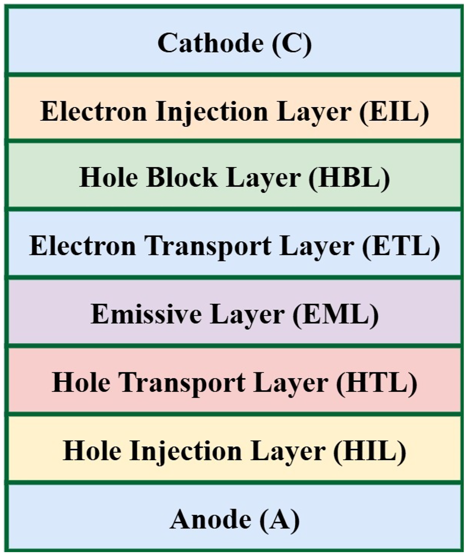
Schematic Diagram of OLED Structure Used in Different Applications.
OLED has various layers situated between a cathode and an anode, there are additional layers designed to enhance charge transmission in between.46,47 The Emissive Layer (EML) is the primary layer of OLEDs, as this is where light production transpires. Upon the application of voltage, electrons are introduced from cathode to the EML, while holes are introduced from anode into conductive-layer. Electrons and holes travel towards one another and merge in the EML to generate excitons. 47 When these excited states revert to a lower energy level, they emit energy as light, a phenomenon termed recombination. The hue of the produced light is dictated by the chemical composition of the light-emitting substance, enabling regulation of the display’s hues. 47 By appropriately designing organic molecules, we may manipulate the energy differential between the excited state and the ground state, enabling emission in the blue, green, and red regions. 47
Ovarian-cancer detection utilizing OLED technology
OLED technology is under development for sensitive ovarian cancer diagnostic devices. OLED biosensors detect certain ovarian cancer markers in body fluids. They function by emitting light from OLEDs as a result of electrical stimulation, indicating the presence of biomarkers. Surface modification of the OLED with antibodies makes signal changes quantifiable if the biomarker is present. This leads to a rapid, cost-effective, early and accurate diagnostic tool for ovarian cancer.
Table 2 illustrates the different applications of OLED detecting ovarian cancer. In 2010, Masilamani et al. 36 developed a non invasive technique to detect cancer based-on fluorescence-characteristics of urine and analyzed Stokes-Shift-Spectra (SSS), Fluorescence-Emission-Spectra (FES). 36 A high-resolution, high-speed light-addressable potentiometric sensor was reported in 2012 by Werner et al. 50 Threm et al. 51 produced cost-effective, high-performance OLED-OPD on one substrate in the same year.
Different applications of OLED in detecting ovarian cancer.

In addition, Marcello et al. 52 created a small biochip with a deep blue OLED for medical diagnosis in 2013. This biochip, with emphasis on fluorescence detection, efficiently excited fluorophore-conjugated antibodies, exhibiting high sensitivity and specificity. Zvarik et al. 53 studied differences in urine fluorescence between healthy humans and women with ovarian cancer. Urine specimens were excited between 250-530 nm, and emission was seen from 270-650 nm.
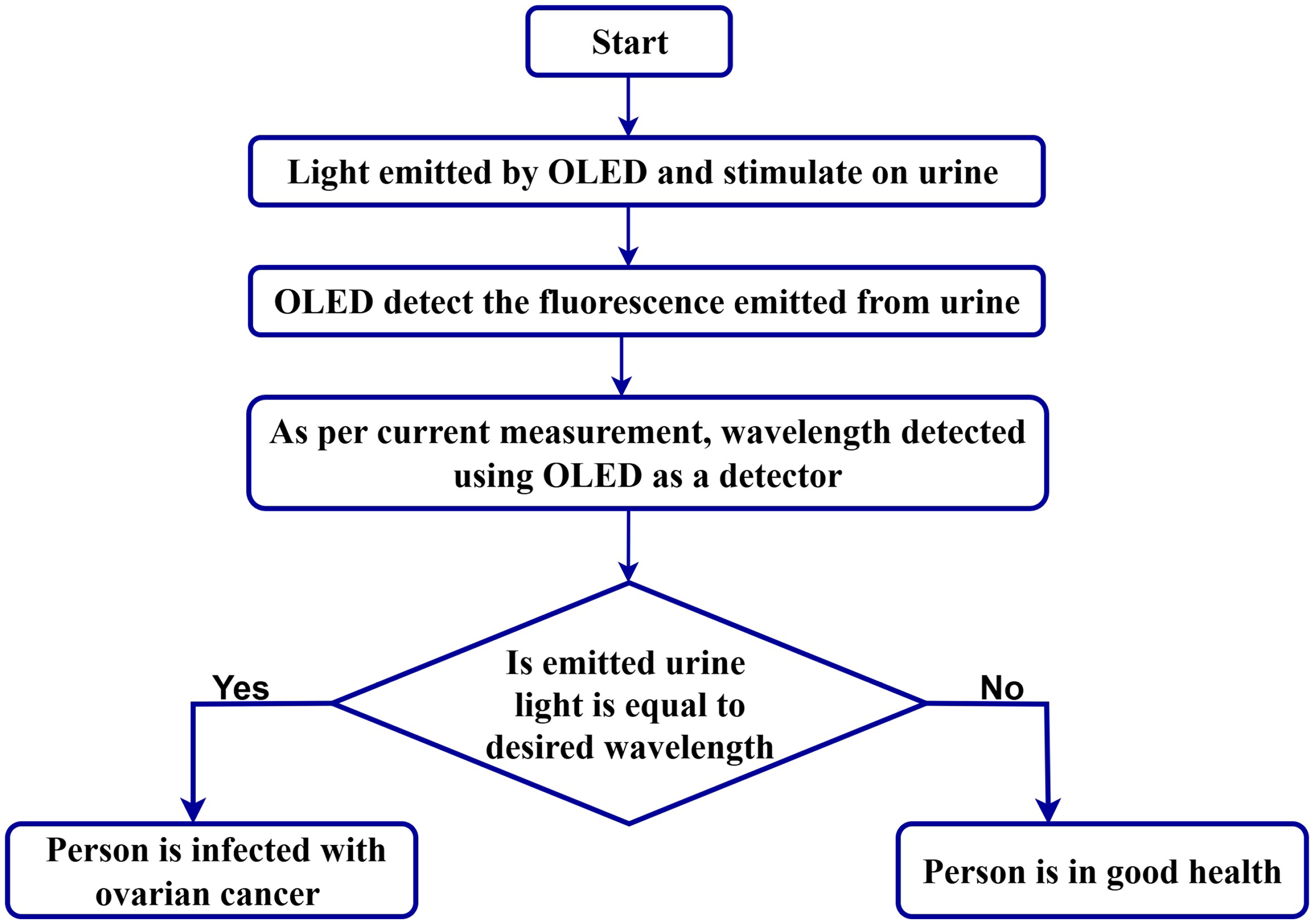
In 2016, Katchman et al. 19 investigated the use of commercial OLED displays for low-cost, multiplexed circulating cancer biomarker detection. The method substitutes optical components with OLEDs, which is ideal for point-of-care. Figure 4 depicts the process diagram of steps involved in detecting ovarian cancer using the fluorescence emission mechanism. A silicon photodiode in photovoltaic mode measures weak fluorescence, allowing accurate identification of low biomarker concentrations essential for cancer diagnosis. 19
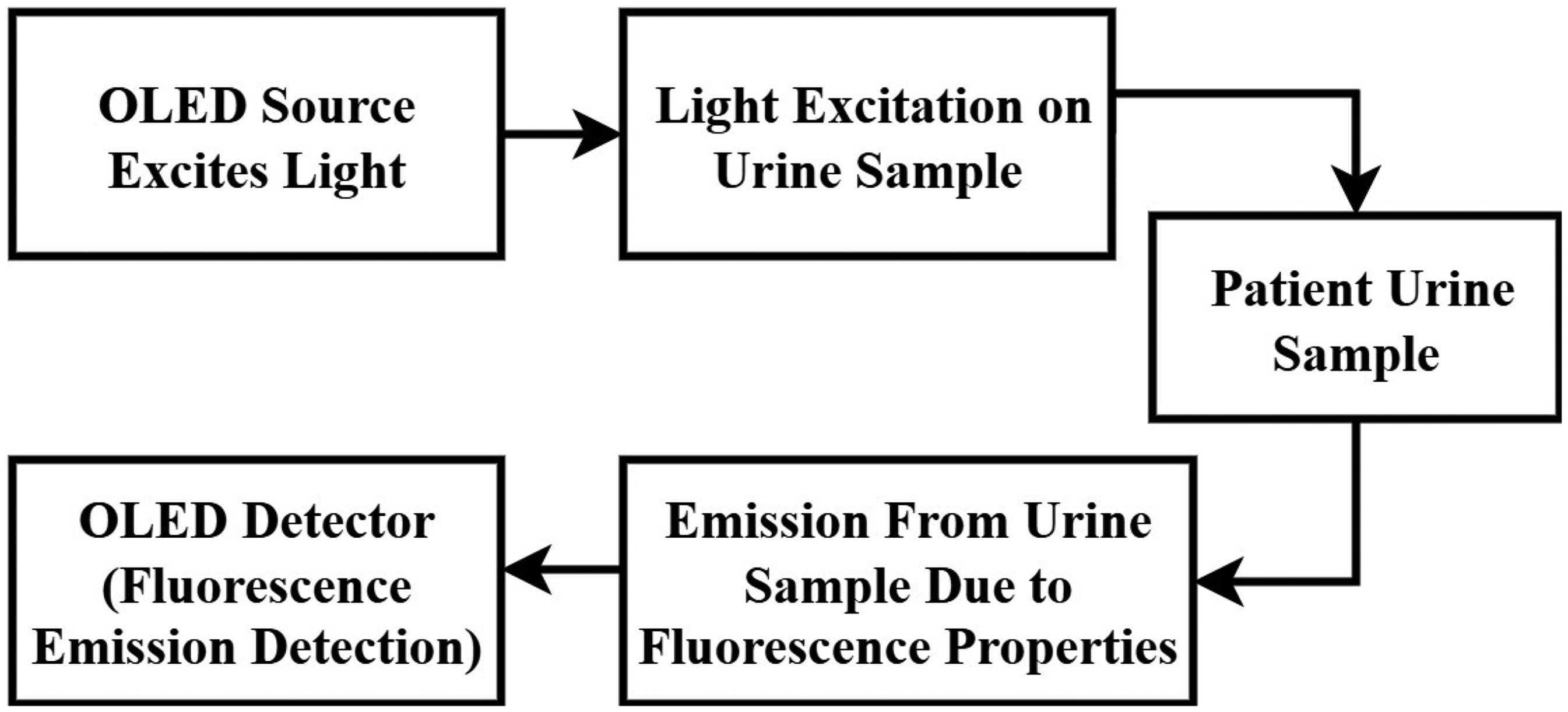
Process Diagram of Steps Involved in Ovarian Cancer Detection Methodology, Where OLED is Used as Source and Detector at the Input and Output Terminal Respectively.
Afterthat, In 2019, Negi et al. 49 suggested a handheld OLED-based approach for the detection of ovarian cancer via fluorescence emission. Here, OLED is used as source and light detector. 49 An OLED light source that fluoresces between the 300-440 nm range stimulates the urine samples, inducing fluorescence. 49 This is followed by detection via another OLED device. The detection OLED generates different values of current depending on the emission of the sample and thereby differentiates between emissions at 420 nm and 440 nm. 49
Further in 2021, Negi et al.
48
suggested a unique triple-hole blocking Layers OLED structure, that was demonstrated an improved luminosity of 25,285 cd/m
Figure 5 illustrates the comprehensive flow chart of the ovarian cancer detection methodology using fluorescence emission. 49 An OLED-based fluorescence sensing system employs OLEDs to excite fluorescent molecules in a sample. 49 The device demonstrated strong responses across various wavelengths, yielding a maximum photocurrent of 93 mA, and can differentiate healthy individuals (420 nm emission, 5 mA cathode current) from cancer patients (440 nm emission, 1 mA cathode current) based on urine fluorescence. 48 For ovarian cancer diagnosis, an OLED violet of 420-440 nm emission is generally used.
Flowchart of Ovarian Cancer Detection Methodology using Fluorescence Emission.
This technology facilitate the creation of portable, versatile, and cost-effective biomedical sensors that are portable and primarily theoretical.4,48,49
Katchman et al. 19 discussed the utilization of flat panel OLED display technology for point-of-care detecting different biomarkers, especially IgG antibodies- various viral antigens and HPV16 proteins, in patient blood samples. This does not provide a direct comparison with biopsy for cancer detection in urine specimens. This immunoassay exhibited comparable accuracy and specificity in identifying blood antibodies against HPV16 E7 when contrasted with the standard laboratory ELISA. Both tests yielded comparable findings for the identification of HPV16 E2 and E7 antibodies. The point-of-care test had a lower sensitivity (59%) than the laboratory ELISA (83%) at 95% specificity. 19 This method enables for multiplexed immunoassays in a small, disposable design, boosting sensitivity and enabling quick detection of several biomarkers. Table 3 illustrates the capabilities and data of clinically tested for OLED-based devices.
OLED-based devices tested on human trials.

In ovarian cancer, although OLED-based fluorescence detection in urine samples may distinguish between healthy and malignant samples, there is an acknowledged necessity for more comprehensive clinical data. 48 In addition to ovarian cancer, OLED-based FLISA have been examined for various-biomarkers in human serum, showcasing its promise for compact and sensitive point-of-care diagnostics. 41 An OLED-FLISA system effectively identified SARS-CoV-2 antibodies in diluted human blood, with one research estimated its sensitivity to be roughly 80 times greater than that of a commercial lateral flow antibody test. 41 This underscores the significant sensitivity attainable with OLED-based systems, which may be enhanced by the optimization of OLED architecture, the management of leakage light, and the selection of suitable fluorescent dyes such as phycoerythrin. 41 However, OLED-based systems for detecting biomarkers remain unsuitable for extensive clinical use and require enhancements in reagent utilization, dimensions, sensitivity, and multiplex testing functionalities. 41
Detection of skin cancer using OLED
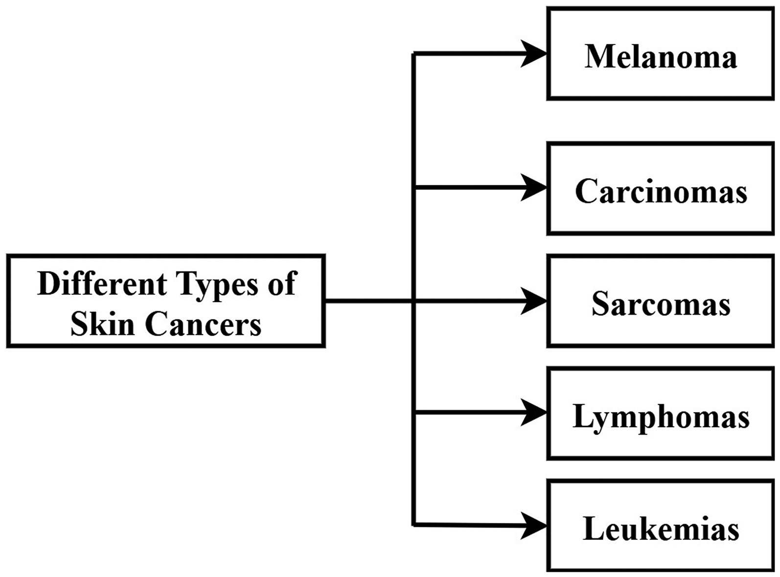
OLED technology enhances skin cancer diagnosis by emitting certain wavelengths of light for improved lesion inspection. This process, in conjunction with AI, provides precise and early detection of different skin cancers, such as carcinoma, sarcomas, and melanoma. The outcome is accurate diagnosis essential for timely treatment. In 2008, Attili et al., 56 presented a low-irradiance, disposable OLED light source for PDT of non-melanoma skin cancers. There were 12-patients having Bowen disease, carcinoma of lesions less than 2 cm, which were histologically confirmed. 56 Red light therapy (550–750 nm, peak 620 nm, 5 mW cmcm2) given twice with aminolevulinic acid, 1 month apart, was beneficial in 7 out of 12 patients after 12 months, median-pain score-1 on Numerical-Rating-Scale (NRS). The different types of skin cancer is illustrated in Figure 6.
Different Types of Skin Cancer.
In 2022, Lian et al. 41 pushed the frontier of point-of-care testing by creating an OLED-based FLISA for identifying SARS-CoV-2 immunoglobulinsin human serum utilizing Phycoerythrin as a fluorescent dye for higher sensitivity. Furthermore, Lian et al. 57 created a compact OLED-based DNA detection system for fluorescence sensing and optimized OLEDs with co-host layer, pulsed voltage operation, employs a Cy3/Cy5 Forster-Resonance-Energy Transfer (FRET) pair to achieve sensitive and accurate DNA/RNA and protein detection, suggesting its suitability for point-of-care diagnostics.
In 2024, Irianto et al. 58 examined the skin cancer identification by integration-region growing techniques with a deep learning algorithm. Further, Lee et al. 59 proved OLED irradiation to be as effective, if not more, as LED in optimizing collagen in-vitro. In addition, OLED exposure in UV-irradiated animal models exhibited decreased wrinkles, enhanced dermal collagen density, and improved wound healing, pointing towards its dermal rejuvenation and tissue repair potential.
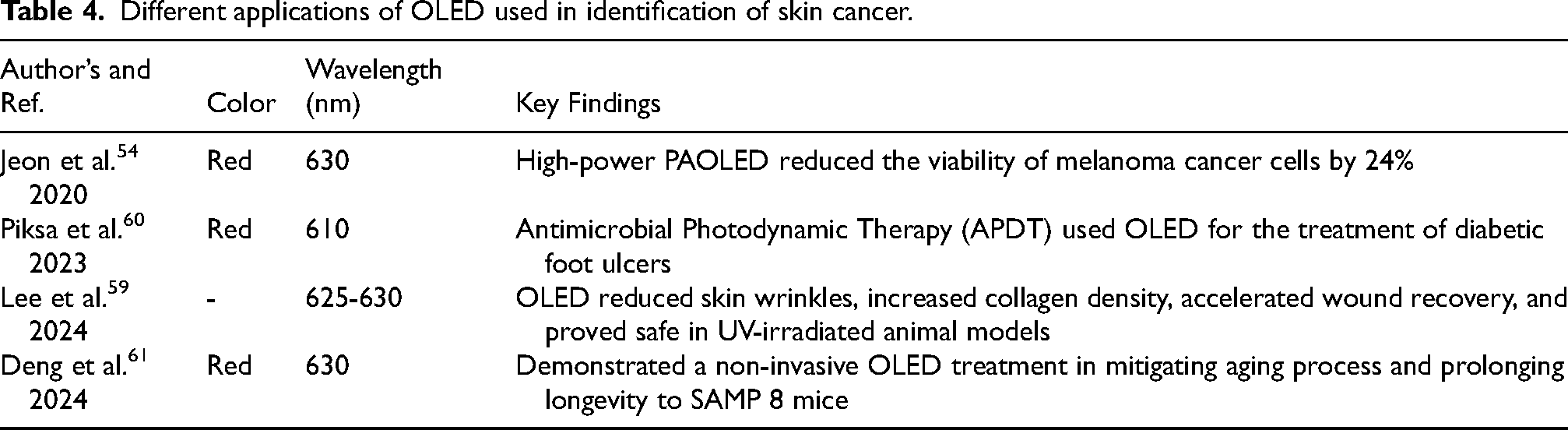
The different applications of OLED used in detecting skin cancer is illustrated in Table 4. Additionally, in 2024 Ramadhan et al. 62 created an elliptical core Photonic-Crystal-Fiber-Sensor predicated upon Surface-Plasmon-Resonance (PCF-SPR) sensor that has a high sensitivity of 24,000 nm/RIU in all four modes. They used Au and TiO2 as plasmonic and adhesive materials, respectively, with the ideal thicknesses. The sensor parameters were optimized using a machine learning algorithm, which produced a remarkably low MSE of 0.00083. 62
Different applications of OLED used in identification of skin cancer.
In 2024, Deng et al.
61
showed that OLED therapy delays aging in SAMP8 mice by promoting stem cell proliferation, migration, and diminishing senescence-associated
Therefore, Red OLEDs with 630 nm wavelengths are utilized as much as possible for the detection of skin cancer because of their combined effectiveness and biological effectiveness. 59
Detection of various cancers using OLED
OLED technology offers a promising approach for cancer diagnostics, especially for Human Papilloma Virus (HPV)-associated cancers like breast, lung, head, neck cancer etc. 19
OLED-immunosensors with their planar sandwich optical structure facilitate multiplexed and affordable detection of HPV protein antibodies such as E2 Katchman et al., 19 which has the potential for extended cancer diagnostics.
In 2015, Guo et al. 66 showed the effectiveness of OLED-PDT in reducing glioma in mice with low intensity and long duration of light, enhancing survival. In 2016, Katchman et al. 19 also demonstrated that OLED technology was able to detect IgG antibodies against viral antigens and HPV16 proteins (E2, E6, E7) in the blood of patients, which can act as cervical cancer biomarkers. FAN et al. 67 in 2016 developed a handheld device based on DPV and a microfluidic paper-based analytical system for quick, sensitive tumor detection with an OLED display. In 2023, Han et al. 64 demonstrated AI can be 95% accurate, making process automated and more effective in detecting defects.
Further in 2025, Tai and Chiu 65 introduced an Active- Plasmonic-Colorimetric (APC) biosensor that combines an OLED and Gold Nano-grating for high-sensitivity using surface plasmon resonance. The sensor identifies changes in the refractive index via wavelength shifts, demonstrates potential for the detection of Neuron-Specific-Enolase (NSE) in lung cancer diagnosis, providing detection range (1–100 ng/mL) with a minimal detection-limit (200 pg/mL). 65 The different types of cancer detection techniques using OLED depicts in Table 5.
OLED applications in the detection process of various types of cancers.

Based on the researches, it is conculded that OLED-based devices in cervical cancer make it easier to identify circulating biomarkers. 19 For anal and oropharyngeal malignancies, OLED technology works with fluorescent biorecognition systems to identify particular cancer biomarkers, increasing detection sensitivity and specificity.68,69. In breast cancer diagnostics, OLED screens aid in the identification of tumor antigen antibodies, facilitating customized cancer detection and management. 19 OLED-enabled optical imaging methods help identify brain cancer by outlining tumor margins with high resolution. 70
For prostate cancer, OLED-based PDT, optical detection technologies are being developed to help in intraoperative evaluation of malignant tissue margins, hence improving surgical results by accurate tumor identification. 4 Based on the research, it is concluded that around 630 nm wavelength, Red color flexible and wavelength tunable OLED has been preferred for early diagnosis of different cancers.
Assessment using photo-dynamics theraphy
Photo-Dynamic Therapy (PDT) targets and eliminates cancerous cells by combining photosensitizers with light radiation. 4 When photo-sensitizer absorbs light energy during a particular wave-length, it acts light sensitive catalyst that excites and passes energy to nearby molecular oxygen, producing Reactive Oxygen Species (ROS), which have a cytotoxic effect and cause tumor cells to be destroyed. 4 Detecting malignant cells using the fluorescence released by Photo-Sensitizers (PS) is commonly referred to as Photo-Dynamic Diagnosis (PDD). 71 It moves into PDT 72 by PDD under increased light intensity.
There are three distinct generations of PS used in PDT. Despite being approved for use in cancer treatments, first-generation PSs like Porfimer Sodium (Photofrin) have some drawbacks, like prolonged photosensitivity of the skin. Further, second generation PSs, like 5 Amino Levulinic Acid (5 ALA) and mTHPC (temoporfin), have higher tissue penetration, reduced skin sensitivity, and enhanced tumor selectivity. 4 As demonstrated by 3-Generation PS that are integrated to molecules by Photofrin linked with monoclonal antibodies, hence enhancing selectivity for cancer cells. 72 Numerous malignancies are treated with PDT. The PDT may employ laser, LED or lamp as light sources. 71
By substituting OLEDs for LEDs, the therapy’s risk is decreased and treatment times can be prolonged since OLEDs don’t need backlights and are sources that emit themselves. 4 In 2009, Attili et al. 56 studied a case utilizing a low irradiance OLED apparatus for Photo-Dynamic Therapy (PDT) in 12-patient with Bowen’s illness with superficial basal cell carcinoma. Individuals recieving medical care received two treatments accompanied by the OLED after aminolaevulinic acid’s use. Seven patients, after follow-up of 12 months, showed complete clearance of their lesions. The research suggested that OLED-PDT is tolerable procedure and potentially expedient alternative to traditional photo dynamic therapy to nonmelanoma skin cancer Attili et al. 56 The different applications of OLED in PDT is depicted in Table 6.
OLED applications in photo-dynamic treatment (PDT).
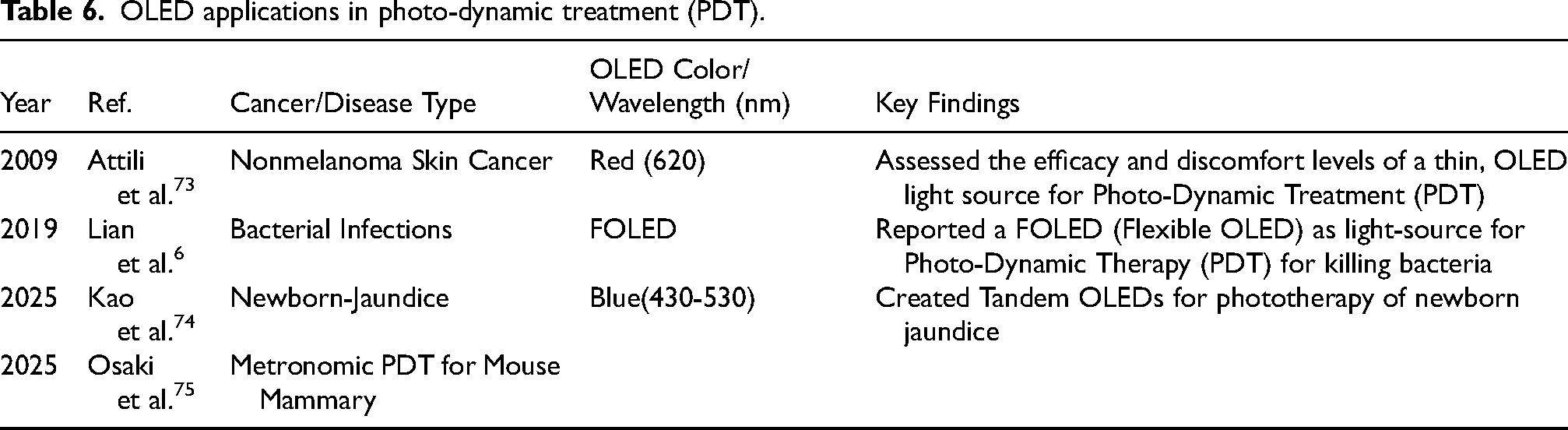
In 2012, Werner et al. 50 demonstrated a light-addressable potentiometric sensor that attained high resolution and improved operational speed. In the same year, 51 documented the manufacture of OLED-OPD on a single-substrate, which decreased development costs and improved performance. Sim et al. 76 suggested the OLED catheter’s potential for treating type 2 diabetes and other diseases. Researcher created a flexible OLED catheter for internal Photo-Biomodulation (PBM) in tubular organs. They tested it on diabetic rats, found that red light delivered via the catheter reduced hyperglycemia, insulin resistance, and liver fibrosis.
In 2018, Lee et al. 77 combined the second generation red, green OLEDs with a small-molecule Organic Photo-Diode (OPD) as onto a flexible substrate, OLEDs and OPDs are used to measure pulse rate by detecting reflected light from blood vessels.
Author optimized the sensor architecture by analyzing light propagation in the skin based on the OLED emission spectra. 77 The monolithic integration of organic devices demonstrates their potential for enabling all-day wearable health monitoring systems due to their form factor advantages and energy efficiency.
Furthermore in 2025, Kao et al. 74 created tandem-OLEDs that include BCzVBi and FIrpic emitters to provide a wide blue emission spectrum (430–530 nm) designed for phototherapy of newborn jaundice. 74 In same year, Osaki et al. 75 compared the therapeutic effects of 5-ALA-mediated metronomic photodynamic-Therapy (mPDT) using red (620nm) and green (542nm) light. While green light produced more localized ROS, red light penetrated deeper, leading to greater overall tumor suppression in vivo. The findings suggested that red light is more effective for 5-ALA-mPDT despite lower singlet oxygen concentration. 75
Additionally, Tiwari et al. 4 focused on the clinical applications of Photodynamic Therapy (PDT) using OLEDs for cancer treatment and diagnosis. The study examined various tumor cases treated with PDT, including its use against antibiotic-resistant infections in diabetic ulcers and the benefits of low-intensity light exposure. The key factors are presented in Table 7 elaborated that the cost factor is high only in case of phosphorescence (2-generation), while light conversion efficiency is low only in case of fluorescence (1-generation).
Impact of OLED generations in PDT applications.

Furthermore, it explored the potential advantages of combining PDT with NIR-OLEDs for improved tumor targeting. 4 The performance and quality of OLED-PDT are determined by both the technology and generating method.
Limitations of light penetration in human tissues during PDT
The penetration of light in human tissues significantly restricts the efficacy of PDT treatment, especially for deep-seated malignancies.
78
This constraints poses a significant challenge to the treatment of internal organs and deep-seated tumors.
79
The impact of various OLED generations in PDT applicaions is depicted in Table 7. The challenges of light penetration in human tissues are as follows:
Overcoming light penetration limitations with OLED-based PDT devices
OLED-based PDT addresses the limitations of light penetration in human tissue by enabling direct and conformal light delivery, offering uniform illumination, and allowing for tunable wavelengths. These characteristics enable more effective treatment of deeper lesions where traditional PDT methods struggle.
Ultra-thin OLEDs have been implanted in animal models to treat deep organ tumors by providing therapeutic light straight to the tumor location.
55
This direct application minimizes energy loss over distance, a significant constraint for external light sources.
71
Itazaki et al.
55
developed a bio-compatible device, for PDT of deep organ cancers. This thin, lightweight device effectively eliminated rat liver tumors via apoptosis with continuous illumination and a photosensitizer, offering a promising new cancer treatment.
55
OLED as biosensor for disease identification
OLED, a subset of solid-state-lighting technology, utilized organic molecules to emit light when an electric current is applied. 47 Since their emergence in the late 1980s, OLED technology has seen significant improvements in device-stability, efficacy, and luminosity. 47 The inherent flexibility of the OLED makes them suitable for various applications, including biosensors for cancer detection and other biological monitoring. OLED-based biosensors function by converting biological events into measurable optical signals. For instance, the process by which glucose oxidase enzymatically oxidizes glucose can be detected through photoluminescence, enabling real-time monitoring of glucose levels, valuable for diabetes management and continuous glucose monitoring systems. 88
Furthermore, OLEDs can serve as both a light source and a detector in biosensing applications, facilitating precise interaction with biological samples and enabling real-time data acquisition. OLED technology has been utilized to detect various disease biomarkers, including antibodies to Human Papilloma Virus (HPV) proteins such as E2, E6, E7, that were relevant to cervical cancer and HPV-associated head, neck-cancer. 19 Additionally, it has been applied in the detection of immunoglobulin A and G antibodies, as well as other dye-conjugated antibodies. 41
The technology has also been explored for detecting biomarkers, that induces Severe-Acute-Respiratory-Syndrome (SARS) using Fluorescence-Linked Immunosorbent Assay. 41 In 2016, Smith et al. 89 explored a novel biophotonic therapy using flexible red OLEDs to stimulate the vagus-nerve in ear, offering non invasive and devoid of pharmacological intervention alternative to treat persistent illnesses, mental health disorders. The tests suggested the 620 nm OLED array can effectively triggered therapeutic responses in optogenetically modified neural tissue.
The main limitations of research were: 1) For deep brain insertion, current optogenetic treatments need invasive surgery. 2) Devices that stimulate the vagus nerve electrically are big and intrusive. and 3) Transducers that are implanted or transcutaneous lack accuracy for certain nerve branches.
In 2019, Jeon et al. 91 reported that Sandwich-Structure OLEDs (STOLEDs) are applicable in versatile disposable and non-disposable wearable medical devices. Their flexibility enables applications on textile and paper surfaces, and these devices are also foldable, washable, and durable.
In skin treatment, red STOLED light is known to increase cell proliferation, migration, and skin repair, thus enabling advances in phototherapy and wound healing. Thereafter, Neonatal jaundice, a common and potentially dangerous condition in newborns, is typically treated with LED phototherapy, which has drawbacks like dehydration and eye damage. 92 In 2022, Choi et al., 92 designed a textile based, blue color OLED that conforms the body. It was emit light at 470 nm, effectively reduced bilirubin levels in lab tests while operating reliably at low temperatures and voltages, suggested safer, wearable alternative for jaundice treatment. The wavelength and output power were the major limitations.
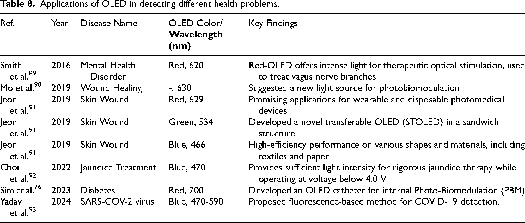
Furthermore, in 2023, Sim et al., 76 developed an OLED catheter for internal Photo-Biomodulation (PBM) to overcome the limitations of light penetration in phototherapeutics. The flexible, biocompatible catheter delivered uniform light via an encapsulated OLED, making it suitable for use in tubular organs. Table 8 demonstrates wavelengths used in various applications of OLED identifing various diseases. Yadav et al. 93 introduced a novel method to detect COVID-19 using a flexible OLED and an integrated Organic Photo-Diode (OPD) system. The technique relied on fluorescence detection within a human saliva sample. The blue OLED was emit light at a 470 nm wavelength to excite the sample, while the OPD measured the resultant fluorescence. Two distinct current levels from the OPD (63.5 mA and 37.2 mA) indicate either a COVID-19 infection or a healthy state, respectively. This OLED-OPD integration offers a potentially rapid and flexible diagnostic tool. OLED has the qualities to detect and monitor different biomarkers. 11
Applications of OLED in detecting different health problems.
The OLEDs have been explored for their potential in fluorescence imaging and photoacoustic imaging, which can offer significant insights into the tumor identification and survelliance of mallignancy. Many researchers have worked on cancer biomarkers used for various health monitoring system, few are depicted in the Table 9.
Various biomarkers used in health monitoring system along with detection limit.

The OLED may be used for cancer detection based on the Fluorescence Sensing System. A fluorescence sensing system compares the fluorescence intensity of a sample against a reference sample. 49 First, the system measures the reference sample’s fluorescence using OLED excitation and a photodiode. 96 Then, the sample containing the target analyte is measured using the same setup. The existence of the target analyte is ascertained by analyzing the disparity in fluorescence signals between the reference and sample. OLEDs, composed several thin organic layers between two electrodes, show promise in PDT. 4
In OLED technology by investigating innovative materials and designs for cancer detection and other diseases, researchers may develop a new non-invasive method that leads to the development of OLED further from display to sensors.
49
Cancer biomarkers that may be detected with OLED technology include antibodies directed against the E2, E6, E7 proteins of HPV-16.
19
These circulating biomarkers are important for the diagnosis of cervical cancers; a research showed that they may be detected using flat-panel OLED technique in conjunction accompained by protein-microarray methods.
19
This demonstrates the efficacy of OLED-based diagnostics in early cancer detection, as the multiplexed diagnosis of these antibodies is accomplished with clinical-level sensitivity.
Immunoglobulin G (IgG) antibody detection to various viral antigens utilizing organic light-emitting diode technology is another important example. The Detection limits for human IgG can be as-low-as 10-pg/mL, this method has shown exceptional sensitivity when applied to patient blood samples.
19
The capacity to conduct multiplexed studies helps in giving full diagnostic profiles,
19
and this high sensitivity is critical for detecting virally-linked malignancies.
Recent developments have shown that Organic Light-Emitting Diode Technology (OLED) has the ability to identify new biomarkers in liquid biopsies, including CTCs and circulating Tumor DNA (cTDNA).
97
Achieving non-invasive cancer diagnostics relies heavily on these biomarkers, which enable continuous tracking of tumor dynamics and treatment effectiveness. These new biomarkers have the potential to transform personalized cancer treatment, and OLED-based detection technologies provide a flexible framework for making them more sensitive.
When it comes to Hepato-Cellular Carcinoma (HCC), one of the well-established tumor markers that may be detected using optical, electrochemical, and mass-based biosensors for monitoring of Alpha-Feto-Protein (AFP).
98
Another biomarker that may be identified with OLED technology is Carcino-Embryonic Antigen (CEA), which has been the subject of much research for a number of malignancies, including colorectal and breast cancers.
67
It has been used for dtection the method may be floureRapid and sensitive detection of CEA levels in blood samples may be achieved with the integration of OLEDs in sensing devices.
67
This aids in diagnosis and the tracking of therapy responses. When used in this way, the small size and high sensitivity of OLEDs greatly aid in the development of efficient cancer management plans. The Figure 7 is highlighting the basic components for detecting breast cancer. Here, OLED is used as Source (which is used to excite light to patient sample and OPD as detector (which detects the extracted emission wavelength from sample of the patient. Generally, the fluorescence and phosphorescence properties of OLED may be used for the light excitation at source. By OLED-OPD integartion and measuring the current or calculating wavelength, we may detect whether person is suffering from breast cancer or not.

Process Flow of Breast Cancer Detection using OLED-OPD Integration. Here, OLED used as Source; Excite Light to Patient Sample and OPD as Detector; Detects the Extracted Emission Wavelength from Patient Sample.
Earlier in 2005, Hofmann et al. 101 invented a microchip to identification for urinary Human-Serum-Albumin (HSA) using thin film OLED as a source of excitation for detecting fluorescence. OLED emitted at 540 nm, excited the albumin blue 580 complex, which emitted 620 nm. They demonstrated that this microchip can detect HSA concentration down-to 10 mg/L, which is sensitive enough for Micro-Albuminuria (MAU) determination.
In 2011, Nakajima et al., 94 created a fluorescence-detection systems for micro-fluidic devices using an OLED as source of light and a CCD as the photodetector. They used this system in a microfluidic device to perform an enzyme linked immuno-sorbent assay to detect Immunoglobulin-A (IgA). The system achieved 16.5 ng/mL limit of detection for IgA, that was sufficient to evaluating stress, and it reduced analysis time and reagent/sample usage compared to traditional methods. Table 10 illustrates the comparison of OLED biosensors with established cancer detection techniques like ELISA, PCR and imaging-based modalities.
Comparison of OLED biosensors with established cancer detection techniques.

In 2016, FAN et al., 67 employed a portable device, Differential Pulse Voltammetry (DPV) for rapid, on-site tumor marker detection. The device, combined with micro-fluidic-paper-based system demonstrated enhanced sensitivity and minimal detection threshold. 67 This system allows for the automatic calculation of Carcino-Embryonic Antigen (CEA) concentration based on antibody-antigen binding. 67
In 2014, Prabowo et al.
95
developed an OLED based movable Surface-Plasmon-Resonance (SPR) sensor, enhanced by Brightness Enhancements Film (BEF) and a Giant Birefringences Optical (GBO) for increased intensity of light and polarization. Hence, the sensor achieved the detection limit RIU
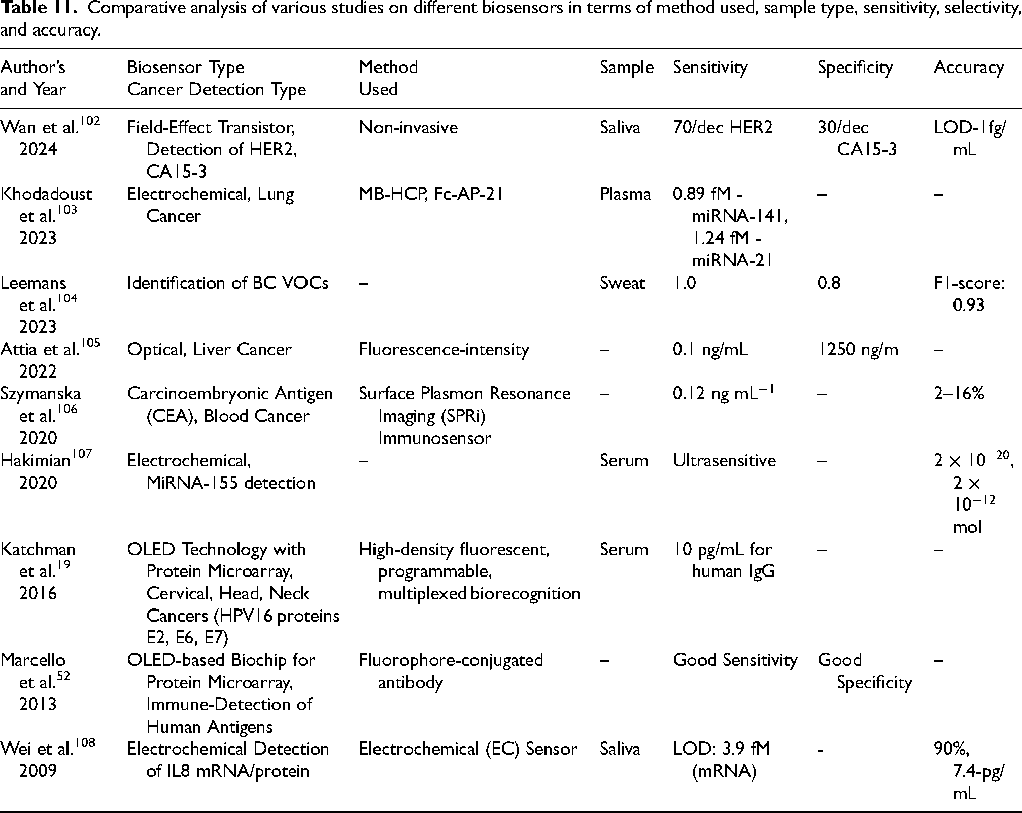
Further in 2022, Lian et al., 41 a Fluorescence Linked Immuno-sorbent Assay was developed for point-of-care testing (POCT). The system used Phyco-Erythrin (PE) as a fluorescent dye and detects antibodies to SARS CoV2, in human-serum. The researcher investigated OLED design to diminish background noise and improve sensitivity for immunodiagnostics. 41 Table 11 depicts the comparative analysis of different researches on biosensors.
Comparative analysis of various studies on different biosensors in terms of method used, sample type, sensitivity, selectivity, and accuracy.
Lian et al. 57 created a small DNA detecting device labeled with dye, OLEDs utilized as source of excitation. The apparatus comprises an OLED light source, an excitation filter, and an emission filter. 57 OLED biosensors have remarkable sensitivity, facilitating the identification of cancer biomarkers. Hence, OLED-based sensors can be designed to detect key cancer biomarkers, leveraging the optical properties of OLEDs to enhance diagnostic accuracy and efficiency.
Multiplexed detection techniques of OLED-based biosensor
A single OLED device can be configured to detect multiple types of cancers simultaneously through multiplexing techniques. 19 This is accomplished by combining several OLEDs in a flat-panel display array with thin film photodiodes, where each OLED/photodiode pair is linked to a particular biomarker identification site, allowing high-density fluorescence detection of different biomarkers in a single patient sample. 19 The incorporation of programmable high-density fluorescent protein microarrays facilitates the concurrent examination of many circulating cancer indicators. 19 Multiplexing in biosensors, particularly those utilizing OLED technology, enables the concurrent detection of numerous biomarkers. 19 Because OLEDs do not require epitaxial growth, it is possible to design adjacent color pixels on the same substrate, allowing for the excitation of several dyes for biosensing. 57 A variety of solutions are utilized to accomplish multiplexing in biosensors i.e., time-division, frequency division, spatial, barcode multiplexing techniques. For OLED-based systems, spatial multiplexing may be achieved by employing an array of OLED pixels combined with sensing element. 109
The techniques illustrated in Table 12 facilitates the development of a unified OLED biosensor platform capable of quickly executing multiplexed cancer detection, providing benefits in sensitivity, specificity, cost-effectiveness, and compactness relative to traditional approaches.19,110 Moreover, forthcoming advancements seek the monolithic integration of OLEDs and organic photodetectors on a singular substrate, perhaps facilitated by inkjet printing, resulting in compact, flexible, and entirely organic integrated sensor platforms. 19
Different multiplexing techniques used in various applications of OLED biosensor.

Comparative analysis of OLED-based detection technique with gold standard techniques
Currently, The gold standard for ovarian cancer diagnosis is based on a histopathology examination. The current gold standard for first screening is a combination of a Trans-Vaginal Ultrasound (TVUS), a CA-125 blood test. 113 As for determining the full scope of the disease, surgical staging is still considered the gold standard. Additional comprehensive clinical data and comparisons with gold standard procedures are clearly required, although fluorescence-based technologies, especially those OLEDs, demonstrate promise for non-invasive and fast detection.36,114 Biomarker assessment, mammography, colposcopy, and computed tomography scans are some of the current cancer diagnostic tools; nevertheless, they can be site-specific, invasive, and costly. 36
A thorough evaluation of fluorescent urine analysis’s performance should be conducted in comparison to these well-established methods, even if it may provide a straightforward, general, and non-invasive approach. 36 Although considering limitations, the sensitivity of ELISA could vary depending on the type and reagents of the test, and certain PCR detection techniques may have limited specificity due to contamination or illegal transcription. 41 It is critical to overcome these constraints by making sure that the upcoming fluorescence-based technologies deliver equivalent or better performance. 41
Moreover, more work is required to improve the sensitivity of OLED-based FLISA, decrease the amount of reagents needed, and allow multiplexed testing, but it has the potential to identify antibodies at concentrations far lower than current lateral flow tests. 41 The accuracy of the sensing data might be compromised by problems such as excessive OLED leakage light, especially when the analyte concentration is low. 41
Disease detection using cutting-edge diagnostic methods like quantitative polymerase chain reaction (qPCR) and Enzyme-Linked Immunoabsorbent Assay (ELISA) is frequently costly, arduous and protracted. 57 This indicates that it is not accessible in primary care. The disparity in healthcare resources between developed and developing nations restricts access to modern diagnostics for individuals in underdeveloped countries. 57 Consequently, it is advantageous to utilize technologies that are cost-effective to manufacture and implement, portable, provide prompt feedback on outcomes, while preserving the device’s sensitivity and robustness without compromise. 57
The comparative analysis of OLED-based detection techniques against gold-standard techniques is illustrated in Table 13. OLEDs are being investigated for fluorescence detection in urine, specifically for ovarian cancer 49 ; however, no direct comparisons with biopsy for cancer diagnosis have been identified. 49
Comparison of OLED-based cancer detection techniques with gold standard methods, including merits and demerits of used techniques.

Conclusion
This paper covers progress in cancer diagnosis and therapy utilizing OLED-based technologies, focusing on biosensor architecture, components, and the efficacy for organic- semiconductors in cancer detection. OLED application used in ovarian and skin cancer detection and photodynamic therapy, highlight their adaptability and promise to improve diagnostic accuracy. The use of OLEDs as biomarkers to identify various medical conditions demonstrates their use beyond cancer detection, as they provide greater sensitivity, real-time monitoring, and non-invasive diagnostic capabilities.
OLED-based biosensors have great potential for early cancer diagnosis, operating as light sources and detectors for non-invasive monitoring of cancer biomarkers, enhancing accuracy and efficiency when compared to traditional diagnostic approaches. The sensitive, flexibility, low-power consumption of OLEDs allow for the development of portable, cost-effective tools suitable for diagnostics and widespread screening, with future research focusing on optimizing OLED technology for various tumor types and investigating novel applications in early cancer detection to improve survival rates.
Moreover, the use of OLEDs enables the advancement of portable and economical diagnostic instruments, thereby increasing accessibility to essential healthcare services worldwide. Subsequent investigations ought to concentrate on improving OLED technology for various tumor types and exploring novel applications in early cancer detection, ultimately shaping the landscape of cancer diagnostics and improving survival rates through timely intervention.
Footnotes
Compliance with ethical standards
The author declares that all procedures followed were in accordance with ethical standards.
Consent to Participate
The author declares their consent to participate in this research article.
Consent for Publication
The author declares their consent for publication of the article on acceptance.
Author Contribution
Authors have made substantial contributions to the conception and design or acquisition of data, analysis, and interpretation of data; have been involved in drafting the manuscript or revising it critically for important intellectual content; and have given final approval of the version to be published. The author has participated sufficiently in the work to take public responsibility for appropriate portions of the content. The author read and approved the final manuscript.
Funding
The author(s) received no financial support for the research, authorship, and/or publication of this article.
Declaration of conflicting interests
The author(s) declared no potential conflicts of interest with respect to the research, authorship and/or publication of this article.