
Abstract
Fungal spores are dispersed through the air but settle and germinate on moist surfaces to form fungal hyphae and conidia, which can develop, digest organic matter, produce mycotoxins, and pose human health concerns. In this study, the hydrophobic and superhydrophobic coatings based on cyanoacrylate adhesive, 1H,1H,2H,2H-perfluorodecyltriethoxysilane (HDFS), 1H,1H,2H,2H-perfluorooctyltriethoxysilane (ODFS), SiO2 and ZnO nanoparticles (NPs) were fabricated and evaluated for their wettability as well as the resistance against molds. All coating samples #1 - #4 were resistant to mold growth when tested by pipetting the spore suspensions on the coating surfaces. In particular, sample #1 composed of cyanoacrylate adhesive, ZnO NPs, and ODFS exhibited the best anti-mold effect against Aspergillus and Penicillium species in fungal spore exposure test. Wettability tests demonstrated that coating sample #1 displays the highest value of water contact angle 159.6° and its contact angle hysteresis does not exceed 8o. Our findings pointed out, for the first time, that there were no mold hyphae colonized on the tested coating surfaces; only the mold spores adhered to the top of the textured structure, but no spore germination was observed. Notably, neither Penicillium sp. nor A. niger spores were able to penetrate inside the porous textured structure of all coatings.
This is a visual representation of the abstract.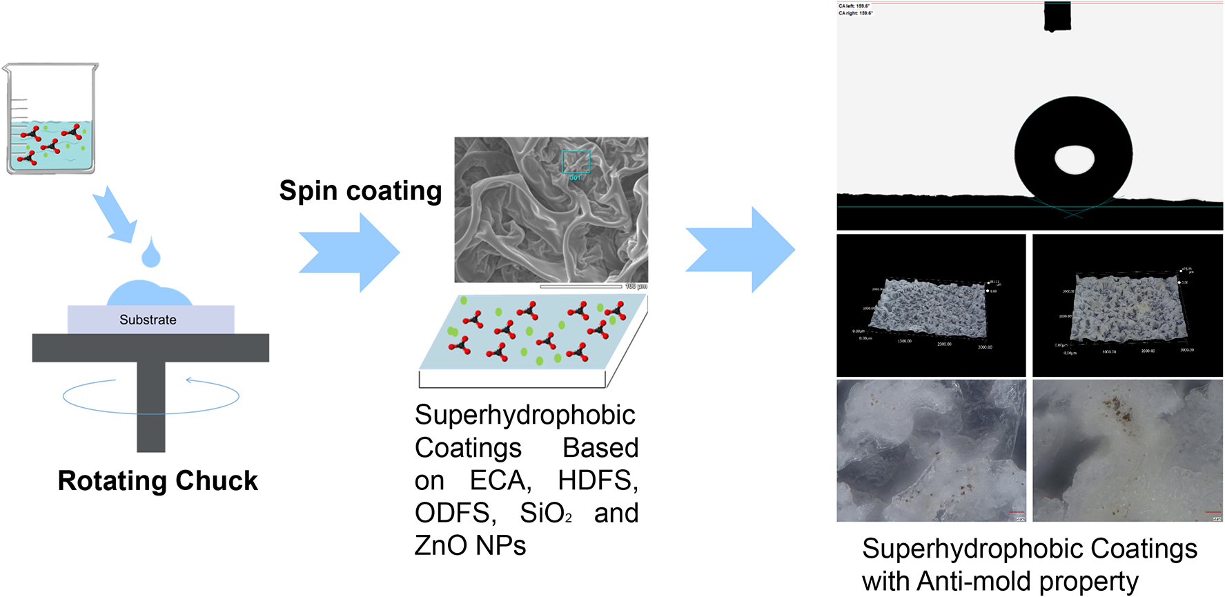
Keywords
Introduction
Species of Aspergillus and Penicillium are mold fungi that grow both indoors and outdoors. In the tropical climate of Vietnam, they cause many concerns to human health and destroy not only the agricultural products such as postharvest products maize, rice, beans, and fruits but also constructing materials and furniture. When the right conditions are met, mold can grow on various material surfaces, producing large amounts of spores, allergens, cell fragments, endotoxins, mycotoxins, and volatile chemical compounds.1–3 Aspergillus niger and A.flavus and Penicillium species are found to be common molds in the Northern parts of Vietnam.1,2 Fungal spores are dispersed through the air but land and remain on surfaces on which they can germinate into fungal hyphae and conidia. 2 Even in cases when there are no obvious water leaks, homes, and buildings can have mold spores through a variety of channels, including seepage through cellar floors and foundation walls, dehumidifiers and air conditioners, wet bathrooms, and potted plants. Mold spores are in the air and can develop and digest whatever they land on a damp surface. 3
In the past, antifungal coatings have been made using organic mold inhibitors (4,5-dichloro-2-octyl-isothiazolone, methylisothiazolinone, chloromethylisothiazolinone, triclosan, chlorothalonil, zinc pyrithione, IPBC (iodopropynyl butylcarbamate)) and inorganic aqueous preservatives (chromate, copper naphthenate, copper oxide, arsenate, tributyltin). 4 These antifungal substances are generally toxic to humans and cause harmful environmental effects.3–5 In 2013, the European Commission proposed concentration limits for 3-iodo-2-propynyl butylcarbamate (IPBC) from 0.06 to 0.45% and zinc pyrithione (ZPT) from 0.025 to 0.25% in dry films and outdoor wood paints/varnishes (Commission Decision (EU) 2014/312/EU). 4 Several main strategies have been currently used to create antifungal coatings on the surface of materials for various functional purposes: 1. Creation of fungicidal surfaces that include an active antimicrobial component; 2. Obtain coatings with their own antifungal activity; and 3. Formation of coatings that prevent the adhesion of fungi to the surface.5–10 In connection with the increase in the resistance of fungi to fungicidal compounds, an important direction is to obtain antifungal superhydrophobic coatings. The water contact angle (WCA) for these coatings is more than 150°, and the wetting hysteresis is less than 10°. 9 Resistance to biofouling and fungicidal activity of their surface is due to the “hanging” of a water drop containing harmful microorganisms on microrelief irregularities with low surface energy. 9 Under such conditions, the drop has a quasi-spherical shape and rolls down from the surface even with a minimal inclination, preventing the adhesion of microorganisms and the formation of a biofilm on it.9,10 To reduce the harmful chemicals and materials and resistance of fungi to fungicidal compounds in paints and coating surfaces, superhydrophobic materials with self-cleaning and anti-mold properties have been developed and used in many countries and various weather regions.5–9 The contact angle is a fundamental property that characterizes the interaction between a liquid and a solid surface. Measuring the contact angle provides valuable insights into the surface properties of materials, including wettability, adhesion, and surface energy. 9
Natural objects such as lotus leaves, rice leaves, rose petals, peanut leaves, butterfly wings, water striders, fish scales, gecko feet, and shark skin all exhibit superhydrophobicity.9–11 The most well-known example is the lotus leaf, which has a hierarchical roughness that contributes to its superhydrophobic and self-cleaning characteristics. This self-cleaning occurrence is referred to as the “Lotus effect” (Nelumbo nucifera). The natural characteristics served as an inspiration for synthesized materials that are created to have properties as superhydrophobic and self-cleaning. 11 In the superhydrophobic research approach, the coating technology plays an important role in material protection in the coastal zone, indoor environment, or under tropical weather conditions. 9 Submarines and other various types of ships should be faster, quieter, and use less fuel due to the water-repellent coating with superhydrophobic materials.10,11 Superhydrophobic coatings can be applied to new or old buildings to repel water, dry fast, reduce dirt, and prevent the growth of mold and mildew. Besides, superhydrophobic coatings can be applied to leather, wood, and fabric in both tropical and indoor environments; they offer protection from water, stains, dirt, oil, pollutants, UV rays, corrosion, oxidation, mildew, and mold resistance.7,12
To make superhydrophobic surfaces on steel, the coating materials such as low surface free energy substances HDFS or ODFS combined with SiO2 NPs were previously investigated and coated on steel grade AISI 430 by a spin coating method. 7 In general, superhydrophobic coatings have abrasion resistance properties, excellent thermal stability, and good acid and alkali resistance properties. 13 Xia et al. used a sol-gel technique to create a superhydrophobic coating of 1H,1H,2H,2H-perfluoroalkyltrichlorosilane (PFDS)-SiO2 on a Eucalyptus wood substrate, giving a large water contact angle (159°). 14 Çakır et al. fabricated superhydrophobic surfaces with 1H,1H,2H,2H-perfluorooctyltriethoxysilane coated on aluminum substrates, giving rise to properties such as moderately glossed surface, low wear rate, and hydrophobicity. 15
This study aims to fabricate the superhydrophobic coatings from cyanoacrylate adhesive, HDFS, ODFS, SiO2 and ZnO NPs, investigate surface morphology and hydrophobicity, and evaluate their resistance against the colonization of molds. The research results open up the potential of coatings with nanoparticles to use in the environment with mold occurrence or under tropical weather conditions.
Materials and methods
Materials
1H,1H,2H,2H-perfluorodecyltriethoxysilane (HDFS) with a purity of 97%, 1H,1H,2H,2H-perfluorooctyltriethoxysilane (ODFS) with a purity of 98%, nanoparticles (NPs) of SiO2 (particle diameter ∼ 10 nm), ZnO NPs (particle diameter <40 nm), triethylamine and hexafluorobenzene (purity 99%), isopropyl alcohol (IP; purity 98%) were purchased from Sigma-Aldrich and used without further purification. Commercial glue “NAVR 505” (Belarus) based on ethyl cyanoacrylate (ECA) was used as cyanoacrylate adhesive. Plates of steel grade AISI 430 (Ra∼0.025 μm, Rz∼ 0.190 μm) with a size of 10 mm × 15 mm were used as substrates for coatings.
Experiment apparatus and procedure
Preparation of samples
Fabrication of coatings
Fabrication coatings with SiO2 nanoparticles: To form the layer, SiO2 NPs suspensions in ECA glue were distributed over the surface of the substrates by static spin coating for 2 min at a rotation speed of 3000 r/min. Previously, SiO2 NPs were dispersed in ECA glue for 5 min at 35 kHz to obtain a suspension with a mass fraction of SiO2 6.6 wt%. Prepared layers of ECA with SiO2 NPs were treated with triethylamine fumes for 1 min to speed up the polymerization process of ECA. Then the obtained coatings were treated with oligomers HDFS or ODFS by dynamic spin coating for 30 s at a rotation speed of 3000 r/min using their solutions in hexafluorobenzene with a concentration of 25 mg/mL. 16
Fabrication coatings with ZnO nanoparticles: To form the layer of ZnO NPs suspensions in IP (5 mg/mL) were distributed over the surface of the substrates by dynamic spin coating for 10 s at a rotation speed of 3000 r/min. This operation was repeated 100 times. Then, a layer of glue was formed on the ZnO layer by static spin coating for 2 min at a rotation speed of 3000 rpm and was treated with triethylamine fumes for 1 min. The formed coatings were treated with oligomers HDFS or ODFS, similar to the coatings containing SiO2.
The coating samples were coded as #1: ZnO-ODFS; #2: ZnO-HDFS; 3#: SiO2-HDFS; and 4#: SiO2-ODFS, respectively.
Characterization of coating surfaces
IR Spectroscopy
Fourier transform infrared (FTIR) spectroscopy measurements were recorded on a Fourier transform infrared spectroscopy (NICOLET IS10, Thermo Scientific, USA) with 32 scans at a resolution of 4 cm−1.
Morphology analysis
The surfaces of treated coatings were evaluated using scanning electron microscopy (SEM; JSM-6510LV and JSM-6000 Jeol, Japan). The steel substrates were coated with a thin evaporated layer of gold to improve the conductivity prior to analyses.
Energy-Dispersive X-ray analysis
Energy-Dispersive X-ray (EDX) analysis is used to analyze the types and the quantity of elemental composition of a sample. The EDX spectrometer EX-230**BU, along with the JSM-6000 SEM Microscope, was used for detecting and analyzing the elemental composition on the surface of the coating samples.
Surface thickness and roughness evaluation
Film thickness measuring instrument German EPK MiniTest 600 device was used in the study. The two primary methods used by this device to measure film thickness were Eddy current method for non-conductive coatings on non-ferromagnetic metal substrates and Magnetic induction method for non-conductive coatings on ferromagnetic metal substrates.
Roughness measurement values were selected as follows: 2.5 mm for the L-filter (λc) and 8 μm for the S-filter (λs). The section length in accordance with standard STN EN ISO 21920–3 (2022) was 12.5 mm for the evaluation length and 17.5 mm for the overall traverse length, which is five times the value of λc. The digital microscope Keyence VHX-970 (Japan) was used as the roughness measurement tool. In the microscope, the VHX-H5 M software tool was utilized to measure roughness. Each image took between five and six minutes to sew together. The size of the generated 3D image was 18 × 18 mm (2880 × 2160 pixels). Then, following the STN EN ISO 21920–3 (2022) standard, the measurement was performed by simply translating lines with a set length or vertical or horizontal profile lines. The Keyence microscope was used to assess the Ra, and Rz parameters independently for each of the five section lengths in accordance with the technical standard.
Contact angle measurement
The instruments (Contact angle measuring instrument OCA 50, DataPhysics Instruments, and DSA100E Kruss, Germany) employed the sessile drop method to measure contact angles, which were measured by means of computer-aided analysis. The water contact angle hysteresis of superhydrophobic samples was defined as the difference between the advancing contact angle and the reducing contact angle. To measure the advancing contact angle, a water drop of 3 µl was placed on the sample, and then its volume was increased to 5 µL. 17 The reducing contact angle was determined after reducing the volume of this drop from 5 to 2 µL. 17
Mold isolation and identification
The molds were isolated by collecting the microorganisms on the material surfaces in indoor environmental conditions in Hanoi city in 2023. The moldy material samples collected in the indoor space were placed directly on the surface of the PDA agar medium containing chloramphenicol (100 µg/mL), carried out under sterile conditions, and incubated at 25°C. From the second to the third day, isolate the colonies that appear, and observe the color and basic morphology to separate each group. Subculture multiple times on the PDA medium to obtain pure mold strains. Every fungal colony in the petri dish was identified as a strain and labeled for future use and storage in slants. The isolated fungal strains, including P, A1, and A2, were subcultured to Petri dishes and incubated at 25°C. After 3–7 days of culture, the characteristics of the colonies of a fungal strain were assessed by microscopic methods.1,18
Microscopic identification of P, A1, and A2 strains according to the characteristics of the hyphal conidia with or without septa, color, smooth or spiny, shape, and color of the spores. The surface morphology of the colonies (granular, horizontal, or vertical mycelium), the color of the upper surface of the colonies (the color of the conidia formed on the surface of the colonies), and the color of the underside of the colony were observed by microscopy. Utilizing scanning electron microscopy (SEM - ZEISS EVO, Germany), the molds were also confirmed in their identification by the observation of their characteristic structures. Standard taxonomic methods were also used to identify these molds based on their micro- and macro-morphological features.
Resistance test of the growth of molds on the superhydrophobic coating surfaces
The bioassays with minor modifications and adapted from ASTM D5590-17 and D3273-16 were used to assess the efficiency of these four coatings to resist mold/ fungal growths.19,20
To determine the resistance of the tested coatings to fungal defacement by accelerated four-week agar plate assay (ASTM D5590-17, 2021), 19 samples #1 – #4 coated on steel substrates were spread out on Petri dishes that had been moistened with phosphate buffer solution (PBS). Then, spores of specific fungi, including A. flavus, A. niger, and Penicillium sp., were added to the plates as a suspension in PBS solution, with a density of 1 × 10⁶ cells/mL (determined by densitometric evaluation). A 100 µL of each spore suspension was pipetted onto the surface of each specimen with dimensions of 10 mm × 15 mm. The suspension spread evenly throughout the specimen surface, and the inoculated samples were incubated at 25 ± 2°C for 4 weeks while being kept at a high humidity level consistently. 21 The specimens were visually assessed for mold growth using the ASTM D5590-17 scale.
In order to assess the relative resistance of coatings to mold on superhydrophobic surfaces based on ASTM D3273-16, 20 the specimens were tested with molds Aspergillus niger, and Penicillium sp in a small environmental chamber under controlled conditions for over four weeks in a repeatable manner. The moisture level of the substrate and the coatings affects the severity and rate of mold growth on the surfaces. Test panel development requires a temperature of 25 ± 2°C and a relative humidity of 50 ± 5% in order to maintain sufficient moisture to facilitate mold growth. Rate the samples for mold growth for 4 weeks on a 0 to 10 rating scale by estimating the percentage of surface defacement (mold appearance area), where 0 represented total defacement and 10 represented no defacement. 20 Besides, after four weeks of testing, the digital microscope called the Keyence VHX-970 (Japan) was used to observe the adhesion of fungal spores on the coating surfaces, and microscope Olympus B061 (Olympus, Tokyo, Japan) was used to observe the mold appearance.
Results and discussion
Surface chemical composition analysis and morphological study
FT-IR spectra and SEM–EDX analyses were performed to verify the transformation of HFDS and ODFS. Figure 1 shows the FTIR spectra of ZnO-ODFS, ZnO-HDFS, SiO2-HDFS, and SiO2-ODFS obtained in the wavenumber range from 400 to 4000 cm−1. In Figure 1, the strong absorption bands at 1010 cm−1 and 855 cm−1 in the material samples were attributed to the Si-O-Si stretching vibration and Si-O bending vibration, respectively, and the peaks at 2851, 2920, and 2987 cm−1 were due to the symmetric and antisymmetric stretching vibrations of –CH. 22 In addition, the Zn–O bond was located in the range from 400 to 750 cm−1, specifically, the broad absorption bands at the regions of 460 cm−1 and 745 cm−1 can be related to the Zn-O and Zn-O-Zn stretching vibrations,23–25 respectively in the ZnO-ODFS, ZnO-HDFS materials. The peaks at 1241–1242 cm−1 and 1141–1151 cm−1 represented the C-F groups. The peaks at 1113 cm−1 for the surface-modified ZnO-ODFS, ZnO-HDFS, SiO2-HDFS and SiO2-ODFS were attributed to the C-F2 group. The peaks at 1350 cm−1 were related to the C-F3 vibration band.26,27 It can be confirmed that oligomers HFDS and ODFS are present on the surface and in the structure of coating samples #1 – #4. In fact, these chemicals are considered to make the coatings hydrophobic. Fluorine-containing coatings have been discovered to have a low surface energy; long fluorocarbon chains, such as HFDS and ODFS, can increase the hydrophobicity of the coating materials.9,15
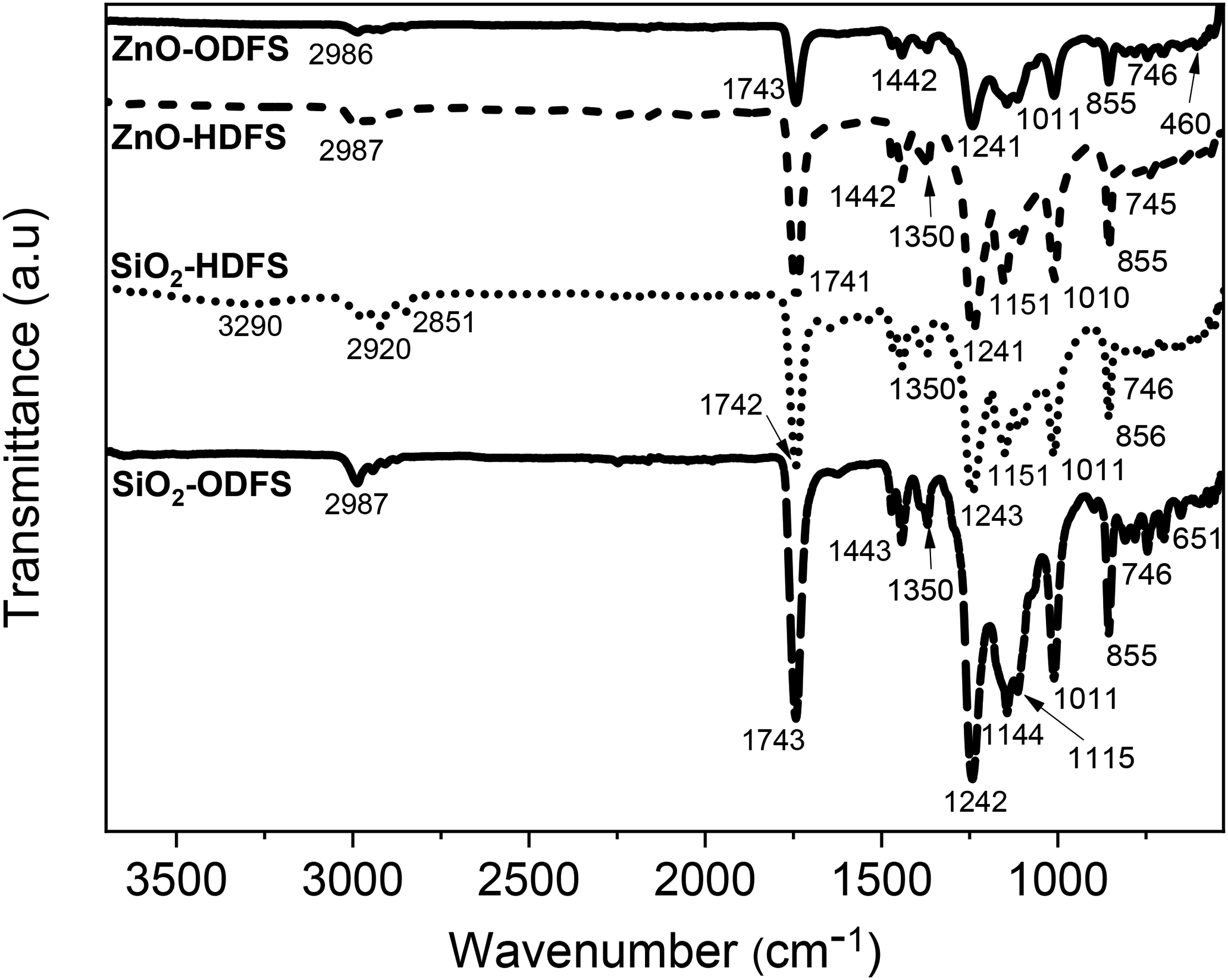
FTIR spectra of the four specimens ZnO-ODFS (#1), ZnO-DHFS (#2), SiO2-HDFS (#3), and SiO2-ODFS (#4).
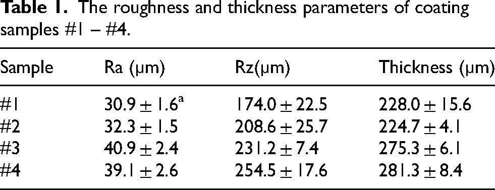
In order to better characterize the surface composition and morphological characteristics of the samples, we conducted experiments using SEM-EDX techniques. Figure 2 illustrates the EDX histograms of the coated surfaces of ZnO-ODFS, ZnO-HDFS, SiO2-HDFS, and SiO2-ODFS, respectively. The SiO2-HDFS and SiO2-ODFS samples consisted of five major elements, namely C, O, F, N, and Si; whereas the ZnO-ODFS and ZnO-HDFS samples additionally contained Zn, as shown in the SEM-EDX results (Figure 2). The SEM image of the hybrid coating containing SiO2 and ZnO shows a homogeneous morphology, and no cracks and fractures were observed on the coating surface. The samples had a porous-textured structure formed during the polymerization of ECA (Figure S1, Supplementary Information). Table 1 presents the parameters of roughness and thickness for the four coating samples #1 – #4. Samples #1 and #2 showed similar thickness values of 228.0 and 224.7 µm, respectively. Samples #3 and #4, which contained SiO2 NPs, showed thicker layers of coatings; the values were 275.3 and 281.3 µm, respectively. Ra and Rz values of #1 and #2 were also similar to each other; they were in the range of 30.9 to 32.3 µm for Ra, and 174.0 to 208.6 µm for Rz, respectively. Compared with coating samples #1 and #2, the roughness parameters of #3 and #4 were larger; they were in a range of 39.1 to 40.9 µm for Ra and 231.2 to 254.5 µm for Rz.
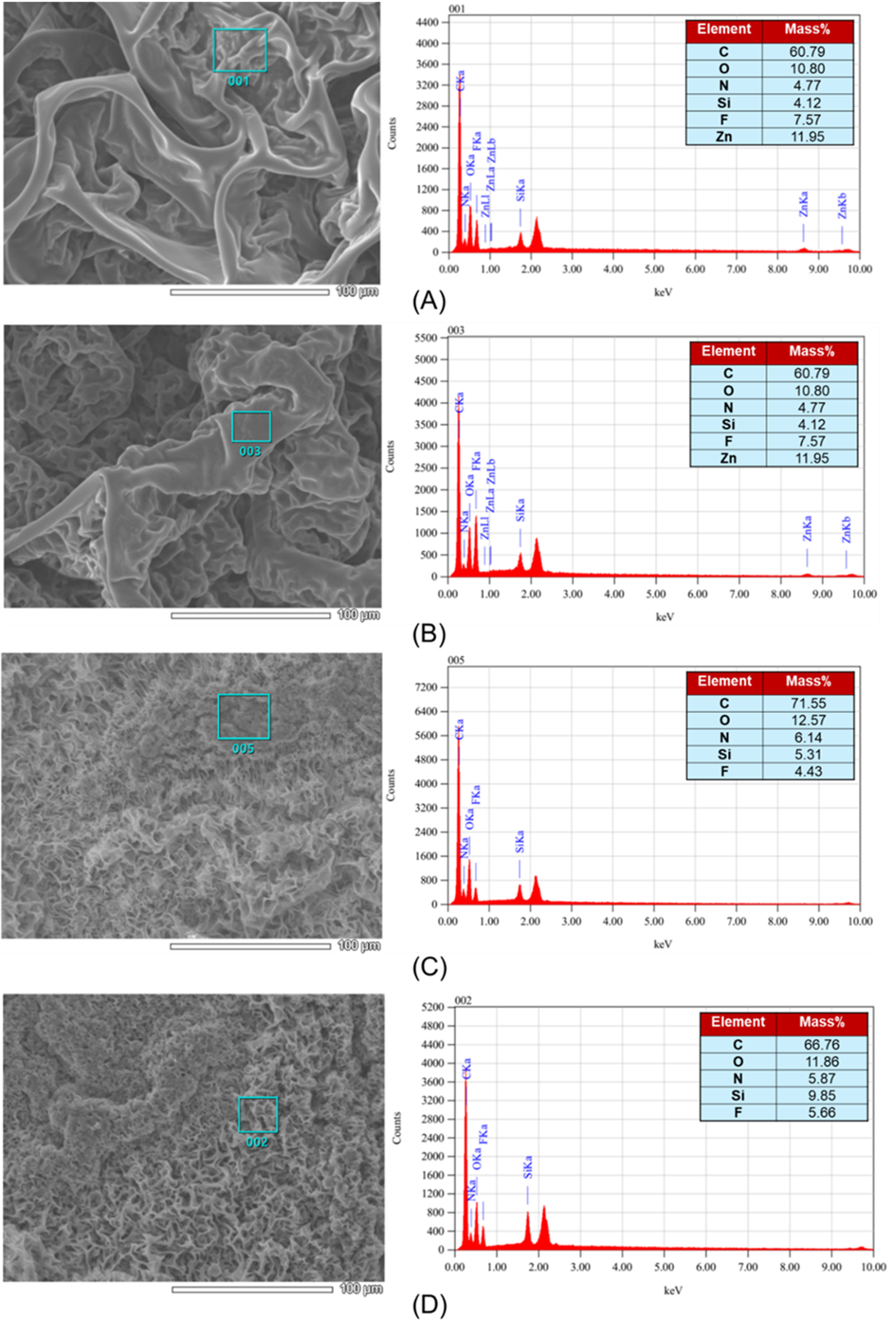
(a) XRD SEM images and EDX spectra of four samples. A: #1 (ZnO-ODFS); B: #2 (ZnO-HDFS); C: #3 (SiO2-HDFS); and D: #4 (SiO2-ODFS).
The roughness and thickness parameters of coating samples #1 – #4.
Wetability analysis
Water contact angle values are indicated for the hydrophobicity of the coating surfaces. In this study, sample #1 displayed the highest value of 159.6°, followed by samples #2 – #4 with values of 144.8, 151.9, and 140.4°, respectively (Figure 3). While uncoated steel surfaces were hydrophilic, demonstrating WCAs of 82.0° (data not shown). This implied that samples #1 and #3 possess superhydrophobic effects. Meanwhile, samples #2 and #4 had weaker hydrophobicity, and #4 indicated the weakest. The appearance of superhydrophobic properties was due to the synergy of micro-roughness induced with SiO2 or ZnO NPs and polymerized ECA, as well as the presence of the HDFS or ODFS oligomers, which lowered the free energy of the treated surface.9,28 Water contact angle values of the samples without treatment with oligomers of HDFS or ODFS did not exceed 120o. It was also revealed that the contact angle hysteresis of composite coatings with WCAs >150 o does not exceed 8o and their surface free energy is less than 6 mJ/m2, which also confirms their superhydrophobic nature.29–33 An increase in the content of ZnO or SiO2 in the coatings leads to their cracking or lowering WCA, and a decrease does not allow obtaining coatings with a WCA greater than 140 o.
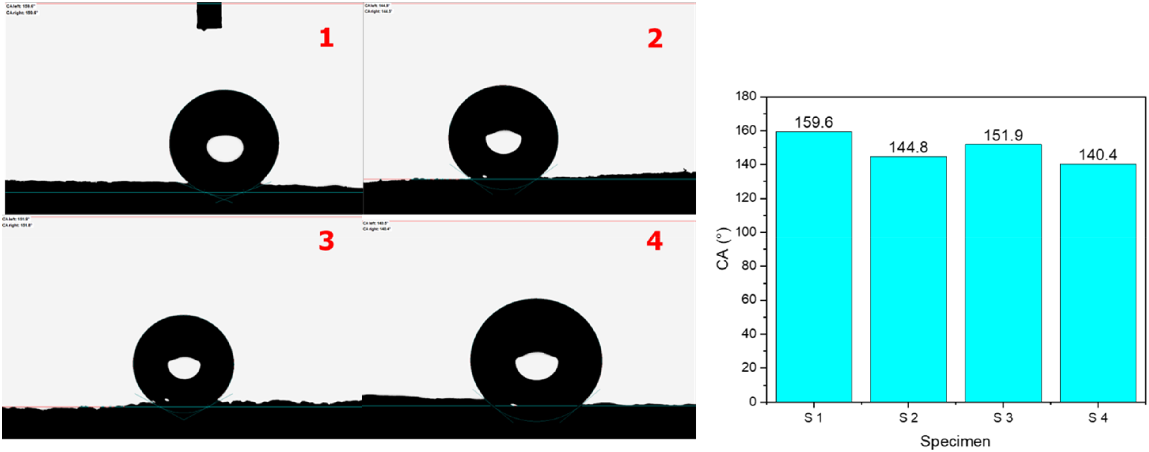
Contact angles of four coating samples #1 – #4.
Mold identification and resistance tests of coatings against mold growth
During the isolation of molds from tropical materials in Vietnam, three fungal isolates P, A1, and A2 were successfully identified as Penicillium sp., A.niger and A.flavus, respectively. Microscopic methods, including SEM had been applied to identify the isolated molds (Figure S2, Supplementary Information). SEM revealed the micromorphology of strain P: conidia were broadly ellipsoidal and connected into chains; conidiophores were strictly verticillate, with stipes having slightly rough walls. According to these observative results, strain P possesses the morphological features of species in the genus Penicillium. 18 Strain A1 belongs to Aspergillus species because it featured spherical conidia, black or black-brown clusters, and smooth or rough mycelia according to the microscopic observations. The SEM images of A1 indicated that the quasi-spherical form of A1 conidia (Figure S2 AN, Supplementary Information) was characterized by equinulated ornamentation on the cell wall surface. 34 When grown in PDA, the fungal morphology of A2 was identical to that of A. flavus; they had dark brown sclerotia and an olive-green tint with a whitish periphery. As shown in Figure S2 AF (Supplementary Information), A. flavus conidiophores also had spherical vesicles, globe-shaped conidia with surface imperfections, and irradiated and uniseriate conidial heads.
In the first type assay experiment, the resistance of four coatings against Penicillium sp., A. niger, and A. flavus was assessed according to ASTM D5590-17. In the anti-mold resistance test, the bioassay results demonstrated an excellent resistance of 4 coating samples against all test molds. After a 4-week assessment, all coating surfaces prevented the growth of Penicillium sp., A.niger, and A. flavus at the same rate of 0, at the inoculum of 106 CFU/mL (Figure S3, Supplementary Information). The results could be explained due to the PDB suspension of the mold spore could not have any adherence with coatings, and the droplets bringing fungal spore and hyphae rolled off the superhydrophobic coating surfaces. According to Lu et al., superhydrophobic surfaces have an inherent ability to clean themselves to make water droplets roll off them, collecting dust, debris, and even fungus spores. 17 This “lotus effect” is driven by the combination of low surface energy materials and surface roughness, which minimizes the adhesion of bacteria as well as fungal spores and hyphae to the surface. Huang et al. also explained that when the superhydrophobic surface is submerged in the bacterial solution, a water film forms spontaneously. 35 This can prevent the surface from coming into contact with the bacteria quickly and, to some extent, lessen bacterial adherence. Besides, the superhydrophobic coatings were self-cleaning, therefore they firmly prevented the accommodation of organic matter, dust, and moisture that are used as nutrients for mold, resulting in poor medium and hard growing conditions for fungi.
In the second type of assay experiment, the resistance of the four coating samples was assessed based on ASTM D3273-16, which is suitable for verifying the performance of superhydrophobic coatings because the uncontrolled fungal or mold development is frequently linked to the defacement of interior settings.20,31 Coatings that are mold-resistant due to superhydrophobic coating surfaces or antifungals were made to prolong the shelf life and improve the aesthetics of interior surfaces. Biocide chemicals, which prevent or hinder the growth of mold and fungus on surfaces, are used in the formulation of these antifungal coatings. Additionally, the utilization of superhydrophobic coatings could lead to reduced amounts of hazardous substances and volatile organic compounds (VOCs) that could be exposed to the atmosphere around.32,36
It is known that adhesion is important for mold spore anchoring, and for triggering germination and infection structure differentiation. 37 In the assay of coating surfaces for samples #1, #2, and #3, there was non-significantly adhesion of Penicillium spores. The coating samples showed the same rating scale of 8 based on ASTM D3273-16 with the defacement of 11.1, 13.0, and 15.5%, respectively (Figure 4(A), (B)). In contrast, coating sample #4 was affected severely by Penicillium spore binding, with a rating of 5 (46.2% of defacement). However, no fungal spore germination and no hyphae penetration, and growth of the mold inside the porous-textured structure of the coating was clearly observed by the digital microscope with a magnification × 1000 (Figures 4(B) (e-h)). This suggested that the walls inside the tubules seemed to have an electrostatic repulsion that repel the fungal spore adhesion.
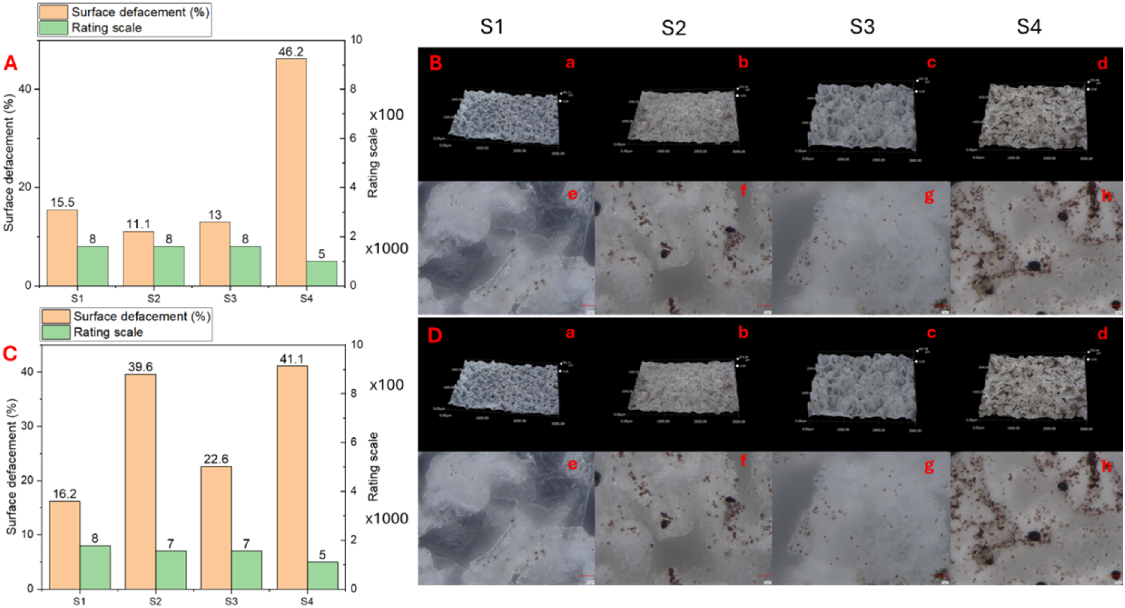
The adhesion of Penicillium sp. (A, B) and Aspergillus niger (C, D) spores on the coating surface of 4 coating samples. Magnifications: × 100 (a-d) and × 1000 (e-h). Rating scales and Surface defacement (%) were calculated according to ASTM D3273-16.
As for the adhesion of A. niger spores, coating samples #1 and #3 showed high resistance for this mold with a rating scale of 8 and 7, which were equivalent to the defacement percent of 16.2 and 22.6% respectively. In contrast, samples #2 and #4 were affected by 39.6 and 41.0% respectively (Figure 4(C)). The data of resistance of sample #2 was a change compared with the test of Penicillium sp., which could be due to the different fungal species with the different adhesive mechanisms of each mold. In summary, samples #1 and #3 showed good resistance against A. niger colonization. As for Penicillium sp. spore adhesion and growth, samples #1, #2, and #3 performed the best resistance. Even though sample #2 was resistant to Penicillium sp. but it seemed to lack effectiveness for A. niger spore adhesion. Besides self-cleaning and water-repellent properties that prevent the coating from mold growth, the resistance mechanism of sample #1 against molds tested may also relate to the generation of electrostatic (repulsion) charges that repel fungal spores. According to Chung et al. (2010), the adhesion force between spores and planar surfaces was significantly influenced by electrostatic forces. 38
By the observation of the spores adhered to the superhydrophobic coatings using a digital microscope with a magnification of ×1000 (Figure 4), the resistance effects of four coatings were also determined. Due to the artificial porous-textured structure and its tops (similar to papillae) on the surface of the coating, it made water repellency similar to lotus morphology. 11 All of the coating surfaces were resistant to the colonization of mold hyphae, and only the spores adhered to the top of artificial papillae, as shown in Figure 4(B) and (D). This phenomenon could be explained by the characteristic of coatings at which no nutrients and moisture on the coating surfaces and that prevents the spores from taking up moisture and starting the germination process. 39 Besides, the occurrence of ZnO NPs and ODFS, and HDFS that contain a high density of F in the molecular structure, plays a role as antifungal additives and helps to inhibit spore germination and hyphae penetration. Especially, ZnO NPs were known as a strong antimicrobial material that is widely used in the coating and paint industry.9,25 In our study, it was noteworthy that there was no single spore of Penicillium sp. or A. niger that could penetrate inside the porous textured structure of coatings. This implied a resistance mechanism that may relate to electrostatic repulsion for fungal spores. In the future, the chemical characterization of sample #1 and its morphology needs further investigation to maximize the other properties, such as UV, thermal stability, and anti-bacterial properties.
Conclusions
In summary, this study successfully fabricated the hydrophobic and superhydrophobic coatings based on cyanoacrylate adhesive, zinc and silicon oxides nanoparticles on the surfaces of steel substrates. The chemical composition and morphology of each coating were characterized by instrumental and microscopic analyses. Samples #1 and #3 displayed the superhydrophobic effects. Of them, sample #1 showed the greatest water contact angle at 159.6°. In contrast, samples #2 and #4 were hydrophobic coatings. All coating samples were resistant to the growth of molds Penicillium sp., A. niger and A.flavus when tested by pipetting spore suspension on the coating surfaces. In the test by exposing fungal spores directly to coating surfaces (ASTM D3273-16), sample #1 (ZnO-ODFS) displayed the best resistance property against mold spore adhesions of Penicillium sp., and A. niger. Coating sample #1 potentially reduced the mold spore adhesion on the top of its textured structure. Moreover, no spore germination or mold growth was observed on the coatings. In this study, for the first time, we observed that no Penicillium sp. or A. niger spores can penetrate inside the porous textured structure of all tested coatings. Our findings also suggested a potential coating composition (#1) with remarkable superhydrophobic and anti-mold properties that could be used for preventing various substrates from mold attachment.
Supplemental Material
sj-docx-1-mgc-10.1177_10241221251390816 - Supplemental material for Anti-mold properties of hydrophobic and superhydrophobic coatings based on cyanoacrylate adhesive, zinc and silicon oxides nanoparticles, and silicon organic compounds
Supplemental material, sj-docx-1-mgc-10.1177_10241221251390816 for Anti-mold properties of hydrophobic and superhydrophobic coatings based on cyanoacrylate adhesive, zinc and silicon oxides nanoparticles, and silicon organic compounds by Kieu Anh Thi Vo, Cuong Van Bui, Anh Son Nguyen, An Quan Vo and Thu Trang Thi Nguyen, Viktoryia Akulova, Ignat Chishankov, Dai Lam Tran, The Tam Le, Tuan Anh Nguyen, Aliaksandr Salamianski, Quang Le Dang in Main Group Chemistry
Supplemental Material
sj-pptx-2-mgc-10.1177_10241221251390816 - Supplemental material for Anti-mold properties of hydrophobic and superhydrophobic coatings based on cyanoacrylate adhesive, zinc and silicon oxides nanoparticles, and silicon organic compounds
Supplemental material, sj-pptx-2-mgc-10.1177_10241221251390816 for Anti-mold properties of hydrophobic and superhydrophobic coatings based on cyanoacrylate adhesive, zinc and silicon oxides nanoparticles, and silicon organic compounds by Kieu Anh Thi Vo, Cuong Van Bui, Anh Son Nguyen, An Quan Vo and Thu Trang Thi Nguyen, Viktoryia Akulova, Ignat Chishankov, Dai Lam Tran, The Tam Le, Tuan Anh Nguyen, Aliaksandr Salamianski, Quang Le Dang in Main Group Chemistry
Footnotes
Acknowledgements
This work was funded by project number QTBY01.07/23-24 from the VAST, Viet Nam; and X23B-001 and 2.15 from NAS, Belarus.
Ethical considerations
Not applicable.
Consent to participate
Not applicable.
Consent for publication
Not applicable.
Author contributions
Supervision: A.S, Q.L.D; Investigation: A.S.N, A.S, I.C, K.A.T.V, Q.L.D, V.C.B; Data curation: A.Q.V, A.S.N, I.C, K.A.T.V; Project administration: I.C, K.A.T.V, T.T.T.N; Validation: A.S, Q.L.D, T.L.T; Conceptualization: A.S, D.L.T, Q.L.D, V.A; Formal analysis: A.S, D.L.T, Q.L.D, V.A; Methodology: A.S, Q.L.D, T.A.N, T.L.T, V.A; Resources: A.S, Q.L.D; Writing – original draft: K.A.T.V, A.S, Q.L.D, T.L.T; Writing – review & editing: A.S, Q.L.D, T.A.N, T.L.T. All authors read and approved the final version of this manuscript.
Declaration of conflicting interests
The authors declared no potential conflicts of interest with respect to the research, authorship, and/or publication of this article.
Supplemental material
Supplemental material for this article is available online.
References
Supplementary Material
Please find the following supplemental material available below.
For Open Access articles published under a Creative Commons License, all supplemental material carries the same license as the article it is associated with.
For non-Open Access articles published, all supplemental material carries a non-exclusive license, and permission requests for re-use of supplemental material or any part of supplemental material shall be sent directly to the copyright owner as specified in the copyright notice associated with the article.