
Abstract
In an effort to correlate the likelihood of in utero transmission of Mycobacterium avium subsp. paratuberculosis (MAP), the causal organism of Johne’s disease, with the test status of the dam, tissues from neonatal calves borne to known test status cows were cultured for the presence of MAP. Tissues from a single calf borne to a test-positive cow shedding large numbers of organisms in the feces were positive for MAP. The detected overall transmission rate was approximately 2% (1/49), and the detected transmission rate in cows that were fecal culture positive and serum enzyme-linked immunosorbent assay suspect or positive was approximately 4.3% (1/23).
Johne’s disease is a bacterial disease of ruminants that is most commonly transmitted via the fecal–oral route, though in utero and milk-borne infection with the causal organism Mycobacterium avium subsp. paratuberculosis (MAP) are also accepted routes of infection. 6 A prior study found viable MAP in tissues of fetuses from 5 out of 28 (18%) clinically normal, heavy fecal shedder cows. 5 The fetuses from 30 clinically normal, fecal culture–positive cows (low or moderate shedders) were all negative for viable MAP. The serologic status of the dam, as tested by agar gel immunodiffusion, complement fixation, and in-house kinetics-based enzyme-linked immunosorbent assay (ELISA), was not associated with fetal infection. In a second study, 5 out of 392 fetuses (1.3%) from “non-randomly” selected cows at slaughter were positive for MAP. 3 The clinical and test status of these cows was not defined. Of the 392 cows sampled in the study, 20 had MAP cultured from ileocecal lymph nodes, and the 5 positive fetuses came from this group of 20 lymph node culture–positive cows. These studies indicate that the likelihood of in utero infection increases as the disease progresses in the dam. Other than the work cited earlier, however, there have been no studies to evaluate the likelihood of in utero infection occurring in a group of cows with a wide range of test results ranging from negative to high positive on a variety of test methods. 5
The current management recommendation for herds trying to minimize Johne’s disease is to avoid feeding milk and colostrum from test-positive cows, but recommendations regarding how to manage the calves born to those cows are vague. Typically, “retaining calves from known positive cows as herd replacements is discouraged.” 2 Unfortunately, such a recommendation does not provide clear guidance for decision making about whether or not to keep a calf born to a cow with a specific category of test results. The project described herein was performed in an effort to determine the likelihood of in utero infection of calves born to cows with a wide range of Johne’s disease test results.
The calves used in the current study were male Jersey calves born on a dairy that had been participating in the National Johne’s Disease Demonstration Herd Project since 2003. Cows in the herd had been routinely tested by fecal culture and serum ELISA at dry off, and historical test results were available. The herd had a fecal culture–positive rate of approximately 7.7% (Table 1). Over a 1-year period, tissues were collected from 49 calves and tested for the presence of MAP by culture or polymerase chain reaction (PCR). Animals of interest were male calves born to cows that had previously had serum ELISA a results >0.25 and/or positive fecal culture b and bull calves born on the same day to test-negative cows. Study personnel were notified by the dairy when appropriate calves were born, and the calves were collected for transportation to the California Animal Health and Food Safety Laboratory (CAHFS; Tulare, CA) within 24 hr of birth. The calves were euthanized with barbiturate solution upon arrival at CAHFS (UC Davis animal use protocol no. 07-12847). Because the removal of calves from the cow and the maternity pens immediately after birth could not be guaranteed, the abomasal and ruminal contents of the calves were evaluated grossly for the presence of milk or milk curd in order to verify the animals had not suckled. If any volume of white fluid or milk curd was found, the animal was removed from the study.
Test results from 1 full year of dry cow testing for Mycobacterium avium subsp. paratuberculosis from herd of origin for the animals in the current study.*
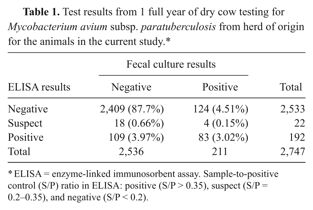
ELISA = enzyme-linked immunosorbent assay. Sample-to-positive control (S/P) ratio in ELISA: positive (S/P > 0.35), suspect (S/P = 0.2–0.35), and negative (S/P < 0.2).
After euthanasia, liver, kidney, spleen, ileocecal valve, ileocecal lymph node, 3 pools of mesenteric lymph nodes (proximal, middle, and distal), and 3 separate ileum samples, for a total of 11 samples, were harvested from each calf. Organ samples were collected first and intestinal samples last to minimize the chances of contamination by intestinal content. The samples were placed on ice immediately, and, when tissue collection had been completed, were frozen at −70°C until testing of the samples could be performed. In addition, postpartum milk, feces, and serum were collected from dams of calves included in the study. Samples were collected from the cows (within 14 days of calving on average), transported to CAHFS, and frozen at −70°C until testing of the samples could be performed.
Serum ELISA testing was performed according to the manufacturer’s instructions. a Serum samples (20 µl) were diluted with the sample diluent (380 µl) and were allowed to incubate at room temperature for 30 min. Diluted serum samples were placed into 2 individual wells of antigen-coated MAP antibody test kit plates and incubated for 30 min at room temperature. The liquid was removed from the wells, and the wells were then washed 4 times with phosphate buffered saline. Following the last wash, the plates were inverted and gently tapped on a paper towel in order to remove excess residual fluid. Horseradish peroxidase conjugate (100 µl) was placed into each well and incubated at room temperature for 30 min. The wash step was repeated. Tetramethylbenzidine substrate solution (100 µl) was placed in each well and incubated for 15 min at room temperature, after which time 100 µl of stop solution was added to each of the wells. The absorbance was measured at 620 nm, and the sample-to-positive control (S/P) ratio was calculated according to manufacturer’s instructions using the mean sample A620.
For fecal cultures, 2 g of feces was mixed with 35 ml of triple deionized water in a sterile 50-ml centrifuge tube and mixed on an orbital shaker at 150 rpm for 30 min. The tube was allowed to stand undisturbed at room temperature for a minimum of 30 min. Using a sterile transfer pipette or autopipettor, a 5-ml aliquot of sample was removed from the top portion of supernatant and added to a tube of half-strength brain heart infusion (BHI) broth with 0.9% cetylpyridinium chloride. The contents were mixed and allowed to stand undisturbed and upright for 18–24 hr in a 35–37°C incubator. The following day, the tube was centrifuged at 3,000 × g for 20 min. After centrifugation, 1 ml of thawed in-house antibiotic mixture (amphotericin, nalidixic acid, and vancomycin) in half-strength BHI was added to the pellet, vortexed to resuspend the material, and then incubated at 35–37°C overnight. The next day, 1 ml of sample was transferred into a culture broth bottle with supplements. b The bottles were incubated for 6 weeks and were monitored for growth by measurement of the headspace pressure. Following incubation, all samples were tested for the presence of MAP by acid-fast staining and PCR for IS900 using methods described previously. 1
Cows from which calves were collected were classified based on historical test results and test results from samples collected after freshening (Table 2). Twenty-one out of 49 were positive by both fecal culture and serum ELISA, 2 were fecal culture positive and suspect on serum ELISA, 5 were positive by fecal culture alone, 1 was positive by serum ELISA alone, and 20 were negative on both test methods. Of the 5 cows that were fecal culture positive but ELISA negative, all had days to positive of 42 or more. Of the 28 fecal culture–positive animals, 6 would have been considered high fecal shedders (<21 days to positive), 4 would have been considered moderate fecal shedders (21–28 days to positive), and the remaining 18 were low or very low fecal shedders (>28 days to positive). The single cow that was ELISA positive but fecal culture negative had an S/P ratio of 0.781 on one sample date, but 2 subsequent ELISA values were 0.168 and 0.058.
Distribution of cumulative testing results for Mycobacterium avium subsp. paratuberculosis for dams of calves used in the current study.*
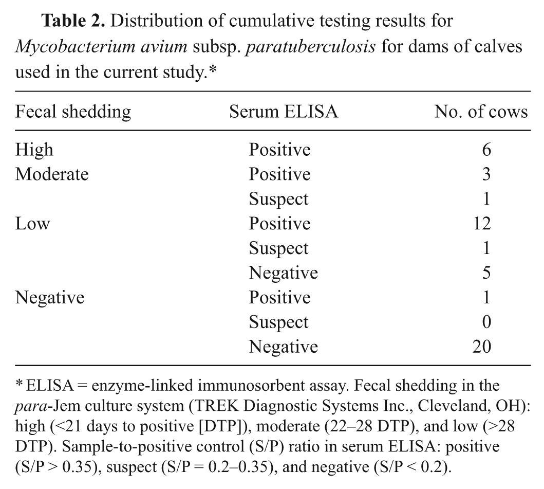
ELISA = enzyme-linked immunosorbent assay. Fecal shedding in the para-Jem culture system (TREK Diagnostic Systems Inc., Cleveland, OH): high (<21 days to positive [DTP]), moderate (22–28 DTP), and low (>28 DTP). Sample-to-positive control (S/P) ratio in serum ELISA: positive (S/P > 0.35), suspect (S/P = 0.2–0.35), and negative (S/P < 0.2).
Testing of calf tissues by culture was performed by the Johne’s Research Laboratory at the University of Pennsylvania (New Bolton Center, Kennett Square, PA). Tissue cultures were performed using methods similar to those described previously. 4 Briefly, 2 g of tissue were added to 25 ml of 0.7% hexadecylpyridinium chloride and homogenized with a stomacher for 2 min. The material was poured into a sterile 50-ml screw-top falcon tube and were left to stand at room temperature for 30 min. Fifteen milliliters of supernatant was removed from the top of the fluid phase, placed in a second falcon tube, and left to stand at room temperature for 3 hr. The sample was centrifuged at 900 × g for 30 min. The pellet was resuspended in 1 ml of an antibiotic mixture (amphotericin, nalidixic acid, and vancomycin) and incubated overnight at 37°C. Following incubation, the sample was vortexed and then inoculated into either 2 flasks of Herrold egg yolk medium or liquid-media tubes. c Liquid-media tubes were incubated for 56 days or until they flagged positive. The flasks were incubated at 37°C for 16 weeks, at which time total colonies were recorded. Colonies from solid media were confirmed as MAP by acid-fast staining, subculturing to determine mycobactin dependence, and by commercial PCR. d Positive liquid-media cultures were confirmed by PCR using the same commercial protocol.
Mycobacterium avium subsp. paratuberculosis was detected in low levels in the tissues from only 1 calf. Two of the ileum samples as well as the ileocecal valve had MAP detected in liquid media at 38, 41, and 46 days of incubation, respectively, and confirmed by PCR. This calf was born to a cow with a fecal culture at freshening that was positive at 16 days incubation indicating a high level of fecal shedding. The cow was also positive by serum ELISA. In the group of calves overall, the apparent infection rate was slightly higher than 2% (1/49). However, in calves born to dams that were positive by fecal culture and serum ELISA suspect or positive, the infection rate was slightly higher than 4.3% (1/23). These values correlate with a previous meta-analysis of the literature that calculated that a herd with a prevalence of 40% would have 3.5–9.3 congenitally infected calves per 100 cows per annum. 6 The results of the study reported herein should be considered in light of the population of cows being more than 50% (16/28 fecal culture–positive animals) low or very low shedders. These findings suggest that calves born to cows shedding small numbers of organisms in the feces are not at significant risk for in utero infection. The ultimate outcome of calves infected in utero with MAP has not been reported but it is presumed that these animals eventually develop clinically evident Johne’s disease.
Only bull calves were used in the current study, but it is considered unlikely that there was any effect of calf gender on the frequency of in utero infection; therefore, any findings in the male calves in the present study should be equally applicable to female calves. A more significant opportunity for misclassification of the calves in the current study could be attributable to the test methods used to find MAP in the calf tissues. Tissues from known infected calves were used as controls; however, the sensitivity of the tissue culture assays is not known, and it is possible more calves were infected with MAP than were detected.
Footnotes
a.
HerdChek M. pt., IDEXX Laboratories Inc., Westbrook, ME.
b.
para-Jem culture system, TREK Diagnostic Systems Inc., Cleveland, OH.
c.
BACTEC MGIT 960 Mycobacterial Detection System, BD Diagnostic Systems, Sparks, MD.
d.
Tetracore Inc., Rockville, MD.
The author(s) declare they do not have any conflict of interest with respect to the research, authorship, and/or publication of this article.
The authors received funding from the Center for Food Animal Health (CFAH), School of Veterinary Medicine, UC Davis, and the Johne’s Disease Integrated Program (USDA-CSREES-NRI 2008-55620-18710).