



Abstract
We evaluated a semi-automated method for DNA extraction, expanded to herbivore feces a gyrA PCR method for the detection of Campylobacter jejuni in canine feces, added an internal positive control (IPC) DNA to specimens before DNA extraction, and multiplexed the gyrA PCR assay with the IPC assay. The addition of IPC DNA and multiplexing of the IPC assay did not affect the performance of the gyrA PCR assay. The method was repeatable with no significant difference in results across 6 d (F = 0.715; p = 0.614 with R2 = 0.818). In a single-laboratory randomized method test evaluating detection in bovine feces, sensitivity was 96.1%, specificity was 100%, and accuracy was 94.1%. In a multi-laboratory randomized method test (M-RMT) in bovine feces, agreement was 90% between laboratories on test results with κ = 0.80 (95% CI [0.64, 0.96]). Comparison of manual DNA extraction kits with a semi-automated extraction method in canine feces found 88.9% agreement, with κ = 0.78 (95% CI [0.64, 0.92]). In a second M-RMT in canine feces, accuracy was 78% with 90% agreement between the laboratories and κ = 0.80 (95% CI [0.64, 0.96]), even though inoculation of the intended fractional level was lower than intended at 45 cfu/g feces. Overall, the gyrA PCR assay was extended to an additional matrix, the inclusion of an IPC DNA and assay was successful, and the gyrA PCR assay for the detection of Campylobacter jejuni provided repeatable results in multiple laboratories.
Keywords
Campylobacter jejuni is one of the top 4 causes of diarrheal disease worldwide, with ~1.3 million people in the United States infected annually.26,27 As a foodborne illness, the source of C. jejuni is primarily contaminated foods of animal origin, including meat, milk, and poultry.25,27,28,33 Exposure to animals that may shed this organism in their feces is also a risk factor for infection, with pet store puppies identified as the source of extensively drug-resistant C. jejuni in 2 U.S. outbreaks (2016–2018 and 2019) that resulted in the infection of 168 people in 25 states.6,7,21,29
Effective response during outbreaks is dependent on the rapid detection of the organism in infected animals and people.
1
Although bacterial culture has long been a gold standard method for detecting C. jejuni, culture can take 5–7 d, requires selective media and microaerophilic conditions, and C. jejuni can be obscured by other bacteria in fecal samples.9,11 PCR methods offer significantly less time to generate results.
35
The use of real-time PCR (
Feces is a unique matrix that often contains inhibitors of PCR.3,8 Complex polysaccharides, glycans, and polyphenolic substances originating from plant material may be present in the feces of herbivores and can inhibit PCR, thereby impacting the ability to detect C. jejuni in feces from these animals.2,14,20 Progress has been made in commercial DNA extraction systems to overcome PCR inhibition. In addition, the inclusion of an exogenous internal positive control (
PCR using DNA extracted directly from feces without prior enrichment was more sensitive for the detection of C. jejuni from canine feces than culture with or without enrichment in a previous study, 17 but a manual DNA extraction kit was used. During the outbreak of extensively drug-resistant C. jejuni from pet store puppies, a semi-automated method of DNA extraction was employed for rapid DNA isolation for rtPCR testing. 21
In this study, we evaluated the impact of several modifications to the method developed for testing canine feces.
17
To improve on the method, we used a commercial exogenous DNA positive control system for the detection of PCR inhibition. Specimens were inoculated with commercial exogenous IPC DNA before DNA extraction. The associated control PCR was multiplexed with the C. jejuni gyrA PCR assay, and the sensitivity of the multiplexed assays was evaluated. A single-laboratory randomized method test (
Materials and methods
Evaluation of semi-automated DNA extraction was initiated in 2019 Jun with the addition of the IPC DNA and IPC control PCR assay in 2020 ( Fig. 1 ; Table 1 ). Following this, individual and M-RMTs were performed (Fig. 1; Table 1).
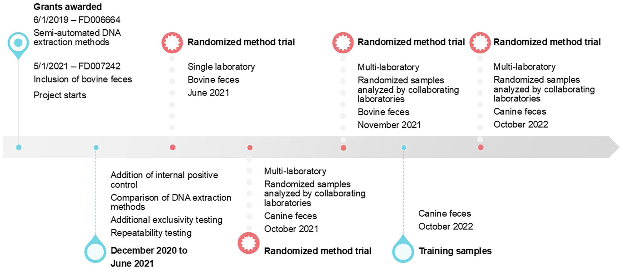
Three-year timeline of the project to evaluate semi-automated DNA extraction and the inclusion of an internal positive control DNA extraction control to the gyrA rtPCR for detection of Campylobacter jejuni in bovine and canine feces.
Organization of samples provided to laboratories for randomized method tests for detection of Campylobacter jejuni in bovine and canine feces.
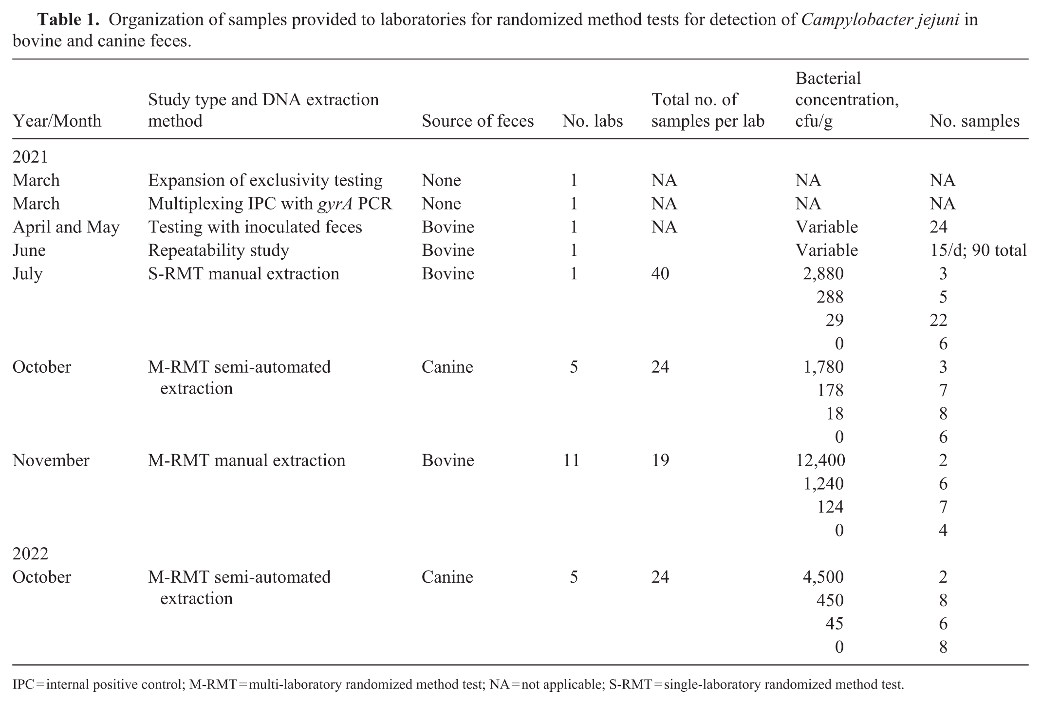
IPC = internal positive control; M-RMT = multi-laboratory randomized method test; NA = not applicable; S-RMT = single-laboratory randomized method test.
C. jejuni gyrA real-time PCR
rtPCR testing was performed (7500 fast real-time PCR system; Applied Biosystems, ThermoFisher). The gyrA primers and probes were obtained from Sigma-Genosys. Positive control DNA was extracted from C. jejuni ATCC 33560T. Negative control DNA was extracted from E. coli ATCC 25922. A no-template negative control of 2 μL of nuclease-free water (Gibco, ThermoFisher) was used. The gyrA rtPCR primers and probe were as published. 13 For each reaction, 2 μL of the DNA sample was combined with 10.6 μL of nuclease-free water, 5 μL of PCR master mix (TaqMan fast virus 1-step master mix; ThermoFisher), 1 μL (10 μM working stock; 0.5 μM final concentration) of forward primer gyrA-F 5′-AAGATACGGTCGATTTTGTTCCA-3′, 1 μL (10 μM working stock; 0.5 μM final concentration) of the reverse primer gyrA-R 5′-CTACAGCTATACCACTTGAACCATTTAATA-3′, and 0.4 μL (10 μM working stock; 0.5 μM final concentration) of the probe gyrA-P 5′-[FAM]TGATGGTTCAGAAAGCGAACCTGATGTTTT[BHQ1]-3′, for a total reaction volume of 20 μL per sample. All reaction mixtures underwent amplification according to the following program: 1 cycle of 50°C for 300 s, 1 cycle at 95°C for 20 s, followed by 45 cycles of 3 s at 95°C and 60 s at 60°C. The positive control was genomic DNA from C. jejuni ATCC 33560T. PCR amplification was reported as the number of PCR cycles at which the amplified product crossed the threshold of detection (Ct). Reactions with Ct values of 14–40 were considered to have detected C. jejuni DNA and were reported as “detected”; reactions in which Ct values were ≥40, or samples in which no signal was detected, were considered not to have detected C. jejuni DNA and were reported as “not detected.”
Inclusion of internal positive control DNA
To control for the presence of PCR inhibitors or inefficiency in DNA extraction, a commercial exogenous DNA (hereafter
DNA extraction
DNA extraction was performed (QIAamp PowerFecal DNA kit, Qiagen; QIAamp PowerFecal Pro DNA kit, Qiagen; MagMAX Pathogen RNA/DNA kit, ThermoFisher; or MagMAX CORE nucleic acid purification kit, ThermoFisher) according to the manufacturers’ instructions. DNA extraction with the MagMAX DNA extraction kits utilized the KingFisher Flex (dx.doi.org/10.17504/protocols.io.8epv5j1b5l1b/v1 19 ), KingFisher Apex, or KingFisher DuoPrime (dx.doi.org/10.17504/protocols.io.14egn286mg5d/v1 18 ) robots (ThermoFisher) following the manufacturer’s protocol. The starting material consisted of 1 mL of fecal slurry described below or 1 mL of C. jejuni in PBS. The resultant DNA was used as the template for the gyrA rtPCR assay. To compare DNA extraction kits and the inclusion of the IPC assay, DNA was extracted from 10-fold serial dilutions of C. jejuni in PBS. The dilutions were plated to trypticase soy agar plates supplemented with 5% sheep blood (blood agar plates; BD), incubated under microaerophilic conditions at 42 ± 2°C for 48 h, and then cfus were manually counted to determine the concentration of bacteria present at each dilution. To evaluate DNA extraction from feces, a fecal slurry was created, as described below.
Exclusivity testing
Inclusivity testing, exclusivity testing, and the specificity of the gyrA primers and probes have been reported. 16 To further test exclusivity, additional Campylobacter species were tested with the gyrA primers and probes. Because of the limited number of Campylobacter isolates available, it was not possible to follow the FDA guidelines 12 to include 50 isolates of the target organism for inclusivity and 30 isolates of non-target organisms for exclusivity testing, but every effort was made to collect isolates for testing. Exclusivity testing was expanded by testing genomic DNA from 4 isolates of C. coli (BAA-370; BAA-371; BAA-1061; ATCC 49941), 2 C. fetus subsp. fetus (ATCC 27374T, ATCC 25936), and 2 C. fetus subsp. venerealis (ATCC 19438T, ATCC 33561T). Previous work tested genomic DNA from C. coli (ATCC 33559 and 1 clinical isolate), C. fetus (a clinical isolate), C. helveticus (ATCC 51210), and Campylobacter upsaliensis (ATCC 43954T and 3 clinical isolates), in addition to 22 other bacteria. 16
Preparation of fecal samples inoculated with C. jejuni
Voluntarily voided, fresh bovine fecal samples were collected and confirmed to be negative for Campylobacter species, including C. jejuni by culture and PCR. Because feces consisted exclusively of voided specimens, this study was considered exempt from the Texas A&M University Institutional Animal Care and Use Committee (IACUC) process. The fecal samples were then mixed using a stomacher to create a uniform matrix. The fecal samples were divided into 8±0.2-g aliquots in 50-mL conical tubes (VWR). Cultures of ATCC 33560T C. jejuni were grown in enrichment broth (Bolton; Hardy Diagnostics) under microaerophilic conditions at 42 ± 2°C for 48 h, diluted in PBS, and used to inoculate fecal samples at ~300,000, 30,000, 3,000, 300, 30, and 3 cfu/g of feces. To confirm C. jejuni cfus, a 100-µL aliquot of the Bolton broth culture was used to create 10-fold serial dilutions in PBS (ThermoFisher) and plated on blood agar plates (BD). Plates were incubated at 42 ± 2°C under microaerophilic conditions for 48 h, manually counted, and averaged across 2 replicates at each dilution to determine the count. Before DNA extraction, 32 mL of PBS was added to each aliquot of inoculated feces to create a fecal slurry.
Method repeatability
To evaluate repeatability of the method, the method originating (
Randomized method tests
For the RMTs, bovine and canine fecal samples were inoculated with a low, medium, or high concentration of C. jejuni or sham-inoculated with PBS as a negative control, packaged, and shipped on ice to the FDA Vet-LIRN program office (
S-RMT
In June 2021, a S-RMT was conducted to evaluate the performance of the gyrA PCR assay to detect C. jejuni in bovine feces using manually extracted DNA. Forty samples (22 low, 7 medium, 4 high concentration, and 7 negative) were shipped from the MO laboratory to the VPO, where a subset of 36 of the samples was randomly selected, anonymous-coded, and shipped back to the MO laboratory for processing and PCR. The selection of a subset of samples for testing was done to ensure that the MO laboratory remained anonymized to sample designations.
M-RMT using manual DNA extraction from bovine feces
A M-RMT was conducted to evaluate the use of the gyrA PCR assay to detect C. jejuni in bovine feces using manually extracted DNA. Eleven laboratories participated. Aliquots of 8±0.2 g of bovine feces were inoculated with C. jejuni at 0, 124, 1,240, and 12,400 cfu/g to represent negative, low, medium, and high concentrations, respectively. Fractional recovery was expected in the samples inoculated with 124 cfu/g. Inoculated fecal samples were packaged on ice with a temperature-monitoring strip for overnight shipment from the MO laboratory to the VPO. On arrival, the VPO checked the temperature-monitoring strip of each package, replaced the temperature-monitoring strip, and shipped specimens overnight to the 11 participating laboratories, including the MO laboratory. Participating laboratories received samples within 2 d following shipment from the MO laboratory. Shipments to the 10 participating laboratories included 19 samples (7 low, 6 medium, 2 high, 4 negative). The MO laboratory provided 2 additional specimens at each concentration, for a total of 27 samples (9 low, 8 medium, 4 high, 6 negative). To ensure that the MO laboratory was anonymized to the number of samples at each concentration, the VPO randomly selected, anonymous-coded, and shipped the originating laboratory a subset of 24 samples (7 low, 9 medium, 2 high, and 6 negative). Analysts at each participating laboratory processed the specimens, performed the rtPCR assay, assessed the amplification curves to confirm the amplification of the PCR product, and reported the Ct values with an interpretation of “detected” or “not detected.” Kappa statistics were calculated to assess agreement between laboratories using an online calculator (described below). To perform these calculations, 5 correctly identified samples (3 medium concentration and 2 negative samples) were removed from the originating laboratory results to balance the sample numbers among laboratories.
M-RMT using semi-automated DNA extraction from canine feces
Two M-RMTs were conducted in October 2021 and October 2022 to evaluate the use of semi-automated methods of DNA extraction. DNA extraction (MagMAX Pathogen RNA/DNA kit; ThermoFisher) was evaluated during October 2021, and DNA extraction (MagMAX CORE nucleic acid purification kit; ThermoFisher) was evaluated during October 2022. During the October 2021 M-RMT, 8±0.2-g aliquots of canine feces were inoculated with C. jejuni at 0, 18, 178, or 1,780 cfu/g to represent negative, low, medium, and high concentrations, respectively. The samples were packaged on ice for overnight shipment from the MO laboratory to the VPO. The VPO shipped specimens overnight to 5 participating laboratories, including the MO laboratory. Shipments to 4 participating laboratories included 24 samples (8 low, 7 medium, 3 high, 6 negative). The originating laboratory provided 2 additional specimens at each concentration, for a total of 32 samples (10 low, 9 medium, 5 high, 8 negative). To ensure that the originating laboratory was anonymized to the number of samples at each concentration, the VPO randomly selected, anonymous-coded, and shipped the originating laboratory a subset of 28 samples (10 low, 7 medium, 4 high, 7 negative). Analysts at each participating laboratory processed the specimens, performed the rtPCR assay, assessed the amplification curves to confirm the amplification of the PCR product, and reported the Ct values with an interpretation of “detected” or “not detected.” Kappa statistics were calculated to assess agreement between laboratories using an online calculator (described below). To perform these calculations, 4 correctly identified samples (1 low, 1 high, 2 negative) were removed from the originating laboratory results to balance the sample numbers among laboratories.
During the October 2022 M-RMT, 8±0.2-g aliquots of canine feces were inoculated with C. jejuni at 0, 45, 450, or 4,500 cfu/g to represent negative, low, medium, and high concentrations, respectively. The samples were packaged on ice for overnight shipment from the MO laboratory to the VPO. The VPO shipped specimens overnight to 5 participating laboratories, including the MO laboratory. Shipments to 4 participating laboratories included 24 samples (6 low, 8 medium, 2 high, 8 negative). The MO laboratory provided 2 additional specimens at each concentration, for a total of 32 samples (8 low, 10 medium, 4 high, 10 negative). To ensure that the MO laboratory was anonymized to the number of samples at each concentration, the VPO randomly selected, anonymous-coded, and shipped the MO laboratory a subset of 29 samples (8 low, 10 medium, 2 high, 9 negative). Analysts at each participating laboratory processed the specimens, performed the rtPCR assay, assessed the amplification curves to confirm the amplification of the PCR product, and reported the Ct values with an interpretation of “detected” or “not detected.” Kappa statistics were calculated to assess agreement between laboratories using an online calculator as described below. To perform these calculations, 5 correctly identified samples (2 low, 2 medium, and 1 negative) were removed from the MO laboratory results to balance the sample numbers among laboratories.
Statistical analysis
For comparison of the gyrA PCR Ct values when multiplexed with the IPC assay or not, a Student t-test for paired samples (p > 0.05) was used. To evaluate the performance of the DNA extraction methods and to determine the agreement between laboratories, kappa statistics were calculated using an online calculator available at http://justusrandolph.net/kappa/.
24
The free-marginal kappa statistic was selected because the analysts were anonymized to the number of positive and negative samples.
32
Kappa values of 0.8–0.9 are considered to have a strong level of agreement.
10
Standard interpretive criteria were utilized.
10
The sensitivity (
Results
Exclusivity testing of Campylobacter species, including 4 isolates of C. coli, 2 isolates of C. fetus subsp. fetus, and 2 isolates of C. fetus subsp. venerealis, confirmed that these organisms were not detected by the C. jejuni gyrA PCR (data not shown).
Addition of the IPC assay to the gyrA PCR assay
A limitation of the original use of the gyrA PCR assay was the lack of an IPC for evaluating DNA extraction efficiency and PCR inhibitors in extracted fecal DNA. To address this issue, we added a commercial IPC DNA to fecal specimens before DNA extraction and evaluated the impact of multiplexing the associated IPC assay with the gyrA PCR
Comparison of DNA extraction methods
Following the prior studies,16,17 the manual DNA extraction kit was modified by the manufacturer from the QIAamp PowerFecal DNA Kit to the QIAamp PowerFecal Pro DNA kit and contained different beads for lysing bacteria.
23
The 2 kits performed similarly with 100% overall agreement with samples >3,000 cfu/mL
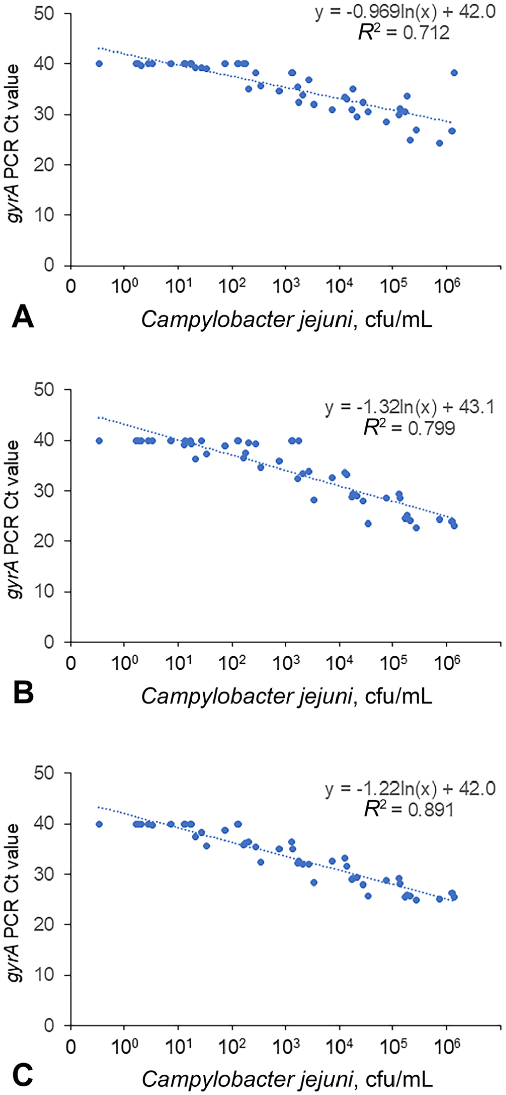
Linear regression of PCR results for DNA extracted with the
PCR results following DNA extraction with the QIAamp PowerFecal DNA kit and the QIAamp PowerFecal Pro kit were also compared to DNA extraction using the MagMAX Pathogen RNA/DNA kit. Overall agreement was 84.7% with κ = 0.69 (95% CI [0.53, 0.85]), which increased to 93.1% agreement with κ = 0.86 (95% CI [0.71, 1.00]) if samples with C. jejuni levels below the fractional level (200 cfu/g feces) were excluded. Linear regression of the plot of Ct values and cfu/mL yielded a linear relationship of y = −1.22 ln(x) + 42.0 with R2 = 0.8910.
Detection of C. jejuni in bovine feces with the gyrA PCR
Eight independent cultures of C. jejuni were used to make eight 10-fold dilution series with 6 dilutions each that were used to inoculate aliquots of bovine feces. DNA was then extracted from the inoculated feces using the QIAamp PowerFecal Pro DNA kit, and the gyrA PCR assay was performed
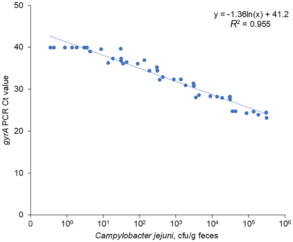
Detection of Campylobacter jejuni in inoculated bovine feces. Linear regression of PCR results from bovine DNA inoculated with various concentrations of C. jejuni.
Repeatability of the assay
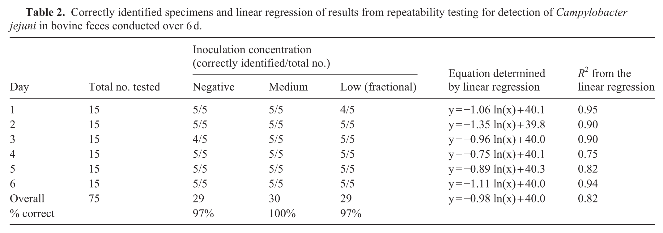
To test the repeatability of the method across multiple days in accordance with FDA guidance,
12
the laboratory tested 5 replicates of 3 levels (negative, medium, and low [fractional] concentrations) of C. jejuni in bovine feces, for a total of 15 samples tested on each day for 6 separate days
Correctly identified specimens and linear regression of results from repeatability testing for detection of Campylobacter jejuni in bovine feces conducted over 6 d.
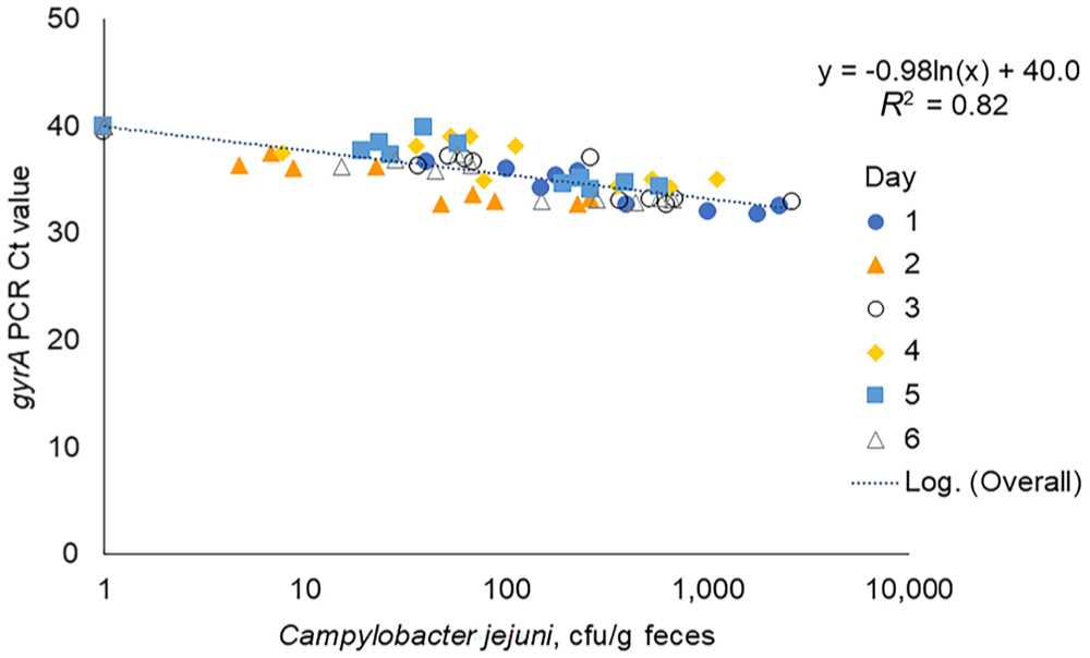
Repeatability of the gyrA PCR in detecting Campylobacter jejuni in bovine feces. The line best fitting the relationship between the cycle at which the PCR reaction crossed the threshold for detection (Ct value) and the number of C. jejuni cfu/g of bovine feces was determined.
S-RMT
In the S-RMT, the laboratory analyst reported the Ct values and an interpretation for each of the 36 tested specimens
Single laboratory randomized method test for detection of Campylobacter jejuni in bovine feces.
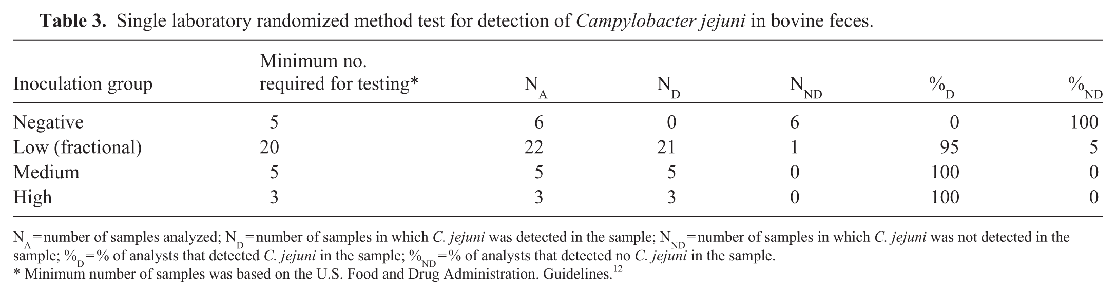
NA = number of samples analyzed; ND = number of samples in which C. jejuni was detected in the sample; NND = number of samples in which C. jejuni was not detected in the sample; %D = % of analysts that detected C. jejuni in the sample; %ND = % of analysts that detected no C. jejuni in the sample.
Minimum number of samples was based on the U.S. Food and Drug Administration. Guidelines. 12
Summary of sensitivity, specificity, and accuracy rates for the randomized method tests for detection of Campylobacter jejuni in bovine and canine feces.
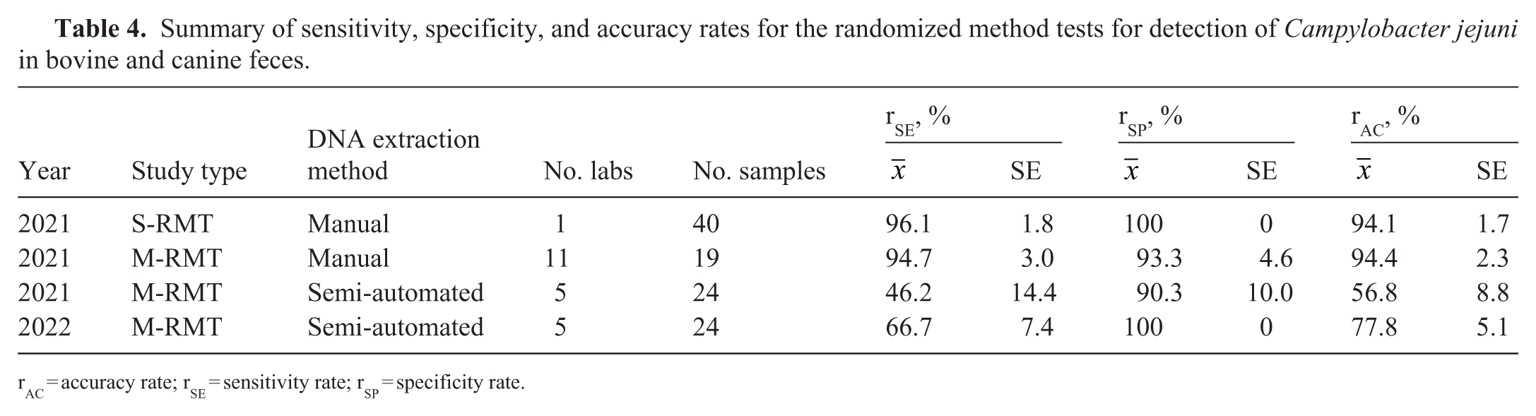
rAC = accuracy rate; rSE = sensitivity rate; rSP = specificity rate.
M-RMT of manual DNA extraction
A M-RMT conducted across 11 laboratories evaluated the use of the gyrA PCR assay for the detection C. jejuni in bovine feces using manually extracted DNA. All laboratories reported no color change in the temperature-monitoring strip for any shipment, indicating that the correct temperature was maintained during shipping. Inoculation concentrations of 0, 124, 1,240, and 12,400 cfu/g were used for negative, low, medium, and high concentration levels. Fractional recovery was expected in samples with 124 cfu/g. A low detection rate was anticipated for the samples inoculated with the low concentration (fractional level) of bacteria, but 6 laboratories correctly identified 100% of these samples as positive and 4 laboratories correctly identified 6 of the 7 fractional samples as positive
Multi-laboratory, randomized method test results for detection of Campylobacter jejuni in bovine and canine feces.
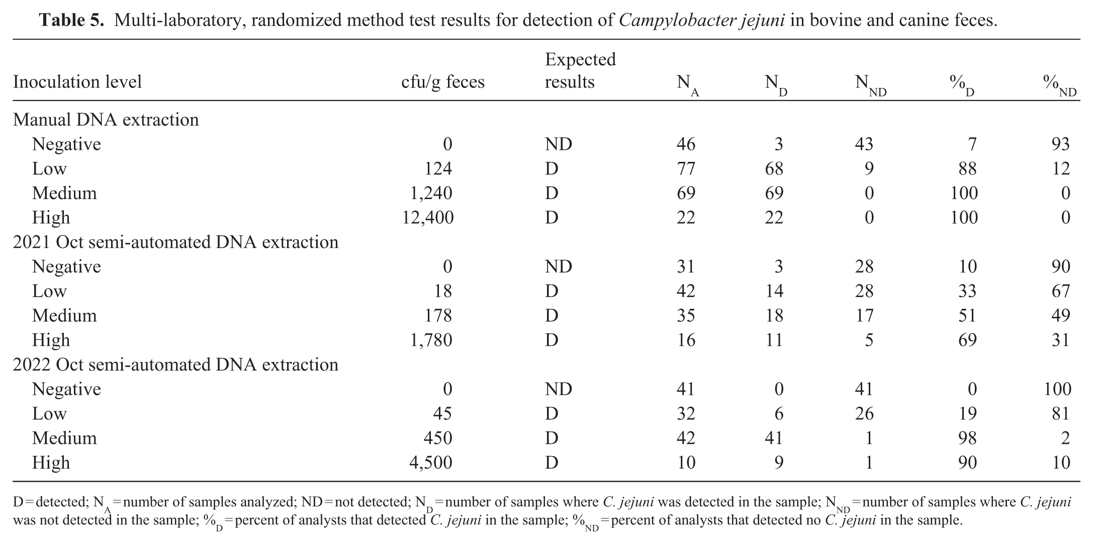
D = detected; NA = number of samples analyzed; ND = not detected; ND = number of samples where C. jejuni was detected in the sample; NND = number of samples where C. jejuni was not detected in the sample; %D = percent of analysts that detected C. jejuni in the sample; %ND = percent of analysts that detected no C. jejuni in the sample.
M-RMTs of semi-automated DNA extraction
A M-RMT conducted across 5 laboratories evaluated the use of semi-automated DNA extraction in the gyrA PCR assay
A second M-RMT with the same participating laboratories was conducted. A training webinar was held before the M-RMT
Discussion
We found that the inoculation level of fecal specimens played a role in the detection of C. jejuni using molecular methods. For all 3 M-RMTs, the inoculation level for the low concentration samples was <200 cfu/g feces, and in 1 case, the October 2021 M-RMT, the level was below the limit of detection (LOD) of 40 cfu/g feces. 16 The fractional recovery level is defined as the proportion of positive responses that fall within 25–75%. 12 The lowest concentration of C. jejuni tested was 18 cfu/g feces in the October 2021 M-RMT and 45 cfu/g feces in the October 2022 M-RMT. C. jejuni was detected in 33% of the October 2021 samples and 19% of October 2022 samples. In the manual DNA extraction M-RMT, the low concentration samples were inoculated with 124 cfu/g feces and were detected in 88% of samples, confirming the importance of inoculation level as a factor in test performance. Together, our study demonstrates that C. jejuni is a difficult organism to detect and use in proficiency testing. In our prior study, we also experienced issues with the detection of C. jejuni during a method test associated with shipping. 16 Additionally, we found that analyst experience played a role in test performance, with an accuracy of 96.4% for experienced analysts compared with 85.8% for a larger group of less-experienced analysts. 16 Despite these issues, the molecular method was more sensitive than culture, with or without enrichment, and the molecular method required less time than culture. 17 Using a molecular method allows rapid detection of C. jejuni, which can be important during an outbreak. 21
One of the long-standing issues with molecular detection of pathogens in patient specimens is the potential presence of PCR inhibitors in feces and body fluids. We reviewed dealing with inhibitors in detail and published best practices for the performance of rtPCR assays in veterinary diagnostic laboratories.30,34 One approach is to use an exogenous IPC, with established acceptable ranges, added to the specimen prior to nucleic acid extraction. Any sample with the IPC not detected or falling outside of the established ranges should be retested with dilution or re-extraction, as we described. 34 Here, the IPC Ct value was 32–38, depending on the DNA extraction kit used. Samples in which the IPC Ct value was >38 were considered to have failed. However, including an IPC has the advantage of controlling extraction efficiency as well as inhibition. In our previous work, we had not included an IPC.16,17 Although we were able to detect C. jejuni from canine feces, we believe that the inclusion of an extraction control and a control for PCR inhibitors is important. Potential differences in the types of PCR inhibitors are also present in feces from patients on different diets (e.g., differences between herbivores, such as cattle, compared with omnivores or carnivores, such as dogs). We found that the inclusion of a commercial exogenous IPC DNA in DNA extraction and multiplexing a PCR assay to detect exogenous DNA did not significantly affect the Ct values of the gyrA PCR assay and allowed the detection of possible inhibition or failure of extraction in a sample when comparing the 2 manual DNA extraction kits. Additionally, we evaluated the ability of the gyrA PCR assay to detect C. jejuni in bovine feces. Cattle may serve as a source of C. jejuni for people through fecal shedding or contaminated milk or meat.25,28,31,33 The gyrA PCR assay performance was similar for DNA extracted from both bovine feces and canine feces.
During the repeatability testing, independent cultures were used for each day. The final concentrations of the bacteria, as determined by culture, varied by day. Samples inoculated with bacteria tested positive on all 6 d. One negative sample tested positive on day 3, although the Ct value was high (39.5). This discrepancy may have come from cross-contamination between samples. Rigorous efforts had been taken to prevent this, including using filtered pipette tips and careful spacing of samples during processing. The R2 value for the data as a whole was 0.818, indicating good agreement across days, although the Ct value varied. Days 4 and 5 had the lowest R2 values and contributed to the reduction in the overall R2 value. One possible explanation for the reduction on these 2 d is analyst fatigue when performing the manual DNA extractions for these assays. Using different cultures to make the specimens for each day was a limitation. An alternative approach would have been to make and inoculate the fecal aliquots at the same time, store them at −80°C, and thaw the aliquots each day before extraction. This was not pursued because of variability in survival in preliminary testing. Similarly, refrigerating samples for 6 d was not pursued. Use of freshly cultured bacteria also created challenges in the M-RMTs. The low inoculation concentrations in the 2 M-RMTs testing semi-automated methods were problematic. Colonies from the inocula were only available for enumeration 2 d after the samples were shipped to the VPO because of sample stability needs.
Overall, we expanded exclusivity testing during our study to include additional isolates of C. coli and C. fetus. The gyrA PCR method was expanded to include the use of an exogenous IPC DNA as an extraction control and the use of a multiplexed IPC DNA assay to detect gyrA PCR inhibition. The gyrA PCR assay performed consistently across multiple laboratories. In general, PCR detection of C. jejuni should be followed up with culture, susceptibility testing, and whole-genome sequencing for the purpose of outbreak monitoring.
Supplemental Material
sj-pdf-1-vdi-10.1177_10406387261454377 – Supplemental material for Matrix extension to bovine feces and evaluation of semi-automated DNA extraction methods for the detection of Campylobacter jejuni in bovine and canine fecal samples using gyrA PCR
Supplemental material, sj-pdf-1-vdi-10.1177_10406387261454377 for Matrix extension to bovine feces and evaluation of semi-automated DNA extraction methods for the detection of Campylobacter jejuni in bovine and canine fecal samples using gyrA PCR by Sara D. Lawhon, Jing Wu, Kristy L. Pabilonia, Zeinab Helal, Guillermo Risatti, Alex Nemethy, Jeremy N. Ray, David Simon, Wanda Tirado, Lijuan Zhou, Paula Bartlett, Ingrid Fernandez, Susan Sanchez, Vanessa DeShambo, Namjung Jung, Carol Maddox, Steven Ensley, Udeni Balasuriya, Keith Strother, Lifang Yan, Candy Zhang, Zhenyu Shen, Shuping Zhang, Sarmila Dasgupta, Shannon Mann, Amar Patil, Laura B. Goodman, Kelly Sams, Jing Cui, Melanie Prarat Koscielny, Qirui Zhang, Yan Zhang, Robin Madden, Akhilesh Ramachandran, Jessica Bower, Li Fang, Deepanker Tewari, Nagaraja Thirumalapura, Corey Zellers, Jake Guag, Sarah M. Nemser and Gregory H. Tyson in Journal of Veterinary Diagnostic Investigation
Footnotes
Acknowledgements
We thank the laboratories and individual analysts who performed the work and patiently offered insights that improved the assays throughout the course of this effort. Of particular note, we thank the members of the FDA Vet-LIRN who organized and shipped test samples. Ms. Alisha Milore at the Pennsylvania Veterinary Laboratory at the Pennsylvania Department of Agriculture provided laboratory support.
Declaration of conflicting interests
The authors declared no potential conflicts of interest with respect to the research, authorship, and/or publication of this article.
Funding
Our work was supported by funding from FDA grants U18FD005013, U18FD006446, U18FD006664, and U18FD007242. Canine feces were provided by Dwayne Schrunk and Dr. Steve Ensley at the Veterinary Diagnostic Laboratory, Iowa State University (Ames, IA, USA) under the contract HHSF223201410256A for diagnostic sample collection and subject matter expertise for Vet-LIRN Proficiency Exercises.
ORCID iDs

Supplemental material
Supplemental material for this article is available online.
References
Supplementary Material
Please find the following supplemental material available below.
For Open Access articles published under a Creative Commons License, all supplemental material carries the same license as the article it is associated with.
For non-Open Access articles published, all supplemental material carries a non-exclusive license, and permission requests for re-use of supplemental material or any part of supplemental material shall be sent directly to the copyright owner as specified in the copyright notice associated with the article.
