



Abstract
Bone marrow mesenchymal stem cell-derived exosomes (BMSCs-Exos) with their molecular cargo have therapeutic potential for pulmonary fibrosis (PF). This research was performed to uncover how microRNA-31-5p (miR-31-5p), carried by BMSCs-Exos, affects PF via modulating IGFBP7. C57BL/6 mice were treated with bleomycin (BLM) to induce PF. Pulmonary function was tested, and fibrotic changes in the mouse lung tissues were examined. Levels of fibrosis-related inflammatory factors, including tumor necrosis factor-α (TNF-α), interleukin (IL)-1β, and IL-6, were tested. Mouse BMSCs were isolated and identified, and BMSCs-Exos were obtained by ultracentrifugation. Exosome morphology was observed by transmission electron microscopy, the surface markers were measured, and the expression levels of BMSCs-Exo marker proteins were assessed. The targeting relation between miR-31-5p and IGFBP7 was assessed, and the expression of both was tested. After modeling, mice exhibited decreased functional residual capacity, lung compliance, inspiratory capacity, vital capacity, total lung capacity, and forced vital capacity. After 14 days of BLM induction, thickening of the main tracheal wall, fibroblast accumulation, immune cell infiltration in lung interstitium, and increased collagen deposition were observed. Elevated levels of TNF-α, IL-1β, and IL-6 were also noted. BMSCs-Exos attenuated BLM-induced PF, and BMSCs-Exo-derived miR-31-5p ameliorated PF in mice. miR-31-5p was shown to target IGFBP7, diminishing both transcript and protein levels. IGFBP7 overexpression reversed the ameliorative impact of miR-31-5p on PF in mice. BMSCs-Exos ameliorate PF development by delivering miR-31-5p to repress IGFBP7.
INTRODUCTION
Pulmonary fibrosis (PF) is a progressive and often fatal lung disorder characterized by excessive extracellular matrix deposition and fibroblast proliferation, ultimately leading to irreversible architectural distortion and respiratory failure. 1,2 As the most common and severe form of idiopathic interstitial pneumonia, idiopathic pulmonary fibrosis (IPF) usually develops progressively, and in most cases, with a median survival of 2–5 years following diagnosis, primarily due to respiratory failure. 3 Current understanding of IPF pathogenesis implicates complex interactions involving genetic predisposition, aging-related processes, and repetitive alveolar epithelial injury that collectively drive progressive fibrogenesis and lung functional decline. 4 Although current antifibrotic drugs such as pirfenidone and nintedanib can delay disease progression, 5 they are largely ineffective at reversing established fibrosis. Therefore, exploring new therapeutic targets and intervention strategies is of great significance.
Mesenchymal stem cells (MSCs) and their derivatives have demonstrated significant potential in the treatment of PF. Preclinical studies have shown that MSCs can alleviate histopathological changes and improve survival in bleomycin (BLM)-induced PF models, 6 with bone marrow mesenchymal stem cells (BMSCs) in particular exhibiting favorable therapeutic effects. 7 More recent research has revealed that BMSCs-derived exosomes (BMSCs-Exos) are involved in the process of PF by regulating the transformation of fibroblasts into myofibroblasts. 8 Specifically, Exos derived from human umbilical cord MSCs can attenuate silicosis-induced PF and improve lung function, 9 as well as alleviate PF and inflammation in mice by modulating monocyte phenotypes. 10
At the molecular level, microRNAs (miRNAs) encapsulated within Exos are considered important mediators in the treatment of IPF. 11 For instance, miR-22 delivered by Exos has therapeutic effects on PF. 12 Notably, miR-31 exhibits abnormal expression in PF, which has been confirmed in both mouse models of PF 13 and lipopolysaccharide-induced embryonic lung fibroblast injury models. 14 Insulin-like growth factor-binding protein 7 (IGFBP7) refers to an extracellular matrix-associated glycoprotein that is highly enriched in activated blood vessels during developmental, pathological, and physiological tissue remodeling 15 and is closely associated with the progression of fibrosis. 16 IGFBP7 has been identified in extracellular vesicles derived from IPF cell lines 16 and its expression is elevated in fibrotic liver tissues, 17 suggesting its potential as a common fibrogenic effector across organ systems.
Despite accumulating evidence supporting the antifibrotic effects of MSC-derived exosomes, the role of miR-31-5p, a key effector cargo, and its regulatory interaction with IGFBP7 remain largely unexplored. In this study, we propose the novel hypothesis that BMSCs-Exos deliver miR-31-5p, which in turn inhibits PF progression by targeting and downregulating IGFBP7. This study aims to systematically elucidate the regulatory function of the miR-31-5p/IGFBP7 axis in PF pathophysiology. Our findings are expected to enhance the mechanistic understanding of BMSCs-Exos in antifibrotic therapy and provide a theoretical foundation for the development of exosome-based precision treatment strategies for PF.
MATERIALS AND METHODS
Ethics statement
The research was conducted with the approval of the ethics committee of Leshan People’s Hospital. Animal pains were minimized as much as possible.
Isolation, culture, and identification of BMSCs
BMSCs were isolated from the femur and tibia of C57BL/6 mice. The extracted cells were suspended in Dulbecco’s Modified Eagle Medium (DMEM) containing 10% fetal bovine serum (Biowest, Nuaillé, France) and penicillin–streptomycin (100 U/mL, Gibco Life Technologies, NY, USA). Medium renewal was performed after 3 days. Cells that reached 80–90% confluence were sub-cultured to a third passage, which was then prepared to a cell suspension (1 × 10⁶ cells/mL, 200 μL). 18
After subculturing to the third–fourth passage, the cultured BMSCs exhibited a typical spindle-shaped morphology and spiral arrangement, indicating uniformity and viability. Fluorescein isothiocyanate-labeled mouse anti-human antibodies (CD29, CD44, CD90, and CD45) were cultured with BMSCs at the third passage and then detected by flow cytometry. 19 Adipogenic and osteogenic differentiation experiments were performed, and then oil red O staining and alizarin red staining were conducted. The stained cells were observed under a microscope (Olympus, Tokyo, Japan). 20
Isolation, extraction, and identification of BMSCs-Exos
BMSCs at third passage were cultured overnight in serum-free DMEM. The conditioned medium of BMSCs (80–90% confluence) was collected and treated with centrifugation to remove cell debris. Then, supernatant was centrifuged at 10,000 g twice and the resulting pellet was resuspended in 25 mM 4-(2-hydroxyethyl)−1-piperazineëthanesulfonic acid (pH = 7.4) and centrifuged again at 10,000 g to obtain Exos, which were stored at −80°C for backup. To further characterize the morphology and size of Exos, 10 μL of Exo suspension was applied onto carbon-coated copper grids and allowed to stand at 25°C for 10 min. After carefully removing the excess liquid with filter paper, the samples were negatively stained with 3% phosphotungstic acid solution for 5 min. The Exo samples were then observed using a 100 kV transmission electron microscope (Krios Rx Cryo-TEM, Thermo Fisher Scientific, USA), and electron micrographs were taken (magnification: 20,000× to 50,000×). Three representative TEM fields were randomly selected, and the diameters of multiple individual vesicles were measured using ImageJ software. Additionally, Western blot was employed to identify the surface antigens CD63 and Alix on BMSC-Exos.
Induction of PF in mice by BLM
Seventy 14-week-old C57BL/6 mice (Charles River Laboratories, MA, USA) were housed in plastic cages and acclimatized for 1 week in a controlled environment (20 ± 2°C, 50 ± 10% humidity, and a 12-h light-dark cycle). All mice were anesthetized with isoflurane and kept under specific pathogen-free conditions. A single dose of BLM (3 U/kg, 50 μL, Shanghai Maokang Biotechnology Co., Ltd., Shanghai, China) was administered intratracheally to induce PF. Mice in the blank control group received an intratracheal injection of 0.9% normal saline (50 μL) and were designated as the normal group (n = 10). 10 The remaining 60 BLM-treated mice were randomly divided into six groups (n = 10 each), including the Model group, Model-Exo group (200 μL of phosphate buffer solution [PBS] containing 20 μg of Exos was injected through the tail vein), Model-Exo negative control (NC) group (25 μg of Exos containing miR-31-5p agomir NC was injected through the tail vein), Model-Exo-miR-31-5p agomir group (25 μg of Exos containing miR-31-5p agomir was injected through the tail vein), Model-Exo-miR-31-5p agomir + overexpression (oe)-NC (25 μg of Exos containing miR-31-5p antagomir and oe-IGFBP7 NC lentivirus were injected through the tail vein), and Model-Exo-miR-31-5p agomir + oe-IGFBP7 (25 μg of Exos containing miR-31–5p antagomir and oe-IGFBP7 lentivirus were injected through the tail vein). All treatments were administered two times a week for 5 weeks. 21 Both the oe-IGFBP7 and control lentiviruses were synthesized and provided by Shanghai Genechem Co., Ltd. (China). The packaging virus and target vector were cotransfected into 293T cells (with a confluence of 80–90%) using Lipofectamine 2000 (11668500, Invitrogen, USA). After 48 h of cell culture, the supernatant was collected, filtered through a 0.45 μm membrane, and then the viral particles were concentrated by ultracentrifugation (25,000 g, 2 h, 4°C). The viral pellet was resuspended in PBS, and the viral titer was determined using qPCR, yielding a final concentration of 1 × 10⁹ TU/mL. In animal experiments, the lentivirus and miR-31-5p antagomir-modified Exos were coinjected via the tail vein into BLM-treated mice. Each injection consisted of 200 μL (containing approximately 2 × 10⁸ TU of viral particles), administered twice a week for 5 consecutive weeks. The mice were euthanized at the end of the experiment, and bronchoalveolar lavage fluid (BALF) and lung tissues were collected. Tissues were either fixed in 4% paraformaldehyde for histological analysis or stored at −80°C for subsequent assays. 22
Determination of pulmonary function
Mice were anesthetized via intraperitoneal administration of ketamine and xylazine. The trachea was intubated and connected to a mandatory lung ventilation system (Buxco Research System; Buxco Electronics, NC, USA). The respiratory rate was set at 150 breaths/min. Functional Residual Capacity (FRC) was determined according to Boyle’s Law. Lung compliance (C chord), inspiratory volume (IC), vital capacity (VC), total vital capacity (TLC), and forced vital capacity (FVC) were measured by the Buxco system. 23
Enzyme-linked immunosorbent assay
Tumor necrosis factor-α (TNF-α), interleukin (IL)−1β, and IL-6 levels were measured. PBS (1 mL) was injected into the lungs for lavage. The supernatant was collected by centrifugation at 700 g. BALF (1 mL) was collected 24 and centrifuged at 300 g. TNF-α, IL-1β and IL-6 levels in the supernatant were measured with enzyme-linked immunosorbent assay (ELISA) kits (SEKH-0047, SEKH-0013, Solarbio, Beijing, China). Absorbance value was recorded at 450 nm. 25
Hematoxylin–eosin staining
Lung tissues were processed into paraffin-embedded sections (4 μm) and stained with hematoxylin and 1% eosin solution. 26 Then, lung tissues were fixed in malol and observed under a BX51 microscope (Olympus). Lung injury was scored as follows: 0 point: normal lung tissue with no pathological features such as inflammation, edema, or hemorrhage; 1 point: mild lung injury with slight inflammatory infiltration and alveolar edema without hemorrhage; 2 points: moderate lung injury with obvious inflammatory cell infiltration, edema, and minor alveolar hemorrhage; 3–4 points: severe lung injury with substantial inflammatory cell infiltration and alveolar edema, accompanied by obvious hemorrhage, necrosis, and other pathological changes. Lung injury was independently scored by two pathologists. 24,27,28 Additionally, images were analyzed using ImageJ image analysis software (http://rsb.info.nih.gov/ij/). The mean linear intercept (MLI) was determined as a measure of the average distance between gas exchange surfaces, following the guidelines outlined in the American Thoracic Society/European Respiratory Society quantitative assessment criteria for lung structure. 29
Masson staining
Lung tissues were made into paraffin-embedded sections (4 μm) and routinely deparaffinized. Cell nuclei were stained with Regaud hematoxylin, followed by Masson-Ponceau red acid fuchsin solution. Afterward, the sections were treated with 1% phosphomolybdic acid aqueous solution and counterstained with aniline blue or light green. The sections were observed under a light microscope (DM 4000B, Leica, Germany), and the optical density values of collagen staining were quantified using Image J v1.8.0 software. 30
Reverse transcription quantitative polymerase chain reaction
Total RNA of the lung tissues was extracted by Trizol (Invitrogen), quantified by NanoDrop2000 spectrophotometer (Thermo Fisher Scientific), and reverse transcribed using the PrimeScript RT kit with gDNA Eraser Kit (Takara, Shiga, Japan). Reverse transcription quantitative polymerase chain reaction (RT-qPCR) was performed using an ABI7500 quantitative PCR instrument (Thermo Fisher Scientific) with the SYBR PreMixExTaq (TliRNaseHPlus) kit (Takara). U6 small nuclear RNA and glyceraldehyde-3-phosphate dehydrogenase (GAPDH) were taken as the internal controls of miR-31-5p and IGFBP7, respectively. The 2−ΔΔCt method was adopted to quantify gene expression. Table 1 lists the sequences of the primer. 19
Primer Sequences for Reverse Transcription Quantitative Polymerase Chain Reaction
GAPDH, glyceraldehyde-3-phosphate dehydrogenase; U6, U6 small nuclear RNA.
Western blot assay
Protein samples were extracted by utilizing radioimmunoprecipitation assay lysis buffer containing protease inhibitors (Beyotime, Shanghai, China), and protein concentrations were tested according to the instructions of BCA kits (Boster, Wuhan, Hubei, China). Samples were fully mixed with the loading buffer and boiled at 100°C, followed by an ice bath and centrifugation. An equal amount of protein was loaded onto 10% sodium dodecyl sulfate polyacrylamide gel electrophoresis gels for separation and then transferred to the nitrocellulose membranes. Membranes were blocked overnight in 5% skimmed milk. Primary antibodies included IGFBP7 (1:500, IPD-ANP11965, Apti Biotechnology, Hubei, China), GAPDH (1:3000, ab8245, Abcam), CD63 (1:1000, ab315108, Abcam), and Alix (1:1000, ab275377, Abcam). After incubation with the appropriate HRP-conjugated secondary antibody (1:1000, Boster), the membranes were developed using ECL detection reagent (Pierce, USA). Images were captured following exposure and development, and band intensities were analyzed by ImageJ software (version 1.8.0, National Institute of Mental Health, USA). GAPDH served as the internal control. 31
Dual-luciferase reporter gene assay
pLUC-IGFBP7 wild-type (WT)/pLUC-IGFBP7 mutant type (MUT) and miR-31-5p mimic/mimic NC were transfected into HEK293T cells via liposome 2000 (Thermo Fisher Scientific). Plasmids were purchased from GenePharma (Shanghai, China). Luciferase activity was detected at 560 nm with a dual-luciferase reporter assay kit (Promega, WI, USA). 24
Statistical analysis
Statistical analysis was implemented using SPSS 21.0 software (IBM, NY, USA) and GraphPad Prism 9.5 (California, USA). Measurement data were expressed by mean ± standard deviation. For normally distributed data with homogeneity of variance, intergroup comparisons were performed using an unpaired t-test. For data analysis among multiple groups, one-way analysis of variance was adopted. A rank sum test was applied to compare data with skewed distribution or variance heterogeneity. Statistical significance was determined with p < 0.05. 32
RESULTS
Successful establishment of PF mice by BLM
BLM-treated PF mouse models were successfully established and assessed to ensure the success of induction. First, pulmonary function was tested: in BLM-treated mice, lower FRC, C chord, IC, VC, TLC, and FVC values were observed (Fig. 1A, B). In Masson’s trichrome staining, collagen content in the interstitial tissue was found to be significantly higher than that in the normal group mice (Fig. 1C). Hematoxylin–eosin (HE) staining (Fig. 1D) revealed that the main tracheal wall of mice thickened 14 days after modeling, with fibroblast and immune cell accumulation in the pulmonary interstitial tissue. Meanwhile, measurements of the MLI showed a significant increase in alveolar spacing in the Model group, further reflecting lung tissue destruction and a reduction in ventilation area (right panel of Fig. 1D). Next, inflammation was evaluated by ELISA (Fig. 1): BLM-treated mice showed increased TNF-α, IL-1β, and IL-6 levels. The above results displayed that the BLM-induced PF model in mice was successfully established.
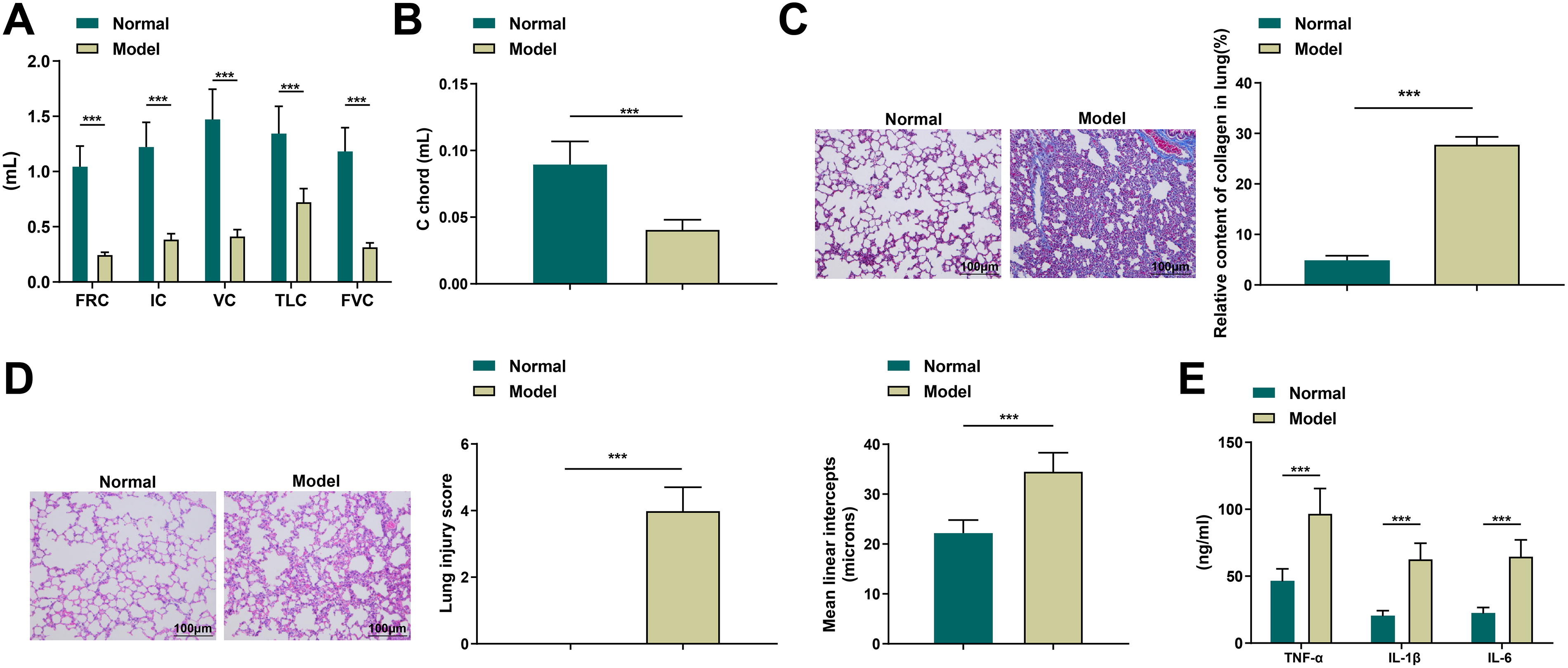
BLM induction reduces lung function in mice.
Identification results of BMSCs and BMSC-Exos
When BMSCs grew rapidly to 90% confluency, the cell clusters displayed an obvious swirling growth pattern, and the cell morphology was long and shuttle-shaped (Fig. 2A). Following lipogenic induction, Oil Red O staining revealed orange-red lipid droplets within the cells under microscopy; after osteogenic induction, Alizarin Red staining revealed obvious red calcium nodule formation (Fig. 2B). Flow cytometry showed positive expression of the surface molecules CD29 and CD9, markers of MSCs, and negative expression of CD45, a marker of hematopoietic cells (Fig. 2C). Under transmission electron microscopy, BMSC-Exos were observed as round or oval membrane-bound vesicles with diameters of 40–100 nm and significant heterogeneity, confirming successful isolation and morphological integrity (Fig. 2D). Western blot results demonstrated (Fig. 2E) that the extracted material positively expressed Alix and CD63 proteins, further confirming the successful extraction of Exos.
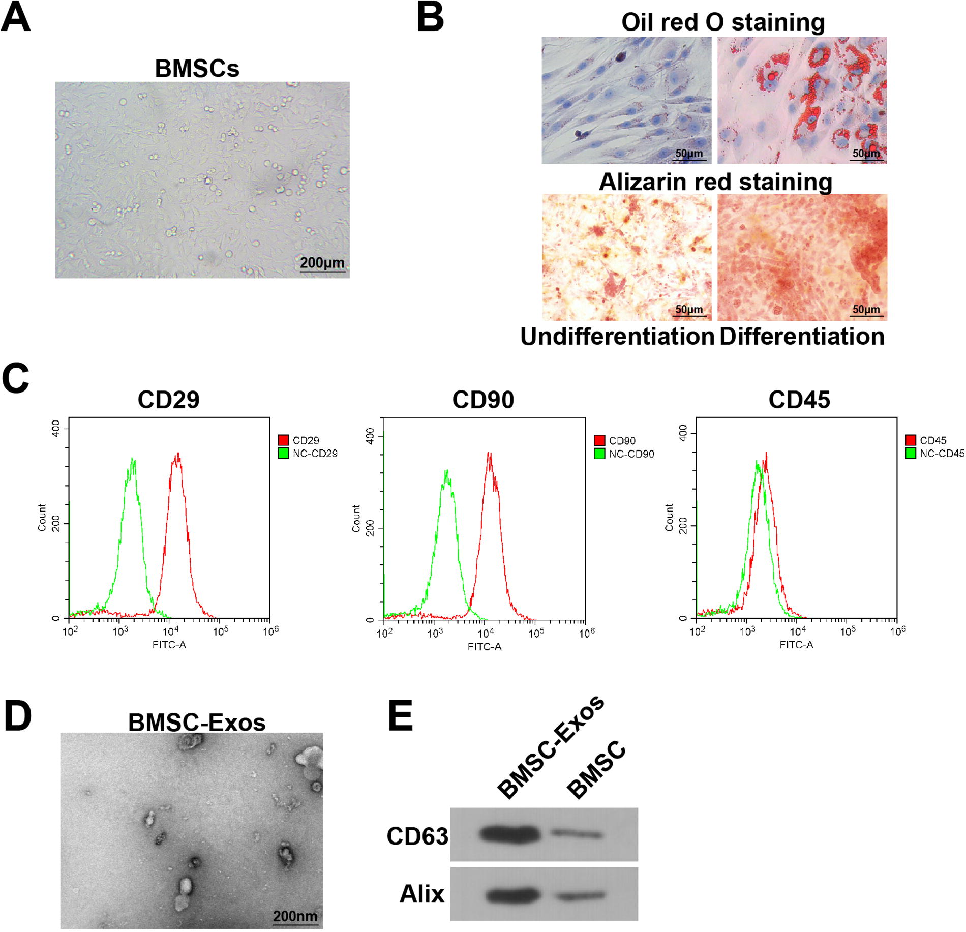
Identification results of BMSCs and BMSC-Exos.
miR-31-5p expression level changes in Exos influence PF in mice
miR-31 has been implied to reduce inflammatory responses and oxidative stress-induced neuronal injury in ischemic stroke, 33 and to mediate the transition from inflammation to re-epithelialization during skin wound healing, suggesting miR-31 has therapeutic potential in injury repair. 34 MSCs-Exos can prevent and reverse the core features of PF. 10 To verify this, the extracted Exos were injected into the model mice (Model-Exo group) and compared with the Model group to investigate whether MSCs-Exos could have therapeutic effects on PF in mice. To further verify the role and mechanism of miR-31-5p in Exos in BLM-induced PF, a miR-31-5p agomir-modified group (Model-Exo-miR-31-5p agomir) and an NC group (Model-Exo-NC) were established based on the Model-Exo group. RT-qPCR results showed that the miR-31-5p expression in the lung tissues of PF model mice was decreased, while its expression was restored in the Model-Exo-miR-31-5p agomir group (Fig. 3A).
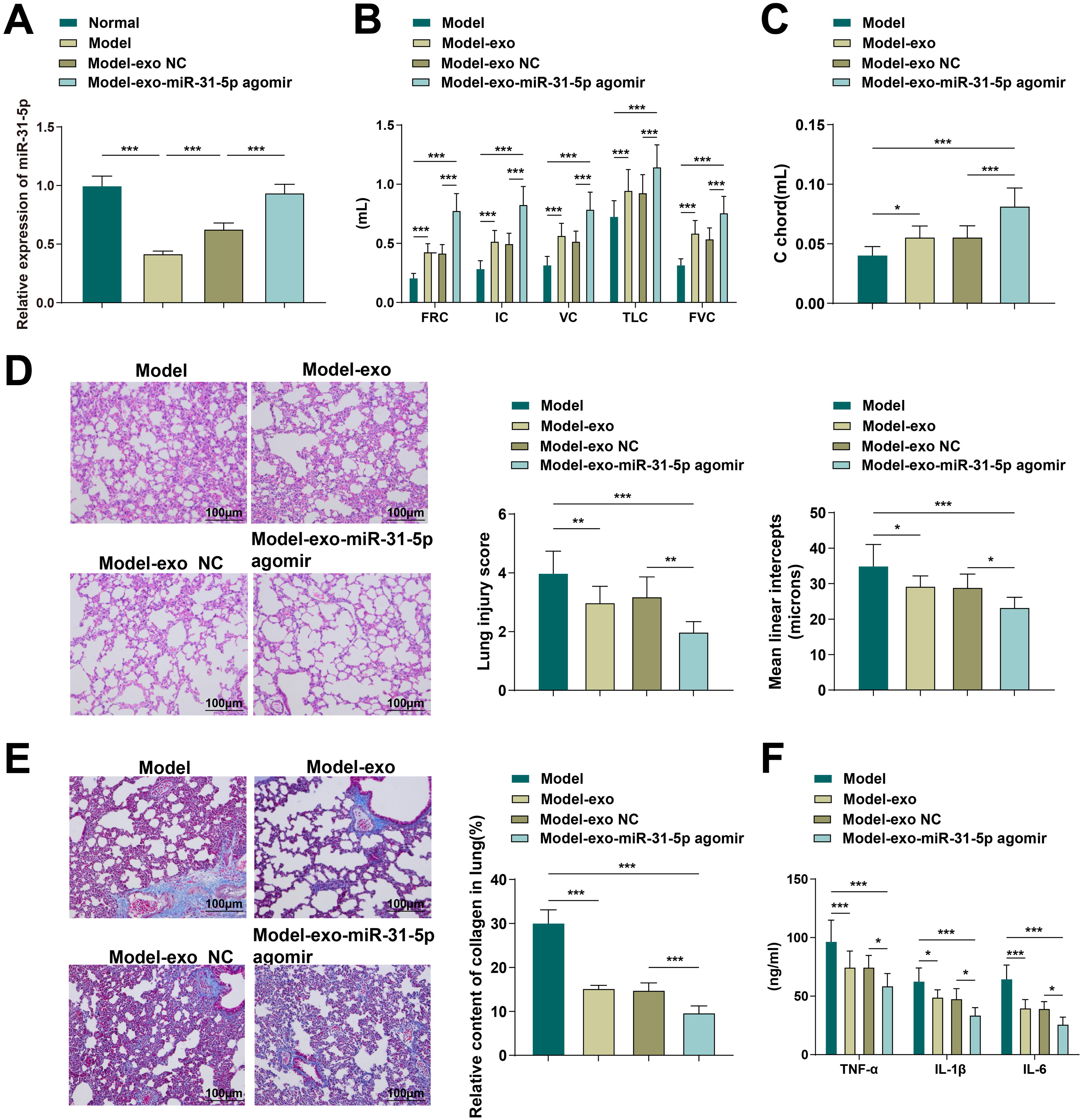
miR-31-5p expression in Exo influences PF in mice.
Lung function assessments revealed that the Model-Exo-miR-31-5p agomir group showed significant improvements in lung capacity (FRC, IC, VC, TLC, FVC) and lung compliance (C chord) compared with the Model, Model-Exo, and Model-Exo NC groups (Fig. 3B, C). HE staining showed improved structural integrity of lung tissue, reduced inflammatory cell infiltration, and significantly decreased lung injury scores and MLI values in the Model-Exo-miR-31-5p agomir group (Fig. 3D). Masson’s trichrome staining indicated a marked reduction in collagen deposition in the lung tissues of this group, suggesting fibrosis alleviation (Fig. 3E). ELISA results further showed that the levels of TNF-α, IL-1β, and IL-6 in the Model-Exo-miR-31-5p agomir group were significantly lower than those in the Model-Exo-NC group (Fig. 3F). These results collectively indicate that BMSC-Exos can effectively inhibit pulmonary inflammation and fibrosis progression, thereby improving lung function, by delivering miR-31-5p. 33,34
A targeting relation exists between miR-31-5p and IGFBP7
A targeting relationship between miR-31-5p and IGFBP7 was predicted through the Starbase website (https://rnasysu.com/encori/) (Fig. 4A). This was confirmed by a dual-luciferase reporter assay, which showed that miR-31-5p bound directly to the 3′UTR of IGFBP7, and the luciferase activity of WT 3′UTR-transfected cells was inhibited, suggesting that IGFBP7 was a direct target of miR-31-5p (Fig. 4B). Furthermore, it was found that IGFBP7 expression levels in PF mice were elevated, and when miR-31-5p was up-regulated, IGFBP7 expression levels were diminished (Fig. 4C, D). The above results display that miR-31-5p targets and suppresses IGFBP7 expression.
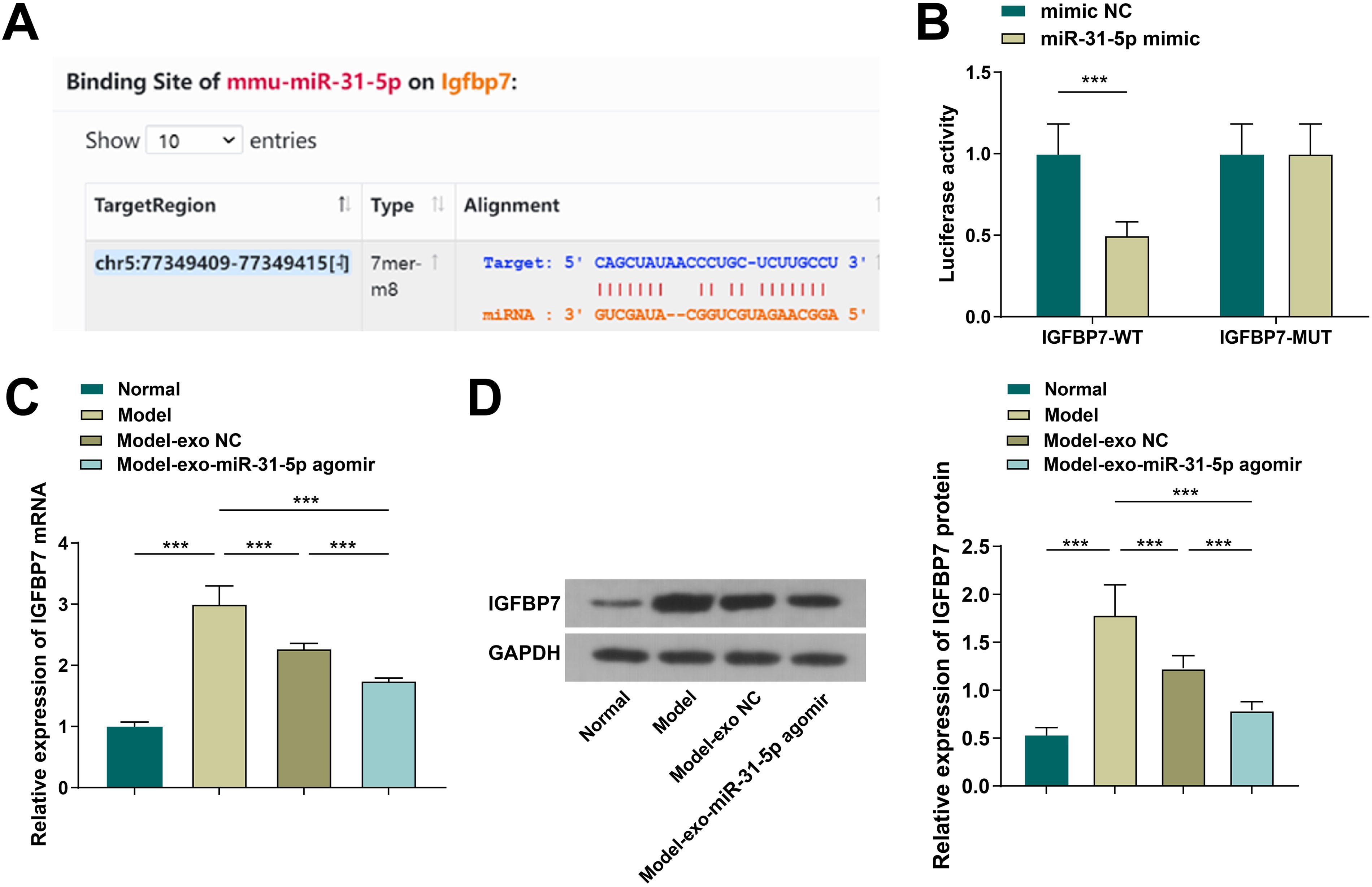
A targeting relationship presented between miR-31-5p and IGFBP7.
BMSCs-Exos affect PF in mice by delivering miR-31-5p to regulate IGFBP7
To further investigate whether BMSC-Exos affect PF via the miR-31-5p/IGFBP7 axis, two additional groups were established: Model-Exo-miR-31-5p agomir + oe-NC and Model-Exo-miR-31-5p agomir + oe-IGFBP7 groups. In the Model-Exo-miR-31-5p agomir + oe-IGFBP7 group, IGFBP7 expression was elevated (Fig. 5A); FRC, C chord, IC, VC, TLC, and FVC were decreased (Fig. 5B, C); lung injury score and MLI values were increased, and PF degree was pronounced (Fig. 5D, E); and TNF-α, IL-1β, and IL-6 levels were elevated (Fig. 5F). Taken together, BMSC-Exos influence PF in mice by delivering miR-31-5p to regulate IGFBP7.
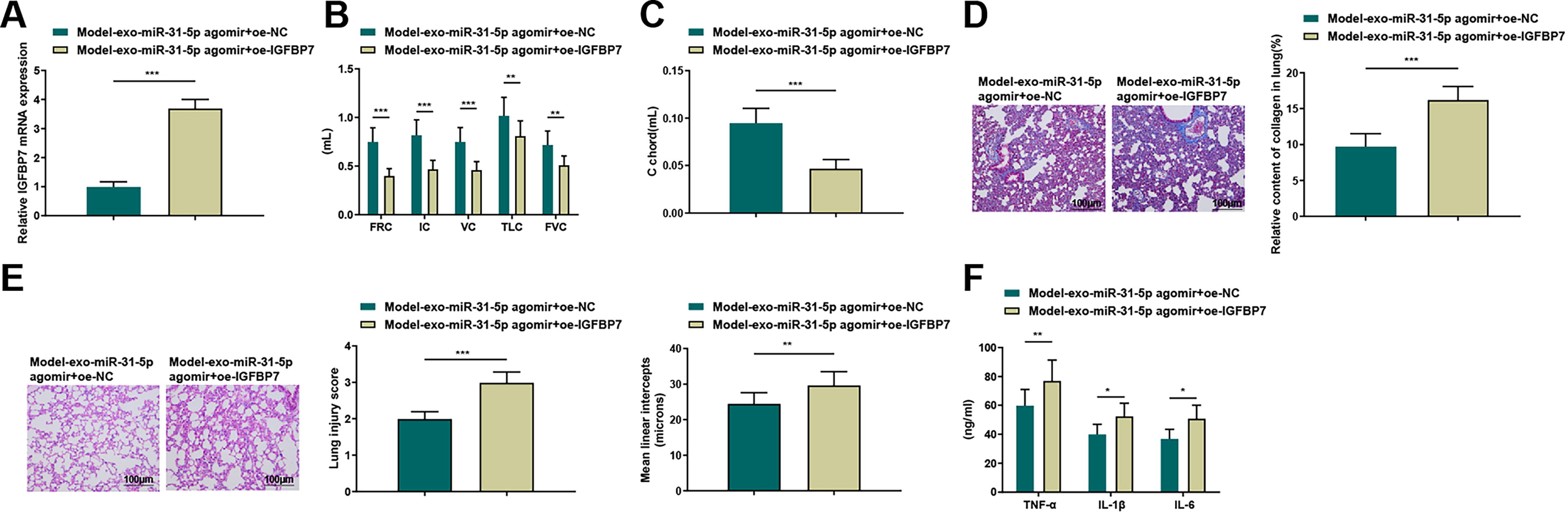
BMSCs-Exos affect PF in mice by delivering miR-31-5p to regulate IGFBP7.
DISCUSSION
IPF is a special form of chronic progressive fibrotic interstitial pneumonia, mainly occurring in the elderly and confined to the lungs. 35 This study investigated the mechanism by which miR-31-5p in MSCs-Exos targets and regulates IGFBP7 to affect PF.
By successfully establishing a mouse model of PF and isolating BMSC-Exos, we systematically examined the expression changes of miR-31-5p in Exos and its therapeutic effects. The results showed that miR-31-5p expression was downregulated during PF progression. Furthermore, BMSC-Exos effectively inhibited lung inflammation and fibrosis progression by delivering miR-31-5p, thereby improving lung function. This suggests that miR-31-5p may be a key effector molecule mediating the antifibrotic effects of MSCs-Exos. This finding is highly consistent with previous studies on the therapeutic effects of MSCs-Exos in lung injury. Multiple studies have demonstrated that MSCs-Exos can prevent or reverse bleomycin-induced PF, 10 ameliorate silicosis-induced lung function impairment, 9 and exert protective effects against hyperoxia-induced alveolar fibrosis. 36 –38 Notably, the protective effects of miR-31-5p extend beyond PF and have demonstrated significant efficacy in other disease models: in a model of vertebral endplate chondrocyte apoptosis, downregulation of miR-31-5p impairs the protective effects of MSCs-Exos 39 ; in cystic fibrosis, restoring miR-31 expression suppresses inflammation by inhibiting cathepsin S activity. 40 Mechanistically, miR-31-5p may exert its therapeutic effects via multiple pathways: in lung fibroblasts, it can alleviate oxidative stress and inflammation by upregulating circANKRD36; 14 and it can also block the profibrotic activity of transforming growth factor β1 and inhibit fibroblast fibrogenesis. 13 These findings not only confirm the conserved protective role of miR-31-5p in different disease models but also highlight its unique therapeutic value in PF. 40
Furthermore, we confirmed a targeting relationship between miR-31-5p and IGFBP7. miR-31-5p was shown to directly bind and suppress IGFBP7 expression. Additional experiments revealed that BMSC-Exos modulated IGFBP7 levels by delivering miR-31-5p, thereby influencing the course of PF in mice. IGFBP7 has been reported to enhance the osteogenic differentiation of BMSCs, 41 and it has also been implicated as a therapeutic target in sepsis-induced acute lung injury, where its suppression alleviated lung dysfunction features in a cecal ligation and puncture model. 42 Other studies have shown that BMSC-Exos overexpressing miR-29b-3p alleviate IPF through targeting FZD6, 43 and human embryonic stem cell-derived exosomes protect lungs from bleomycin toxicity via the miR-17-5p/Thbs2 axis. 21 These studies collectively suggest that the therapeutic actions of exosomal miRNAs through targeting specific signaling molecules represent a common mechanism, although the regulatory networks formed by different miRNAs and their targets are highly specific. This study focuses on uncovering the novel therapeutic axis of miR-31-5p/IGFBP7 in PF.
To shortly summarize, exosomal miR-31-5p alleviates PF in mice by targeting and suppressing IGFBP7 expression. This study provides the first evidence of the involvement of exosomal miR-31-5p in PF pathogenesis and offers new insights into its molecular mechanism. However, several limitations must be acknowledged. First, this study was conducted in a murine model, and its findings require validation in human tissues or patient-derived systems. Second, the specific lung cell types targeted by Exos were not identified, limiting our understanding of their cellular interactions. Third, although IGFBP7 was confirmed as a direct target of miR-31-5p, other downstream pathways may also contribute to the observed effects.
In conclusion, our findings provide new mechanistic insights into how BMSC-derived exosomal miR-31-5p mitigates PF via IGFBP7 suppression. The novelty of this study lies in the identification of miR-31-5p as a key effector mediating the antifibrotic effects of BMSC-Exos, the discovery and validation of IGFBP7 as a novel target, and the elucidation of a new regulatory mechanism for PF improvement through miR-31-5p-mediated IGFBP7 inhibition. This opens new avenues for PF treatment and supports further development of exosome-based miRNA delivery strategies. Future studies should focus on employing cell-specific labeling to trace the targeting of Exos in lung tissue, constructing conditional IGFBP7 knockout models to confirm its role in specific cell types, developing targeted miR-31-5p delivery systems to enhance therapeutic precision, and exploring potential synergistic interactions with other known antifibrotic pathways.
Footnotes
AUTHORS’ CONTRIBUTIONS
L.Z.: Finished the study design. K.Q.: Finished the experimental studies. C.Z.: Finished the data analysis. J.W.: Finished the manuscript editing. All authors read and approved the final version of the manuscript.
AUTHOR DISCLOSURE STATEMENT
The authors declared that they have no conflicts of interest regarding this work.
FUNDING INFORMATION
No funding was received for this article.