



Abstract
In gene therapy using adeno-associated virus (AAV) vectors, treatment-induced anti-AAV antibodies pose barriers for re-administration of the same or different AAV serotype vectors. We aimed to investigate whether the administration of AAV5, AAV8, or AAV9 in Cynomolgus monkeys resulted in the formation of cross-reactive antibodies. To achieve this, we developed a Biacore SPR-based total binding antibody (TAb) assay to identify anti-AAV antibodies in monkey plasma and assess the cross-reactivity of these antibodies against AAV5, AAV8, or AAV9 vectors on a sensor chip. AAV5, AAV8, and AAV9 vectors were immobilized onto the surface of a CM5 sensor chip on Fc2, Fc3, and Fc4 flow cells, respectively, using amine coupling, while Fc1 served as a reference. Plasma samples flowed through four channels, followed by injecting anti-monkey IgG and IgM antibodies to determine the immunoglobulin (Ig) isotypes. We analyzed TAb against the AAV serotypes in the plasma using a Biacore-based TAb assay 29 days after administration to evaluate the anti-AAV antibody responses. The TAb detected by the Biacore-based assay showed cross-reactivity between antibodies against AAV8 and AAV9; however, there was minimal cross-reactivity between antibodies against AAV5 and those against AAV8 or AAV9. Both IgG and IgM TAb were detected at 29 days post-dosing, and the antibody profiles determined by both the Biacore and ELISA platforms were comparable. The Biacore assessment confirmed the absence of cross-reactivity of anti-AAV5 antibodies against AAV8 and AAV9 vectors, and vice versa. This absence of cross-reactive antibodies against a specific AAV serotype indicated the possibility of re-administering a different AAV serotype.
Keywords
INTRODUCTION
Gene therapy using adeno-associated virus (AAV) vectors has emerged as a promising approach for treating various genetic disorders, including hemophilia, muscular dystrophy, and retinal diseases. AAV vectors are favored for their ability to deliver therapeutic genes to target cells with high efficiency and minimal pathogenicity.
The immune response to AAV vectors is complex and involves both innate and adaptive immunity.1–3 Upon infection by wild-type AAV, the host immune system can recognize the AAV capsid proteins as foreign, producing preexisting neutralizing antibodies (NAb) and total binding antibodies (TAb). NAb can directly inhibit the infectivity of the AAV vector by preventing it from entering target cells. At the same time, TAb can bind to the vector and facilitate its clearance from the circulation. Treatment-emergent antibodies pose a significant barrier to re-administration of the same or different AAV serotypes after administration of AAV vectors. 4 Both NAb and TAb have been used to prescreen patients for AAV-mediated gene therapy.5–10
Re-dosing with AAV vectors can be challenging due to the development of treatment-emergent immunity in patients who have previously been treated with AAV vectors.11,12 If the patient is re-dosed with the same AAV serotype, the treatment-emergent NAb can block the vector’s uptake and transduction, reducing efficacy. Using alternative AAV serotypes or capsid variants not recognized by pre-existing NAb can be one of the strategies.
Understanding the cross-reactivity of anti-AAV antibodies is crucial for developing strategies to mitigate immune responses and enable the successful re-administration of AAV vectors.13,14 The lack of cross-reactivity would suggest that different AAV serotypes could be used sequentially without being neutralized by pre-existing antibodies.
To address this challenge, we aimed to evaluate the cross-reactivity of anti-AAV antibodies in cynomolgus monkeys following intravenous administration of AAV5, AAV8, or AAV9 vectors. We employed a surface plasmon resonance (SPR)-based TAb assay using the Biacore system to detect anti-AAV antibodies in monkey plasma and assess cross-reactivity. The SPR-based assay offers several advantages, including label-open-access detection, automated operation, rapid analysis in real-time, and high specificity. 15
In this study, we immobilized AAV5, AAV8, and AAV9 vectors onto a CM5 sensor chip surface and analyzed plasma samples from AAV vector-injected cynomolgus monkeys for the presence of anti-AAV antibodies. We also evaluated the total antibody (TAb) responses using an ELISA assay and compared the antibody profiles determined by the Biacore SPR-based TAb assay and ELISA platforms.
MATERIALS AND METHODS
Materials
HBS-EP+ buffer (10 mM HEPES, 150 mM NaCl, 3 mM EDTA, 0.05% Surfactant P20) was purchased from Cytiva (Cat# BR-1006-69). Biacore T200 and Series S Sensor Chip CM5 (Cat# BR-1006-68) were purchased from Cytiva (Marlborough, MA) and used for biosensor analyses. An amine coupling kit was purchased from Cytiva (Cat# BR100050, Cytiva). Goat anti-monkey IgG (Cat# AAI42) antibody was purchased from Bio-Rad (Hercules, CA). Goat anti-monkey IgM (μ-chain specific) antibody (SAB3700778) was purchased from Millipore Sigma (Burlington, MA). Non-specific binding (NSB) Reducer (Cat# BR100691) was purchased from Cytiva.
Animals and AAV administration
Monkeys were housed in groups of 2–3 per cage by dosing group. They were all males between 2.5 and 3.5 years old and weighed between 2.9 and 4.5 kg. There were no group differences in age, weight, or sex.
Cynomolgus monkeys were intravenously administered with AAV5, AAV8, or AAV9 vectors as described before. 16 The Institutional Review Board approval number was DN20027. Plasma samples were collected on day 29 post-administration for analysis. Animals were grouped based on the AAV serotype and dose received. The AAV5 group included animals 1101 to 1103, each dosed at 1.14 × 10¹⁴ vg/kg. The AAV8 group consisted of animals 2101 and 2102, also dosed at 1.14 × 10¹⁴ vg/kg. Two groups received AAV9 vectors: animals 3101 to 3103 received 1.14 × 10¹⁴ vg/kg, while animals 4001 and 4002 received a higher dose of 2 × 10¹⁴ vg/kg. Animal ID numbers were used consistently to identify treatment groups in the data figures and analyses.
Biacore-Based TAb Assay
We developed a Biacore-based TAb assay to detect anti-AAV antibodies directly. AAV5, AAV8, and AAV9 vectors in Na acetate buffer with pH 4.5 were immobilized on the dextran matrix of the CM5 sensor chip to a level of ∼10,000 response units (RU) on Fc2, Fc3, and Fc4, respectively, using an amine coupling kit. Fc1 served as a reference and was subtracted from Fc2, Fc3, and Fc4, respectively. Plasma samples were diluted 100-fold in HBS-EP buffer containing 1× NSB. They were flowed across the four flow channels, one sample per cycle, followed by injection of anti-monkey IgG and IgM antibodies, each at 10 μg/mL in HBS-EP buffer, to determine isotypes. Following the regeneration of the sensor chip, the next sample was injected into the sensor chip.
Chemiluminescent enzyme-linked immunosorbent assay (ELISA)
TAb titers against the AAV serotypes were evaluated using ELISA assay.16,17 Plasma samples were diluted 10,000-fold and analyzed for TAb as relative luminescence units (RLU) on day 29 after administration. Goat anti-monkey IgG antibody was used as a detection antibody.
RESULTS
Assessment of dilution linearity of plasma samples
To determine how the dilution of plasma samples affected the measurement of specific interactions, the plasma sample from Monkey 2102, which was administered with AAV8, was diluted in HBS-EP buffer containing NSB reducer. The dilution ranges varied from 1:100 to 1:800. These diluted samples were injected into the AAV8-immobilized flow cell (Fc3) and the blank immobilized Fc1. Figure 1a provides a schematic representation of AAV5, AAV8, and AAV9 vectors immobilized on flow cells Fc2, Fc3, and Fc4, respectively. Figure 1b shows a two-fold increase in RU when comparing the dilution ratios of 1:800 to 1:400, 1:400 to 1:200, and 1:200 to 1:100. Figure 1c shows linear regression of dilution of plasma samples vs. Biacore binding RU with R2 = 0.998 and p = 0.0011.
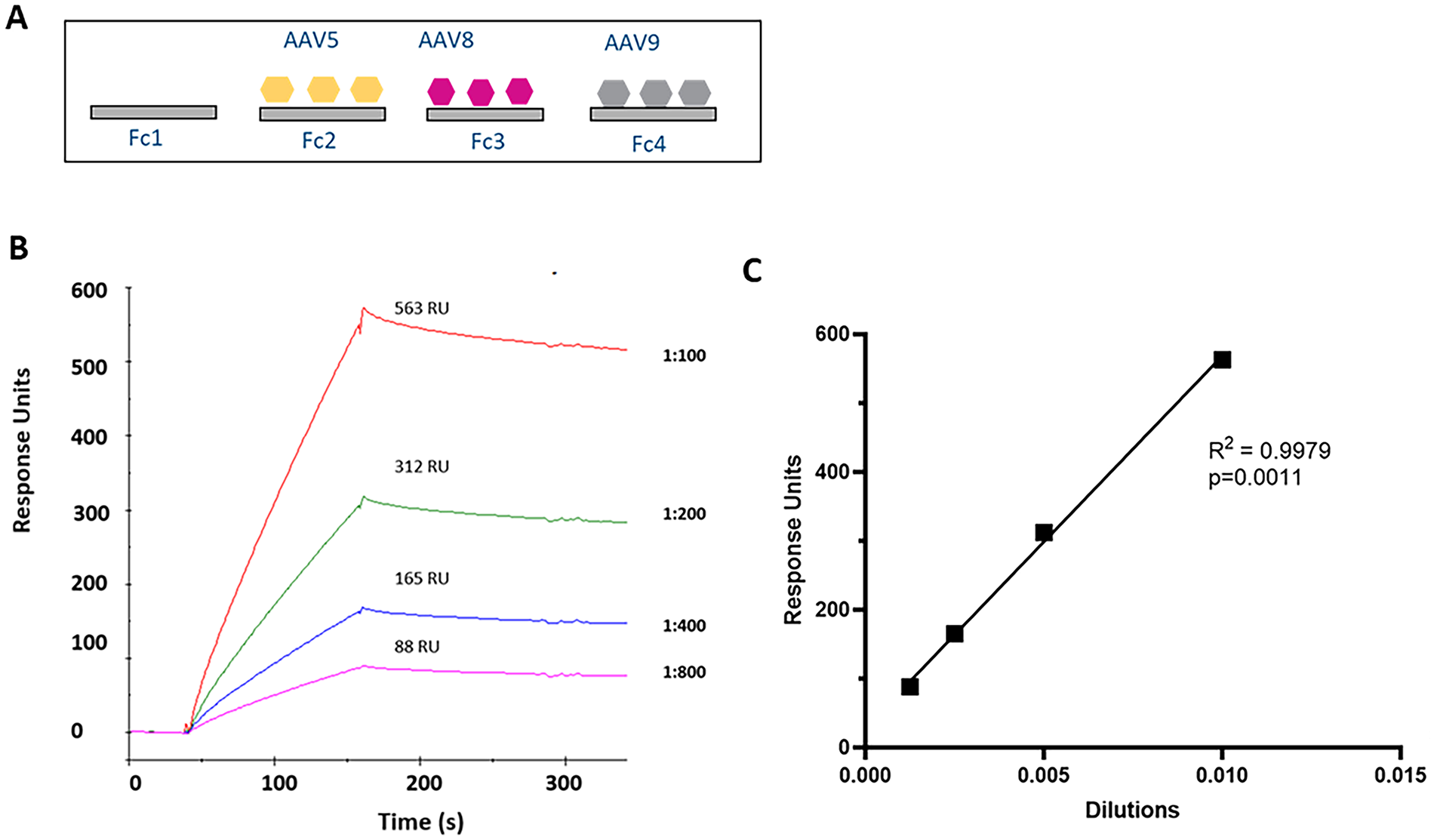
Assessment of sample dilution linearity.
An AAV5-immobilized sensor chip detects AAV5 antibodies from monkeys administered with AAV5 vectors
Figure 2 demonstrates that the AAV5-immobilized sensor chip detected plasma AAV5 antibodies in monkeys administered with AAV5 vectors, but not in those who received AAV8 or AAV9 vectors. This indicates minimal cross-reactivity between AAV5 vectors immobilized on the sensor chip and the plasma antibodies induced from AAV8- and AAV9-injected animals. Both IgG and IgM antibodies were identified as part of the anti-AAV5 response, using anti-IgG and anti-IgM antibodies, respectively.

Detection of anti-AAV5 antibodies in monkey plasma by Biacore after administering the AAV5 vector. Plasma samples from Monkeys 1101 to 1103, which were administered with AAV5 vectors, were analyzed using Biacore. The samples were injected onto the AAV5 sensor chip, one sample per cycle. After the association and dissociation phases, anti-IgG antibodies were injected, followed by anti-Anti-IgM antibodies. Additionally, plasma samples from monkeys that received AAV8 and AAV9 vectors were analyzed, but no response units were detected on the AAV5 sensor chip. The labeled animal ID numbers on curves refer to plasma samples from animals treated with AAV vectors, the same as for Figs 3 and 4.

Cross-reactivity of anti-AAV9 antibodies against immobilized AAV8 vector on sensor chip by Biacore. As expected, the plasma anti-AAV8 antibodies from monkeys 2101 and 2102, which were administered with AAV8 vector, were detected on the AAV8 sensor chip. Plasma samples from monkeys 3101 to 3103 and 4101 to 4102, which were administered with AAV9 vectors at different doses, were also injected onto the AAV8 sensor chip, one sample at a time, in a cycle. Plasma anti-AAV9 antibodies from AAV9-injected monkeys were detected on the AAV8 sensor chip due to cross-reactivity. Furthermore, plasma samples from monkeys 1101 to 1103, which were administered with the AAV5 vector, were loaded onto the AAV8 sensor chip, and the Biacore AAV8 sensor chip did not detect anti-AAV5 antibodies in these plasma samples.
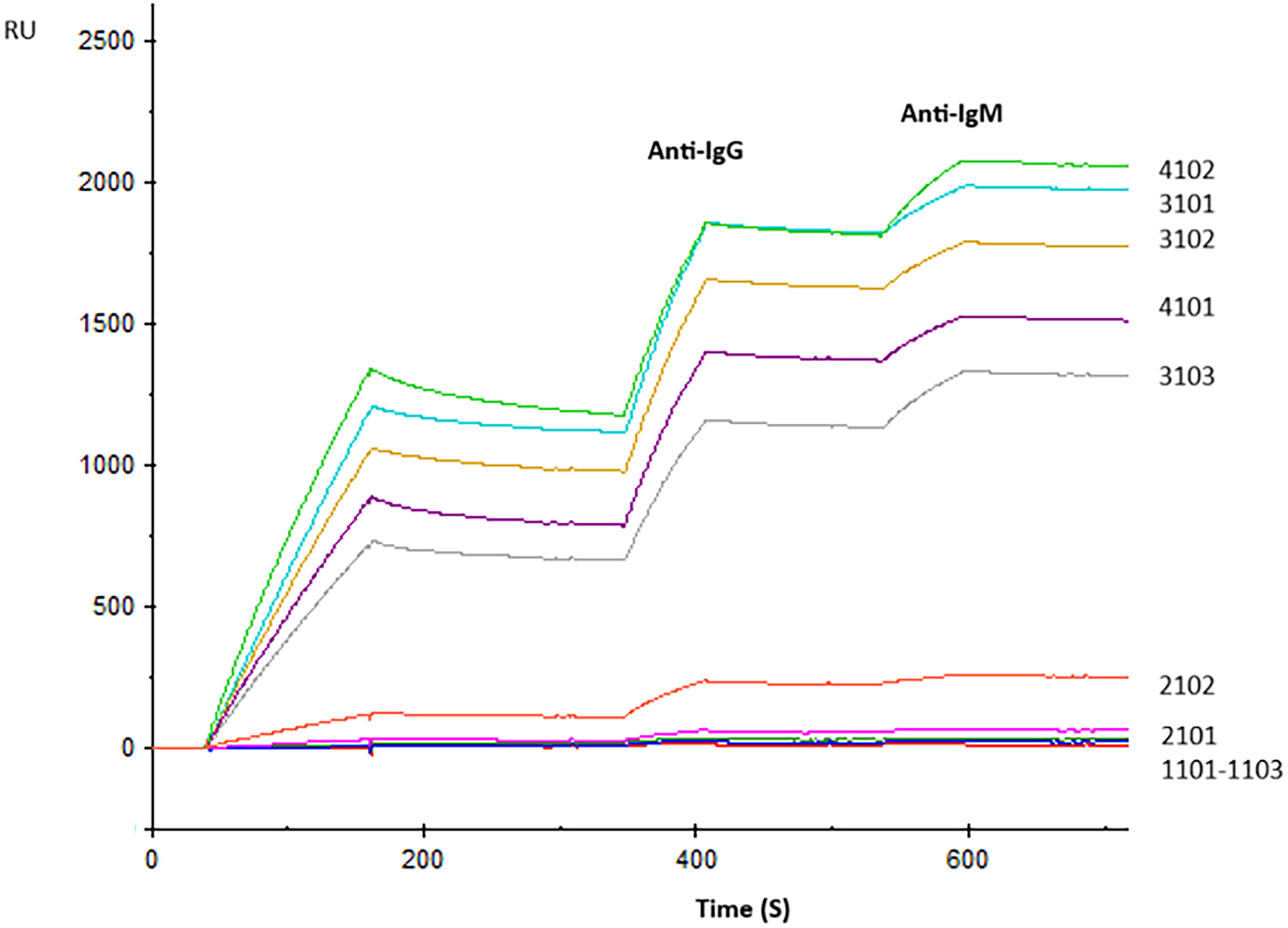
Detection of Anti-AAV9 Antibodies by Biacore and Assessment of Cross-Reactivity. Plasma samples from monkeys 3101 to 3103 and 4101 to 4102, which were administered with AAV9, were loaded onto the AAV9 sensor chip, one sample per cycle. Then, the anti-IgG and anti-IgM antibodies were injected. The AAV9 sensor chip detected anti-AAV8 antibodies from the plasma sample of monkey 2102, which was administered with AAV8, but it did not detect antibodies from monkey 2101. The AAV9 sensor chip did not identify anti-AAV5 antibodies in plasma samples from monkeys administered with AAV5 (animals 1101 to 1103).
An AAV8-immobilized sensor chip detects AAV8 antibodies and exhibits cross-reactivity with AAV9 antibodies
The AAV8-immobilized sensor chip detected AAV8 antibodies in monkeys that were administered with the AAV8 vector (Fig. 3). AAV8 vectors also exhibited cross-reactivity with anti-AAV9 antibodies from monkeys that received AAV9 vectors, with binding levels reaching approximately one-third of those observed for anti-AAV8 antibodies binding to AAV8 capsid. (Fig. 3). Both IgM and IgG antibodies to AAV8 and AAV9 were identified. However, the sensor chip did not demonstrate any cross-reactivity with AAV5 antibodies from monkeys who were given the AAV5 vector (Fig. 3).
AAV9-immobilized sensor chip detects AAV9 antibodies and exhibits cross-reactivity with AAV8 antibodies
The AAV9-immobilized sensor chip detected AAV9 antibodies in monkeys who had been administered with AAV9 vectors at two different doses, Group 3101 to 3103 receiving 1.14E14 vg/kg, and Group 4101 to 4102 receiving 2E14 vg/kg (Fig. 4). We did not find a difference in antibody response units between the two AAV9 groups. Furthermore, the chip exhibited cross-reactivity with AAV8 antibodies from monkeys treated with AAV8 vectors. Both IgM and IgG antibodies to AAV8 and AAV9 were identified. However, the AAV9 chip did not detect cross-reactivity with plasma AAV5 antibodies induced by monkeys receiving AAV5 vectors.
Comparison of AAV antibodies detected by Biacore and ELISA
The detection of AAV5, AAV8, and AAV9 antibodies was compared using Biacore and ELISA. We measured the RU from the Biacore analysis during the peak of the association phase in 10 monkey plasma samples. The response units in the anti-IgG and anti-IgM binding were not included. The data collected from this analysis are shown in Figure 5a, which quantifies the antibody responses against the different AAV serotypes. In addition, we conducted an ELISA to assess the antibody levels in the monkey plasma samples receiving the same AAV serotypes. The RLU obtained from the ELISA results is shown in Figure 5b. This analysis allows for a comparative study of the antibody profiles detected by both methods. Anti-AAV antibody cross-reactivity was not assessed using the ELISA format. Figure 5c shows the correlation between ELISA and Biacore measurements for each sample. Pearson’s correlation coefficient (r) was 0.88, and the associated p value was 0.0008. The strong positive correlation supported the consistency between the two assay platforms.
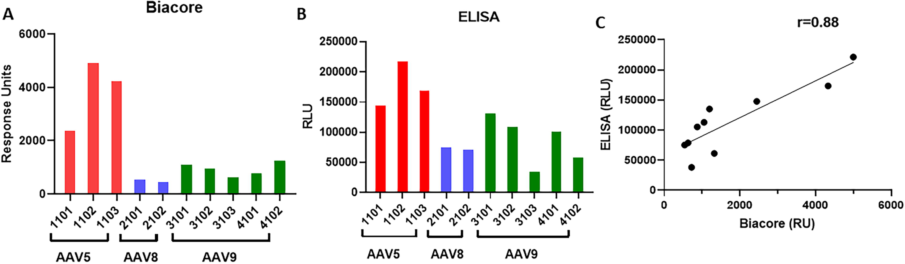
Comparison of anti-AAV antibodies analyzed by Biacore and ELISA. Plasma samples collected on Day 29 from monkeys administered with either AAV5, AAV8, or AAV9 were analyzed by Biacore
DISCUSSION
The AAV-immobilized sensor chip represents a new tool for detecting antibodies specific to AAV serotypes, which is crucial for understanding immune responses in animal models. In this study, monkeys injected with AAV5, 8, and 9 vectors showed a marked presence of AAV5, 8, and 9-specific antibodies in their plasma. This finding is shown in Figures 2, 3, and 4, where the sensor chips identified these antibodies.
The AAV5 sensor chip did not detect AAV antibodies in monkeys administered with AAV8 and AAV9 vectors, indicating a significant level of specificity. This low cross-reactivity was consistent with 58% identity between the capsid VP1 amino acid sequences of AAV5 and AAV8/9. 18 Cross-reactivity of the anti-AAV8 with AAV9 vector and that of the anti-AAV9 with AAV8 vectors were consistent with 93% identity between capsid VP1 amino acid sequences of both serotypes. 18 Both IgG and IgM antibody classes were identified in response to the administration of AAV5, AAV8, and AAV9. Using anti-IgG and anti-IgM antibodies for their detection demonstrated the effectiveness of the sensor chip in distinguishing between these antibody classes. When screening pre-existing AAV NAb from human or minipig serum samples, some samples tested positive for several AAV serotypes.19,20 This suggests either co-infection with several wild-type AAV serotypes or cross-reactivity. 21 Our data suggest that cross-reactive antibodies only partially bind to closely related AAV serotypes. As shown in Figure 3, anti-AAV9 antibodies from AAV9 vector-injected monkeys exhibited measurable cross-reactivity to AAV8 capsid immobilized on the Biacore sensor chip, with binding levels reaching approximately one-third of the response observed for anti-AAV8 antibodies from AAV8-injected animals binding to the same AAV8 capsid. These findings indicate that while cross-reactivity exists, it is generally of lower magnitude compared with serotype-specific responses. Therefore, when antibody titers against multiple AAV serotypes are comparable in a given plasma sample, this pattern may more likely reflect prior co-infection with multiple distinct AAV serotypes rather than cross-reactivity alone.
The Biacore-based assessment has provided insights into the cross-reactivity of anti-AAV antibodies following the intravenous administration of AAV5, AAV8, and AAV9 vectors in monkeys, in which pre-existing AAV antibodies tested negative for each AAV serotype. 16 The results confirmed that anti-AAV5 antibodies did not cross-react with AAV8 and AAV9 vectors, and conversely, AAV8 and 9 antibodies did not cross-react with AAV5 vector. This suggests that re-administration of a different AAV serotype may be possible, depending on the routes of delivery.
The minimal cross-reactivity observed between AAV5 antibodies and AAV8/9 serotypes is significant. It suggests that sequential administration of different AAV serotypes could be a viable strategy to overcome immune responses and maintain therapeutic efficacy. This finding is consistent with previous studies that have reported limited cross-reactivity among specific AAV serotypes, thereby supporting the potential for using different serotypes sequentially to achieve sustained therapeutic outcomes.22,23
Furthermore, the analysis revealed that both IgG and IgM antibody types were part of the anti-AAV5 immune response. This was confirmed through the use of anti-IgG and anti-IgM antibodies. The presence of both IgG and IgM indicates a robust immune response, indicating that the administration of AAV5, 8, and 9 elicited a strong and specific antibody production in the tested monkeys. This insight is important for understanding the immune dynamics associated with AAV vector administration and can inform future therapeutic applications involving these vectors.
The Biacore SPR-based TAb assay proved to be a reliable method for detecting and characterizing anti-AAV antibodies. This assay offers several advantages over traditional ELISA methods, including label-open-access detection and high specificity. In direct-binding ELISA assays, the concentration and specificity of the secondary antibody play a critical role in determining background signal, sensitivity, and dynamic range. Careful optimization is required to minimize NSB while adequately detecting low-abundance antibodies. 9 Bridging ELISA assays rely on a balanced ratio between capture and detection AAV vectors, and assay performance can be influenced by the degree of labeling on the AAV vectors. 10
The Biacore assay does not require species-specific secondary antibodies, allowing for more direct and unbiased comparison of anti-AAV antibody responses across different species, including mice, NHPs, and humans. In addition, SPR provides real-time, label-open-access measurement of binding kinetics, offering insight into antibody presence and affinity. These features make SPR a valuable platform for assessing total anti-AAV antibody responses in preclinical and clinical studies. The comparable antibody profiles determined by Biacore and ELISA platforms validated the robustness of the Biacore assay. By providing a comprehensive assessment of anti-AAV antibody responses, the Biacore assay can help identify optimal strategies for vector re-administration. 14
In conclusion, the Biacore SPR-based TAb assay offers a reliable and efficient method for detecting and characterizing these antibodies, supporting the potential for re-administration of different AAV serotypes in gene therapy applications.
AUTHORS’ CONTRIBUTIONS
Y.D. designed and performed the experiments. All authors contributed to the article and approved the submitted version. G.W. contributed to initial method development. G.W. was a summer intern from the University of Pennsylvania.
Footnotes
ACKNOWLEDGMENTS
The authors thank Uma Kavita for conducting the ELISA experiment.
AUTHOR DISCLOSURE STATEMENT
The author declares that the research was conducted without other commercial or financial relationships that could create a conflict of interest.
FUNDING INFORMATION
No funding was received for this article.
DATA AVAILABILITY STATEMENT
The data supporting this study’s findings are available on request from the corresponding author.