



Abstract
CLN5 disease, caused by mutations in the CLN5 gene, is a form of neuronal ceroid lipofuscinoses (Batten disease). Patients suffer progressive motor dysfunction, vision loss, seizures, and dementia, leading to premature death. Here, we report a preclinical study of AAV9-mediated gene therapy in a Cln5−/− mouse model. Single-dose AAV9 carrying human CLN5 driven by the CAG or human synapsin 1 promoter (hSYN) was administered via intracerebroventricular injection into neonatal and juvenile Cln5−/− mice. Treatment efficacy was evaluated by assessment of neurodegeneration, neuroinflammation, locomotor function, and survival. AAV9 expressing CLN5 driven by the hSYN promoter significantly alleviated neurodegeneration, improved biochemical and glycosphingolipid profiles, neuropathological and locomotor function, and extended lifespan of the Cln5−/− mice. However, gene transfer employing the CAG promoter demonstrated limited therapeutic efficacy. Furthermore, delayed intervention in juveniles provided superior therapeutic response compared with early neonatal intervention and normalized lifespan. Finally, blood plasma neurofilament light that is significantly elevated in the Cln5−/− mice is restored to normal wildtype levels following treatment. These results indicate that brain-directed adeno-associated virus (AAV) gene therapy could be a promising treatment strategy for CLN5 disease and efficacy might be monitored using a noninvasive blood plasma biomarker.
Keywords
INTRODUCTION
The neuronal ceroid lipofuscinoses (NCLs) are a group of fatal inherited neurodegenerative diseases with an incidence of approximately 1–3 per 100,000 live births.1,2 Currently, 13 genetic types of NCLs sharing similar clinical and pathological features have been identified (CLN1-8, 10–14), 3 resulting from variation in different genes (NCL mutation database, https://www.ucl.ac.uk/ncl-disease). A pathological hallmark of NCLs is the accumulation of endolysosome-derived storage material in cells throughout the body, particularly neuronal cells of the brain, which leads to widespread neurodegeneration in the brain, retinal degeneration, and neuroinflammation. Affected children progressively develop blindness, motor degeneration, epilepsy, and dementia, eventually reaching total dependency and premature death. Medications mainly involve symptom management, and to date, there is only one clinically approved treatment, a targeted enzyme-replacement therapy for CLN2 disease. 4
CLN5 disease is biallelic and caused by homozygous or compound heterozygous mutations in CLN5, which encodes a soluble lysosomal lumen protein, CLN5. A recent study has elucidated the function of CLN5 as the bis(monoacylglycero)phosphate (BMP) synthase that is essential for conversion of lysophosphatidylglycerol to BMP in the late endosomes/lysosomes. 5 Patients typically have an onset of disease in late infancy, but cases with adolescent or adulthood disease onset have also been reported.6–8 Symptoms include vision loss, motor dysfunctions, ataxia, epilepsy, and cognitive impairment, with variable rates of disease progression leading to death around 14–36 years of age.6,9 To date, there is no approved targeted treatment for CLN5 disease.
Several mammalian animal models mimicking CLN5 disease are currently available.10–15 The Cln5 knockout (Cln5−/−) mouse model was generated in 2004 (14) by insertion of a Neo cassette into exon 3 of the mouse Cln5 gene resulting in a frame shift and an early stop codon. The Cln5−/− mice present accumulation of autofluorescent storage materials in the brain and retina, and ultrastructural analysis reveals a mixture of lamella profiles including curvilinear and fingerprint patterns, 14 as in the human disease. 16 Further characterization of the model demonstrates early onset of brain astrogliosis from 1 month of age and relatively late onset of neurodegeneration from 4 months of age, which first occurs in the cortex and subsequently progresses into the thalamus. 17 Although progressive vision loss has been confirmed, full characterization of potential motor dysfunctions of this Cln5−/− mouse model is still required.
As a monogenic disorder, CLN5 disease is an attractive target for gene therapy as CLN5 is a soluble lysosomal protein and can be secreted by mammalian cells when it is overexpressed.18–22 This allows a potential mechanism for cross-correction and may enhance the efficacy of gene therapy. Viral vector-mediated gene therapy studies for CLN5 disease utilizing the CLN5 Borderdale sheep model have been reported.23–26 The most recent studies published in 2023 showed that intracerebroventricular (ICV) administration of an scAAV9 vector carrying the ovine Cln5 gene driven by the CBh promoter resulted in long-term efficacy and safety.23,26 Survival of the CLN5 sheep was extended, disease progression was delayed, and brain neuropathology was attenuated, particularly when the sheep were treated at presymptomatic and early-symptomatic disease stages. Moreover, ICV and intravitreal administration of the gene therapy provided long-term efficacy for both the brain and retinal aspects.
Here, we conduct a systematic and in-depth study to optimize gene therapy for CLN5 disease. We constructed two AAV9 vectors, both carrying the human CLN5 gene but driven by two different promoters (the human synapsin 1 [hSYN] vs. the synthetic CAG promoter in which the cytomegalovirus enhancer is fused to the promoter of the chicken β-actin gene) and compared the efficacy following neonatal and juvenile ICV administration. The results showed that the vector employing the hSYN promoter produced superior therapeutic effects. Unexpectedly, delayed intervention at the juvenile stage with the AAV9.hSYN.CLN5 vector resulted in the highest efficacy with restoration of normal lifespan. Furthermore, we show that the neurofilament light (NfL) blood plasma biomarker for neurodegeneration is elevated in the untreated mice but completely normalized following gene therapy with AAV9.hSYN.CLN5.
MATERIALS AND METHODS
Plasmid and viral vector production
The human CLN5 cDNA (NCBI RefSeq NM006493.2) was cloned into an adeno-associated virus (AAV) construct containing the promoter (human synapsin I hSYN or the synthetic CAG promoter), the Woodchuck Posttranscriptional Regulatory WPRE Element, and human growth hormone polyadenylation signal sequence, flanked by AAV2 inverted terminal repeats. Human Embryonic Kidney 293T cells were triple transfected with a 1:1:3 ratio of the inverted terminal repeat (ITR)-containing plasmid carrying the transgene cassette, a helper plasmid expressing AAV2 Rep and AAV9 Cap and a third construct containing the adenovirus helper functions (HGTI), using 3.5 mg/mL DNA polyethylenimine MAX (Polysciences, Inc. Warrington, PA, USA). Cells were harvested at 72 h post-transfection and lysed by three times of freeze–thawing cycles (−80°C to 37°C) in 1× Tagment DNA (TD) buffer. Vectors were purified by size exclusion on a Sephacryl S300 column, followed by anion-exchange chromatography using a POROS 50 HQ column (GE Healthcare). Vectors were washed in 1× phosphate-buffered saline (PBS) with 1 mM MgCl2 and 2.5 mM KCl (PBS-MK) and concentrated using Vivaspin 4 (10 kDa) concentrators (Vivaproducts, Littleton, MA). Viral genome (vg) titers were determined by quantitative real-time PCR using a probe-based assay annealing to the ITR of the genomic plasmid.
Animal welfare and ICV injection
All animal work was approved by the UK Home Office for the conduct of regulated procedures under license (Animal Scientific Procedures Act, 1986) and followed the Animal Research Reporting of In Vivo Experiments (ARRIVE) guidelines and recommendations. A loss of >15% body weight or signs of any serious health issues were defined as a humane end point. Mice were maintained in group-housed ventilated cages, on a 12 h light/dark cycle with access to water and food ad libitum. Mice were randomly allocated to experimental groups. A total of 100 mice were used in the study: n = 79 (male 41 and female 38) for Cln5−/− mice and 21 (male 10 and female 11) for wildtype (WT) C57BL/6J mice. Neonatal ICV injection was carried out as previously described. 27 At postnatal day 0–1, Cln5−/− neonates were injected with 5 × 10¹¹ viral vector genomes via bilateral ICV injection targeting the anterior horn of the lateral ventricle using a 33-gauge needle (Hamilton, Reno, NV, USA). For delayed intervention (termed “juvenile”), Cln5−/− mice at 4 weeks of age were treated via single ICV injection of 1 × 10¹² viral vector genomes with a stereotaxic frame under isoflurane anesthesia. Age-matched WT and untreated Cln5−/− mice were employed as controls. Mice were euthanized at 6 months of age for short-term study, or at their humane end point (defined as 15% loss of maximum body weight or signs of paralysis, spasticity, or unconsciousness for more than 4 h) or 18 months of age for long-term study, by transcardial perfusion with PBS under terminal isoflurane anesthesia, and brains, major visceral organs, and plasma samples were collected.
Behavioral analysis
Mice were weighed bi-weekly, and behavioral tests were conducted monthly from 2 months of age.
For open field test, the mouse was placed in the center of a square plexiglass chamber of 27 × 27 cm2 and allowed to freely explore the chamber for 5 min as a session. Animals were filmed from the top of the chamber. The analysis of the tests was performed using ANY-maze Behaviour Tracking Software v. 4.99 (Stoelting, Dublin, Ireland).
For accelerating rotarod analysis, tests were conducted with a Touchscreen Rota Rod (Panlab, Harvard apparatus, Cambridge, UK). The mice were first habituated to the rotating rod a week prior to the first test, at a constant speed of 4 rpm for up to 5 min, or until they fell if mice were unable to run longer. For each test, five runs were acquired per mouse, with appropriate rest (>1 min) between runs. Rotation was electronically set for acceleration from 4 to 40 RPM across 5 min with increases at even intervals.
For automated gait analysis, tests were conducted with the CatWalk system (Noldus, Wageningen, The Netherlands), where mice were filmed from underneath walking a minimum of three times across the walkway stage of the device. Runs were assigned and analyzed using the CatWalk XT software v9.1 (Noldus) to assess footprint, stride, and various parameter measurements.
For the foot-fault test, the mouse was placed on a rodent metal mesh sheet elevated 20 cm from a surface. The animal was allowed to walk around the device, and the run was recorded from underneath the animal for 1 min. The number of total steps and the number of foot faults, defined as misplacement of a paw slipping through the grid, were manually counted.
Brain histological staining
Following transcardial perfusion and brain dissection, one hemisphere of the brain was fixed in 4% paraformaldehyde for 48 h at 4°C and then transferred into PBS containing 30% sucrose for cryoprotection and long-term storage at 4°C. Hemispheres were then cryo-sectioned at −20°C using a cryostat microtome, and serial brain sections were collected at 40 mm thickness.
Immunohistochemical staining was used for neuronal and glial analyses. Endogenous peroxidase activity was depleted by incubating sections in 1% H2O2 in Tris-buffered saline (TBS) for 30 min followed by three times washing in TBS. Endogenous nonspecific protein binding was blocked by incubation in 15% normal serum (Sigma) in TBS-T (TBS with 0.3% Triton X-100) for 30 min. Sections were incubated overnight at 4°C in 10% normal serum in TBS-T with primary antibodies for NeuN (1:500, MAB377, Millipore, MA, USA), Glial Fibrillary Acidic Protein (GFAP) (1:2000, MAB3402, Millipore, MA, USA), or CD68 (1:2000, MCA1957, AbD Serotech, Hemel Hempstead, UK). Following three washes in TBS, sections were incubated in 10% normal serum in TBS-T with biotinylated secondary antibodies anti-mouse, anti-rabbit, or anti-rat IgG (1:1000, Vector Laboratories, Inc., Burlingame, CA, USA) for 2 h at room temperature. Staining was visualized using Vectastain avidin–biotin solution (ABC, Vector Laboratories) and 3,3′-Diaminobenzidine (Sigma). Sections were then mounted on slides, air-dried, dehydrated in ethanol, cleared in histoclear (National Diagnostics, Hessle, UK), and finally coverslipped with DPX (VWR, East Grimstead, UK). Representative images were captured using a live video camera (Nikon, DS-Fil, Melville, NY, USA) mounted onto a Nikon Eclipse E600 microscope.
Immunofluorescent staining was used for the analysis of transgene expression and subunit c of mitochondrial ATP synthase (SCMAS) analysis. Sections were first mounted on slides, and endogenous nonspecific protein binding was blocked by incubation in 15% normal serum (Sigma) in TBS-T (TBS with 4% Triton X-100) for 60 min. Sections were incubated for 2 h at room temperature in 10% normal serum in TBS-T with primary antibodies for CLN5 (1:100, ab170899 Abcam), NeuN (1:100, MAB377, Millipore), or SCMAS (1:100, ab181243, Abcam), followed by secondary antibody incubation with appropriate fluorescent Alexa-Fluor-labeled IgG secondary antibodies (Alexa-Fluor goat anti-rabbit 488, Invitrogen A-11008, 1:200; Alexa-Fluor goat anti-mouse 546, Invitrogen A-11003, 1:200). After washing in TBS, lipofuscin autofluorescence was quenched with TrueBlack (Biotium, USA), and sections were coverslipped with Fluoromount G with 4′,6-diamidino-2-phenylindole (Invitrogen). Slide-scanned images at 10× magnification for immunofluorescent stained sections were collected using a Zeiss Axioscan Z1 (Zeiss Microscopy Deutschland GmbH, Oberkochen, Germany).
Quantitative analysis of immunohistology
Quantification of neurons was carried out on NeuN-stained serial brain sections using StereoInvestigator software (MBF Bioscience, Williston, VT, USA) on a Nikon Optiphot microscope (Nikon) attached to a Q-Imaging Model 01-MBF-2000R-CLR-12 camera (MBF Bioscience). Neuronal counts within the S1BF and Ventral Posteromedial and Ventral Posterolateral (VPM/VPL) regions were estimated using the optical fractionator probe. Using a 100× objective, NeuN-stained cells were counted. A border was traced around the region of interest, a grid was superimposed (grid size: S1BF, 225 × 225 mm2; S1BF Layer-V, 125 × 125 mm2; VPM/VPL, 175 × 175 mm2), and neurons were counted within a series of 50 × 50 mm2 counting frames arranged according to the sampling grid. A coefficient of error of 0.05–0.1 was achieved for all the counts indicating sufficient sampling efficiency. 28
Levels of GFAP, CD68, and SCMAS staining were measured by quantitative thresholding image analysis as previously described. 29 For the neonatal gene therapy study, 10 nonoverlapping images per section were captured for each region of interest using a live video camera (Nikon, DS-Fil) mounted onto a Nikon Eclipse E600 microscope at 40× magnification with constant light intensity. For the juvenile gene therapy study, slide-scanned images at 10× magnification for all stained sections were collected using a Zeiss Axioscan Z1 (Zeiss Microscopy Deutschland GmbH, Oberkochen, Germany), and regions of interest were then defined. Images were analyzed using Image-Pro Premier (Media Cybernetics, Cambridge, UK), where immunoreactivity is measured using a constant threshold that is applied to all images for each respective antigen. Data are presented as the mean percentage area of immunoreactivity for each region.
Glycosphingolipid analysis
Following transcardial perfusion and brain dissection, one hemisphere of the brain was snap frozen on dry ice and stored at −80°C until analysis. Frozen brain hemispheres were weighed and homogenized in ddH2O. Protein concentration of homogenates was assayed using Pierce BCA Protein Assay (Life Technologies), and samples were set up for extraction containing equal amounts of protein. Glycosphingolipids (GSLs) from brain homogenates were extracted and purified with chloroform:methanol (1:2, v/v) and C18 chromatography columns (Telos, Kinesis, UK). GSLs were dried down under nitrogen and digested with recombinant endoglycoceramidase I (rEGCaseI, custom synthesized by GenScript). Released glycans were fluorescently labeled with anthranillic acid (2AA). Purified 2AA-labeled oligosaccharides were separated and quantified by normal-phase high-performance liquid chromatography (NP-HPLC). 30 Individual GSL species were identified by their glucose unit values, calculated using the HPLC Empower software with a homopolymer dextran ladder. Results were normalized to total protein content.
Plasma NfL analysis
Blood sample was collected in an Ethylenediaminetetraacetic acid tube via cardiac puncture prior to transcardial perfusion, and plasma was then extracted via centrifugation and stored at −80°C until use. The plasma NfL levels were determined after being diluted 40× using single molecule array technology utilizing the Quanterix (Billerica, USA) NfL Advantage kit according to the manufacturer’s instructions on the HD-X platform at the UCL DRI Biomarker Factory. Intraplate coefficient of variation for quality control samples was <5%.
Statistical analysis
Data plotting and analysis were performed using Graphpad Prism software (version 9.1.1). Data were represented as means ± standard error of the mean. Statistical analysis was conducted using a one-way or two-way analysis of variance (ANOVA) depending on the analysis, followed by Dunnett’s multiple comparison tests. Mouse survival data were analyzed with Kaplan–Meier estimate and a Mantel–Cox test. Statistical significance was assumed for p < 0.05.
RESULTS
Widespread expression of CLN5 in the brain of Cln5−/− treated mice
We constructed AAV9 vectors carrying the human CLN5 gene, utilizing either the neuron-specific human synapsin 1 (hSYN) promoter or the ubiquitous synthetic CAG promoter. Both vectors also incorporated the human growth hormone polyA and the woodchuck hepatitis virus posttranscriptional regulatory element sequences. Neonatal Cln5−/− mice (postnatal day 0–1) were treated with either AAV9.CAG.CLN5 or AAV9.hSYN.CLN5 via bilateral ICV injections at a dose of 5 × 10¹¹ vg. CLN5 expression in the mouse brain was verified at 6 months of age through rostrocaudal sectioning of the brain and using immunofluorescent staining with a CLN5-specific antibody. Widespread CLN5 expression was detected throughout the whole brain of both injected groups (Fig. 1a), suggesting the two vectors using different promoters were both capable of driving strong CLN5 expression in the brain. Endogenous levels of CLN5 could not be detected in control WT brains using the antibody. No CLN5 staining could be detected in control untreated Cln5−/− mice. Costaining with NeuN antibody, a commonly used neuronal marker, showed that both vectors mediated strong neuronal CLN5 expression in various regions such as the cortex, hippocampus, and thalamus (Fig. 1b). As expected, compared with the AAV9.hSYN.CLN5-treated mice, AAV9.CAG.CLN5 treatment resulted in more evenly distributed CLN5 expression in the brain due to expression not limited to neurons only.
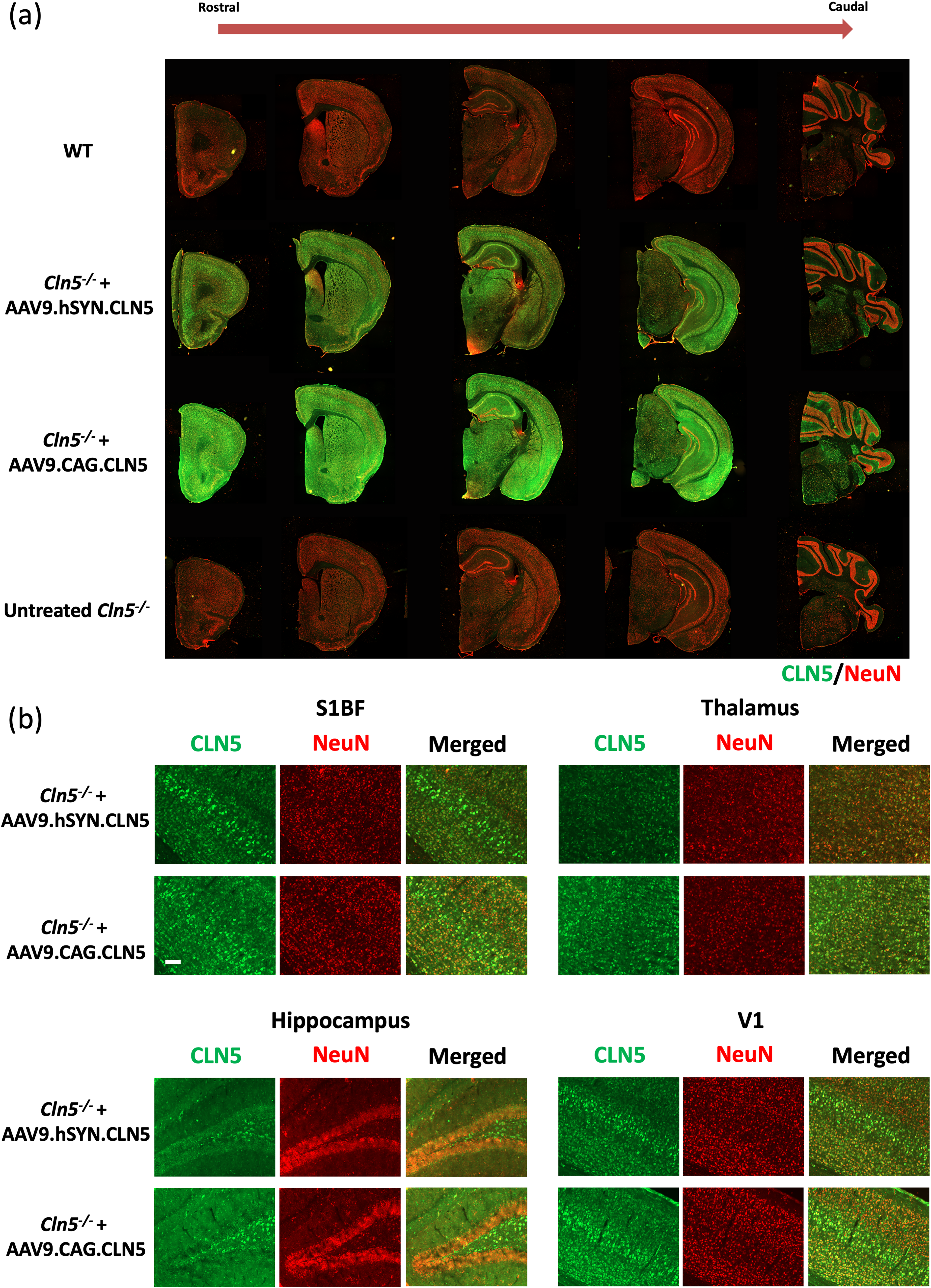
CLN5 overexpression in the brain from neonatally treated Cln5−/− mice.
Optimal survival and locomotor performances in Cln5−/− mice following neonatal treatment using AAV9.hSYN.CLN5
Survival of the two treated groups and two control groups (untreated WT and Cln5−/− mice) was monitored (Fig. 2a) up to maximum 18 months of age based on our institutional animal ethics requirements, with a humane end point defined as loss of >15% body weight or any other serious health issue. Untreated Cln5−/− mice (n = 12) exhibited a median lifespan of 12.35 months, and no mice survived beyond 14 months of age. All WT mice (n = 8) survived up to 18 months of age, except one that demonstrated unexpected severe weight loss and had to be euthanized at 17.5 months. The Cln5−/− mice that received AAV9.hSYN.CLN5 (n = 9) exhibited a significantly improved survival (Mantel–Cox test, p < 0.0001); six out of nine mice in the group did not deteriorate to the humane end point and survived to 18 months of age. In contrast, mice receiving AAV9.CAG.CLN5 (n = 8) did not show significantly improved survival compared with untreated Cln5−/− mice (p = 0.4).

Neonatal AAV9-hCLN5 gene therapy results in improvement of survival and locomotor function of Cln5−/− mice.
Body weight was monitored bi-monthly from 1 month till 18 months of age or the humane end point (Fig. 2b). The Cln5−/− mice, both treated and untreated, initially showed normal development and maturation, with comparable weight gain to WT mice. As the animals aged, WT mice continuously gained weight, whereas untreated Cln5−/− mice and those receiving AAV9.CAG.CLN5 seemed to plateau at a slightly lower level for a period of time before they progressively declined until reaching the humane end point. This was particularly striking for the AAV9.CAG.CLN5-treated group. AAV9.hSYN.CLN5-treated mice that lived a normal lifespan exhibited significantly lower body weights for the latter duration of their lifespan as compared with the WT controls.
Consistent with the survival analysis results, AAV9.hSYN.CLN5 treatment also resulted in better therapeutic efficacy than AAV9.CAG.CLN5 in improving locomotor functions of Cln5−/− mice. To evaluate the level of motor coordination, a rotarod test was conducted monthly from 2 months of age (Fig. 2c). In comparison with WT mice, all the Cln5−/− mice, treated and untreated, seemed to start with a lower performance, which then further declined with age. However, the decline in the AAV9.hSYN.CLN5-treated group was delayed; at 12 months of age, the performance of AAV9.hSYN.CLN5-treated mice was significantly better than untreated and AAV9.CAG.CLN5-treated Cln5−/− mice. Moreover, the foot-fault test, another behavioral analysis to assess motor coordination, also showed that AAV9.hSYN.CLN5-treated mice performed better at 12 months of age than the untreated mutant group, whereas AAV9.CAG.CLN5-treated mice did not show any improvement compared with untreated animals (Fig. 2d). Another observed behavioral phenotype of the Cln5−/− mice was hyperactivity, as shown in the open field analysis (Fig. 2e, f). Compared with WT mice, untreated Cln5−/− mice showed an age-dependent increase of total distance traveled in an open field, which was almost double WT values from 8 months to 11–12 months of age. The hyperactivity was significantly lowered in both treated groups, albeit still higher than the WT group. Last, gait analysis was carried out monthly using a CatWalk XT semiautomated gait analysis system (Fig. 2g, h). The Cln5−/− mice maintained a relatively normal walking speed and step sequence pattern for the duration of their lifespan (Fig. 2h; Supplementary Fig. S1). However, the print position, a gait regularity parameter defined as the distance between the forefoot print and the hindfoot print in one step cycle, was observed to progressively increase with age in Cln5−/− mice (Fig. 2h), suggesting an age-dependent decline in the ability of the mice to swing their hindlimbs to reach their forelimb placement in a step cycle, even though the walking speed was still maintained at a comparable level to that of WT mice. Both treated groups exhibited significant improvement in the print position as compared with untreated Cln5−/− mice, and particularly so for the AAV9.hSYN.CLN5-treated group that showed long-term normalization to WT levels. Overall, AAV9.CAG.CLN5 treatment had limited efficacy, whereas the AAV9.hSYN.CLN5-treated mice showed significant long-term improvements in various locomotor function analyses as compared with untreated Cln5−/− mice. No significant differences in locomotor function or responses to therapy were observed between male and female mice.
Significant alleviation of neurodegeneration in Cln5−/− mice following neonatal treatment using AAV9.hSYN.CLN5
To investigate efficacy of the neonatal gene therapy to prevent neurodegeneration, treated Cln5−/− mice and untreated WT and Cln5−/− controls were culled at 6 months of age and the brains harvested for histological analysis. A group of age-matched AAV9.CAG.GFP-treated Cln5−/− mice provided an additional control. According to behavioral and locomotor function analysis, 6-month-old Cln5−/− mice were still at a relatively early-middle stage of disease with only minor behavioral changes such as hyperactivity. Serial brain sections were immunohistochemically stained with a NeuN antibody, which is commonly used as a neuronal marker. Using optical fractionator stereology, neurons were counted in the VPM/VPL region of the thalamus and the S1BF region of cortex. At 6 months of age, although we did not observe significant neuronal loss in the S1BF region (Supplementary Fig. S2), untreated Cln5−/− mice showed significantly decreased neuronal counts in the VPM/VPL region compared with WT controls (Fig. 3a, b; p < 0.001), with an average decrease of 26%. Neuronal counts from the AAV9.hSYN.CLN5-treated Cln5−/− mice indicated a complete rescue of neurodegeneration in the thalamus, showing no significant difference from the counts in WT controls. However, the AAV9.CAG.CLN5 treatment did not result in significant amelioration in neurodegeneration compared with untreated Cln5−/− mice. As expected, the neuronal count estimates from the Cln5−/− mice treated with an AAV9 GFP vector showed no significant difference compared with the estimates from untreated Cln5−/− mice.

Amelioration of neurodegeneration in AAV9.hSYN.CLN5 neonatally treated Cln5−/− mice.
Although neurodegeneration was prevented in the 6-month-old AAV9.hSYN.CLN5-treated Cln5−/− mice, neuroinflammation was not improved. Immunohistochemistry with antibodies against GFAP and CD68 was used to respectively label fibrillary astrocytes and microglia to investigate astroglia- and microglia-mediated inflammatory response. Quantitative thresholding analysis was performed in the VPM/VPL region of thalamus and the S1BF region of cortex (Fig. 3c–f). In both brain regions, particularly in the thalamus, extensive astrogliosis and microgliosis was observed in the untreated Cln5−/− mice. Although the AAV9.hSYN.CLN5 neonatal gene therapy rescued neurodegeneration at 6 months of age, no significant improvement in astrogliosis or microgliosis was observed in the treated mice. In the thalamus, which exhibited more severe astrogliosis in 6-month-old Cln5−/− mice, quantification of GFAP showed similar level of immunoreactivity between all treated groups (AAV9.hSYN.CLN5, AAV9.CAG.CLN5, or AAV9.CAG.GFP) and the untreated Cln5−/− mice, whereas, in the cortex, increased GFAP immunoreactivity was observed in the treated, AAV9.hSYN.CLN5 (p < 0.0001), and AAV9.CAG.GFP-treated mice (p < 0.05) compared with the untreated animals (Fig. 3c, d). Analysis of microgliosis with CD68 labeling revealed similar results. Gene therapy using either AAV9.hSYN.CLN5 or AAV9.CAG.CLN5 did not lead to any significant changes in CD68 immunoreactivity in either region (Fig. 3e, f). Additionally, the AAV9.CAG.GFP-treated group showed significantly higher CD68 immunoreactivity than the untreated Cln5−/− mice.
Delayed intervention using AAV9.hSYN.CLN5 on survival and locomotor function of Cln5−/− mice was therapeutically beneficial
We investigated the potential efficacy of delayed intervention in treating Cln5−/− mice. As neurodegeneration was found to be significantly ameliorated only by hSYN.CLN5 but not CAG.CLN5 following neonatal AAV9 gene therapy, the AAV9.hSYN.CLN5 vector was employed for the delayed intervention study. Juvenile mice were treated at 4 weeks of age via ICV injection at a dose of 1 × 10¹² vg. Survival of the treated mice was monitored and compared with untreated Cln5−/− and WT mice (Fig. 4a). The delayed gene therapy still resulted in a significant increase of lifespan (Mantel–Cox test, p < 0.0001). Seven of eight treated mice survived till 18 months of age, a better survival than the neonatal gene therapy with the same vector. Notably, the one treated mouse that was euthanized at 12.7 months of age reached the humane end point because of a severe skin infection and did not show dramatic neurological symptoms associated with end-stage untreated Cln5−/− mice. For the duration of their lifespan, body weight was monitored bi-weekly (Fig. 4b). Similar to the WT mice, treated Cln5−/− mice gained weight with aging but plateaued earlier (two-way ANOVA, p < 0.0001 for the main effect between WT and treated Cln5−/−, p < 0.0001 for the main effect of age, no significant interaction between age and experiment group).

Delayed intervention with AAV9.hSYN.CLN5 results in restoration of survival and long-term improvements in locomotor function of Cln5−/− mice.
The delayed gene therapy with AAV9.hSYN.CLN5 also resulted in improved therapeutic effects on the locomotor dysfunction of Cln5−/− mice. The rotarod test was conducted monthly from 2 months of age, and the foot-fault test was conducted at 12 months of age to evaluate motor coordination (Fig. 4c, d). Throughout the lifespan of the Cln5−/− mice administered with delayed gene therapy, we did not observe the typical dramatic age-dependent impairment in rotarod performance of the Cln5−/− mouse model toward the end stage of disease. The treated mice exhibited significantly better rotarod performance than untreated Cln5−/− mice (Fig. 4c). Up to 18 months of age, the treated mice maintained their capacity of running the rotarod task although some differences to the WTs were measured (two-way ANOVA, p < 0.0001 for the main effect of experiment group between WT and treated Cln5−/−, p < 0.01 for the main effect of age, no significant interaction between age and experiment group). The foot-fault test also showed that the impairment in the untreated mutant group at 12 months of age was rescued by the delayed treatment with AAV9.SYN.hCLN5 (Fig. 4d). Similar to the neonatal AAV9.hSYN.CLN5 treatment, hyperactivity revealed by the total travel distance in open field analysis was still observed in the delayed treatment group compared with WT mice. However, this was steady throughout their lifespan and did not increase with age, as observed in the untreated Cln5−/− mice (Fig. 4e, f). Finally, gait analysis also demonstrated normalization to WT levels at 12 months of age (Fig. 4g) and throughout the lifespan following delayed treatment with AAV9.hSYN.CLN5 (Fig. 4h). Taken together, the juvenile gene therapy with AAV9.hSYN.CLN5 resulted in significant long-term improvements across all the locomotor function analyses that were conducted, and particularly for the rotarod test, the delayed treatment group demonstrated maintained capacity of performing the task. This suggests better therapeutic effects than the neonatal gene therapy with the same vector, which still showed significantly impaired rotarod performance with aging.
Delayed intervention using AAV9.hSYN.CLN5 significantly attenuated both neurodegeneration and neuroinflammation in the brain of Cln5−/− mice
We investigated effects of the delayed gene therapy on neurodegeneration and neuroinflammation. Cln5−/− mice administered with AAV9.hSYN.CLN5 via ICV administration at 4 weeks of age were harvested for histological analysis at 6 months of age, as well as age-matched untreated Cln5−/− and WT control mice. Serial brain sections were immunohistochemically stained with NeuN to label neurons, and neuronal count was estimated using stereology in the VPM/VPL region of the thalamus and the S1BF region of the cortex (Fig. 5a). As we did not observe significant neuronal loss in the all-layer S1BF region of Cln5−/− mice at 6 months of age previously (Supplementary Fig. S2), in this stereology analysis we only focused on Layer-V of the S1BF cortex in this stereology analysis, the internal pyramidal layer mainly comprising projecting pyramidal neurons, to distinguish potential early-stage neurodegeneration in the cortex. Stereological analysis revealed significant neuronal loss in the VPM/VPL and the S1BF Layer-V regions of the Cln5−/− mouse brain. Similar to the neonatal gene therapy with AAV9.hSYN.CLN5, delayed intervention also significantly rescued neuronal loss in the VPM/VPL region of Cln5−/− mice (p < 0.001), with neuronal counts normalized to levels that showed no significant difference with the WT neuronal estimates. In the S1BF Layer-V region, we did not observe a significant difference in neuronal counts between the treated and untreated Cln5−/− mice, but there was still a trend toward reduced neuronal loss in the S1BF Layer-V region of the delayed treatment group (p = 0.086; Fig. 5a).

Amelioration of neurodegeneration and neuroinflammation in the delayed intervention group with AAV9.hSYN.CLN5.
In contrast with neonatal gene therapy showing no improvement in neuroinflammation, the delayed gene therapy with AAV9.hSYN.CLN5 resulted in a significant amelioration of astrogliosis and microgliosis. Immunohistochemistry against GFAP and CD68 revealed that, in the VPM/VPL region, delayed AAV9.hSYN.CLN5 treatment significantly reduced both GFAP and CD68 immunoreactivity (Fig. 5b–e). In the S1BF region, only the CD68 immunoreactivity, but not GFAP, was significantly decreased in the treated mice.
Subunit c of ATP synthase (SCMAS) is one of the specific lysosomal storage body proteins in various forms of NCLs, including CLN5 disease.16,31–35 We investigated accumulation of SCMAS in the brain of Cln5−/− mice and whether delayed gene therapy could make any potential improvement (Fig. 5f, g). Immunofluorescence studies using an SCMAS antibody showed that, compared with WT mice, untreated Cln5−/− mice demonstrated significantly increased SCMAS accumulation in both the VPM/VPL and the S1BF region. Additionally, in the VPM/VPL region, the accumulation was more severe than in the S1BF region, which is consistent with other neuropathological parameters measured in this study. The delayed treatment group showed significantly reduced SCMAS accumulation in the VPM/VPL region compared with the untreated mutant group. However, in the S1BF region, no significant improvement of SCMAS accumulation was observed in the treated mice.
Overall, with respect to neuropathology, both neonatal gene therapy and delayed intervention using AAV9.hSYN.CLN5 significantly prevented neurodegeneration in Cln5−/− mice by 6 months of age. However, in contrast with the neonatal treatment, the delayed intervention also demonstrated significantly ameliorated neuroinflammation.
GSL changes in the Cln5−/− mouse brain are corrected by delayed gene therapy using AAV9.hSYN.CLN5
GSLs are highly expressed in the central nervous system and play an important role in various biological processes. 36 Metabolism of GSLs largely relies on endolysosomal pathways and has been found to be dysregulated in various lysosomal storage disorders such as Gaucher disease and Tay–Sachs disease. We evaluated whether the brains from Cln5−/− mice demonstrated alteration of GSL profiles and whether delayed gene therapy using AAV9.hSYN.CLN5 could correct this. Brain hemisphere homogenates from 6-month-old Cln5−/− mice treated with AAV9.hSYN.CLN5 via ICV administration at 4 weeks of age, and age-matched untreated WT and Cln5−/− controls were analyzed for GSL levels (n = 5 per group). In total, there were 16 GSL peaks from HPLC detected in the brains, of which 9 species were identified and fell into o- (LacCer, GA1), a- (GM3, GM2, GM1a, GD1a), and b- (GD1b, GT1b, and GQ1b) series GSLs (Fig. 6a). Total GSL levels did not show any significant difference across different groups and did not identify outlier values (Fig. 6b). Four of the nine GSL species (GM1a, GD1a, GD1b, and GT1b) were particularly highly enriched in the brain (>700 pmol/mg protein) but did not show any significant changes in the Cln5−/− brains (Fig. 6b). The other five GSL species were of relatively low abundance in the brains (<300 pmol/mg protein). However, among these lower-expression GSLs, GM2 and GM3 were found to be significantly increased in the untreated Cln5−/− brains, which was around 2-fold higher to that observed in WT brains (Fig. 6c). Importantly, increases of both GSL species were corrected in the treated Cln5−/− brains with no significant difference to control WT levels (Fig. 6c).

Increased levels of GM2 and GM3 in the Cln5−/− mouse brains are corrected by the delayed intervention with AAV9.hSYN.CLN5.
Increased plasma NfL chain levels in Cln5−/− mice were corrected by delayed gene therapy using AAV9.hSYN.CLN5
NfL chain is emerging as a commonly recognized biomarker of neurodegeneration. We explored whether blood plasma NfL levels could also be a potential biomarker for CLN5 disease and to assess whether this responded to, and reflected, the efficacy of the gene therapy. Plasma samples were collected from 6-month-old mice that were harvested for the above histological and biochemical analysis in the delayed intervention study. Plasma NfL levels were measured using an ultrasensitive single molecule array assay (SIMOA). At 6 months of age, in comparison with WT controls (mean plasma NfL of 34.09 ± 9.77 pg/mL), the untreated Cln5−/− mice exhibited significantly higher levels of plasma NfL (214.08 ± 33.28 pg/mL), corresponding to an average of around 6-fold increase (Fig. 7). However, the delayed gene therapy-treated Cln5−/− mice demonstrated significantly lower levels of plasma NfL that were restored to WT levels, suggesting a complete normalization of this blood plasma biomarker.
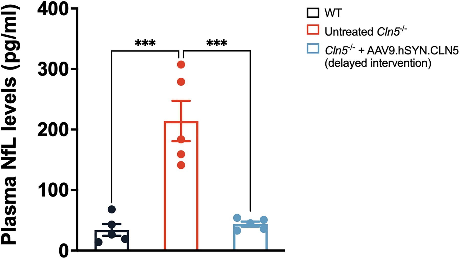
Increased plasma NfL levels in Cln5−/− mice are corrected by the delayed intervention with AAV9.hSYN.CLN5. Plasma samples were collected from 6 months old Cln5−/− mice treated with AAV9.hSYN.CLN5 and age-matched WT and untreated Cln5−/− mice, and NfL levels were measured using an ultrasensitive single molecule array assay (SIMOA). Results show a sixfold increase of the NfL level in the untreated Cln5−/− plasma, which is normalized to WT levels by the delayed intervention with AAV9.hSYN.CLN5. N = 5 per experimental cohort. Data are represented as means ± SEM with individual data. One-way ANOVA with Dunnett’s comparison. ***p < 0.001. ANOVA, analysis of variance; NfL, neurofilament light; SEM, standard error of the mean; WT, wildtype.
Long-term therapeutic benefits on neuropathology and GSL accumulation by delayed gene therapy with AAV9.hSYN.CLN5 in the long-surviving 18 months Cln5−/− mice
Since we observed that lifespan of Cln5−/− mice was normalized by the delayed gene therapy to at least 18 months (based on our institutional animal ethics requirements), we next examined levels of neuropathology in those long-surviving mice at 18 months of age. The brains from end-stage untreated Cln5−/− mice (around 12 months), 18-month-old gene therapy-treated Cln5−/− mice, and age-matched WT mice were collected. Immunohistochemistry and immunofluorescence with antibodies against CD68 and SCMAS were performed to respectively assess neuroinflammation and lysosomal storage in the brain. The results showed that the delayed gene therapy led to long-term amelioration of microglial activation and SCMAS storage in Cln5−/− brains. In the VPM/VPL region, end-stage untreated Cln5−/− mice (around 12 months of age) displayed significant CD68 and SCMAS positive staining, whereas the 18-month-old gene therapy-treated Cln5−/− mice exhibited comparable levels of CD68 and SCMAS to those in age-matched WT mice (Fig. 8a–d). In the S1BF region, the 18-month-old-treated Cln5−/− mice showed no significantly different levels of CD68 and SCMAS compared with end-stage untreated Cln5−/− mice (Fig. 8a–d). Interestingly, although the quantitative levels were similar, biodistribution of the CD68 and SCMAS pathology in the S1BF region is different between the long-surviving treated and the end-stage untreated Cln5−/− mice. In the treated mice, the neuropathology tended to be concentrated in deep cortical layers, whereas in end-stage untreated Cln5−/− mice pathology was more evenly distributed in the cortex (Fig. 8a, c), which may represent variable vulnerability and responsiveness of different cell types to the disease/treatment.

Sustained therapeutic benefits on neuropathology and GSL accumulation by the delayed intervention with AAV9.hSYN.CLN5.
In the short-term study at 6 months of age, we observed early biochemical correction of GM3/GM2 accumulation by the delayed gene therapy, and thus we also ran GSL analysis on brains from those aged mice (end-stage untreated Cln5−/− mice and 18-month-old gene therapy-treated Cln5−/− mice and WT mice) to investigate long-term durability and efficacy. Compared with levels of aged WT mice, GM3 and GM2 were significantly higher in untreated end-stage Cln5−/− brains (Fig. 8e). Despite being analyzed at an older age, the treated Cln5−/− mice exhibited significantly lower GM3 and GM2 levels in the brain compared with untreated end-stage Cln5−/− mice, suggesting a sustained therapeutic effect on the GM3/GM2 accumulation. However, the treated Cln5−/− mice still showed significantly elevated GM3 and GM2 levels compared with age-matched WT mice.
DISCUSSION
CLN5 disease is a lethal condition caused by loss of functional CLN5 expression and currently there is no effective therapy available. In this study, we evaluated the therapeutic efficacy of an AAV9-mediated gene therapy administered via ICV injection into a transgenic mouse model of CLN5 disease. As neurodegeneration is the primary cause of death in CLN5 patients, we prioritized the brain as the primary target of the treatment. The rationale for ICV delivery was aiming to achieve widespread brain expression of the therapeutic transgene via a less invasive surgical approach compared with multipoint intraparenchymal injection and with a lower dose of vectors compared with the intravenous delivery route. Furthermore, we compared the efficacy of an AAV9-mediated gene therapy approach administered at different stages of development and utilizing different promoters driving expression of the therapeutic human CLN5 gene. We chose to compare the widely used synthetic CAG promoter that drives strong ubiquitous transgene expression with the human synapsin I (hSYN) promoter that is a neuronal specific promoter.
As CLN5 disease is a progressive neurodegenerative condition, with characteristic storage inclusions visible before birth, 37 theoretically the earlier the intervention can be administered to patients, the more effective the therapy will be in preserving the brain. On this basis, we initially administered AAV9-mediated gene therapy (hSYN.CLN5 or CAG.CLN5 vectors) via ICV administration to neonatal Cln5−/− mice. Long-term monitoring of behavioral performance and lifespan showed that only the hSYN.CLN5 vector provided significant improvement, whereas the CAG.CLN5 vector exhibited comparatively limited efficacy. Neurodegeneration analysis also supported the behavioral and lifespan results; the neuroprotection is more prominent in the hSYN.CLN5-treated mice than in the CAG.CLN5-treated animals. This was unexpected considering the widespread neural cell type CLN5 expression driven by the CAG promoter. A possible reason is that strong CLN5 overexpression might be accompanied by some potential toxicity. Particularly, excessive CLN5 accumulation has recently been found to cause NCL-like cellular phenotypes in vitro and associate with pathogenesis of another NCL subtype, CLN14 disease.38,39 The dose used in our neonatal gene therapy is 5 × 10¹¹ vg per brain, which is relatively high considering the volume of the neonatal mouse brain. Our immunohistology analysis confirms supraphysiological levels of CLN5 expression throughout the treated mouse brain, which may result in cellular toxicity. Moreover, secretion of the target protein offers opportunity for cross-correction through mannose-6-phosphate receptor uptake by other cells not expressing the protein, which may further amplify the potential effect of the gene therapy. Secretion of CLN5 protein when overexpressed in cells has been observed in previous research in various model systems including Dictyostelium and mammalian cells.19,21,22,40–42 However, excess extracellular CLN5 in the brain might be a potential microenvironmental trigger of an inflammatory response. This is supported by our astrogliosis and microgliosis assessment that does not show a reduction in inflammation in various brain regions examined in the neonatal gene therapy, even in the hSYN.CLN5 vector-treated mice that exhibit significant prevention of neurodegeneration. Alternatively, expression of hSYN.CLN5 is restricted to neurons, 43 whereas transgene expression from CAG.CLN5 is ubiquitous, which in the case of AAV9 is both neuronal and astrocytic. 44 Such supraphysiological expression of CLN5 in astrocytes might be detrimental. Thus, although neurons have gained CLN5 expression in the CAG.CLN5-treated mice, potential neuroprotection may be counteracted by the toxicity resulted from astrocytic CLN5 overexpression, leading to limited therapeutic efficacy. Furthermore, using hSYN.CLN5 to target only neurons may lead to a limited uptake of secreted protein by other cells including astrocytes, thereby avoiding toxicity in such cells.
The significant efficacy in neonatal gene therapy with the hSYN.CLN5 vector is encouraging and promising. However, neonatal gene therapy is not easily achievable in the clinical setting in the absence of newborn screening. Therefore, we investigated a delayed intervention where the gene therapy using hSYN.CLN5 was delivered at 4 weeks of age, which represents a juvenile stage of mouse development. It should be emphasized that the Cln5−/− mice are still at a presymptomatic stage at this age with no behavioral phenotype observed, although some pathology has been detected in the brain previously. 17 Unexpectedly, compared with the neonatal gene therapy, the delayed intervention with the hSYN.CLN5 vector exhibited superior therapeutic efficacy. The improvement in locomotor function and lifespan was more prominent compared with the neonatally administered mice. With respect to pathology, in addition to ameliorating neurodegeneration, the delayed gene therapy resulted in the prevention of neuroinflammation by 6 months of age, which was not observed in the neonatal gene therapy. Analysis of SCMAS also showed significantly reduced accumulation, although not to WT levels, as the neurons may have already begun to accumulate this storage material by the stage of intervention. However, correction of neurodegeneration and neuroinflammation suggests likelihood of a halt of the disease progression. Pathological analysis on those aged treated Cln5−/− mice surviving to 18 months of age also showed significantly reduced neuroinflammation and SCMAS accumulation compared with untreated end-stage Cln5−/− mice, indicating long-term durability of the therapeutic efficacy. The reasons for better efficacy from the delayed intervention compared with neonatal gene therapy with the same vector are not clear. The dose in the delayed gene therapy is doubled compared with that in the neonatal gene therapy. However, the size of the mouse brain is significantly larger at 4 weeks of age, and the cellular efficiency of AAV transduction throughout the brain via ICV administration is always higher in the neonatal brain. Therefore, the level of CLN5 expression achieved in the delayed gene therapy is likely to be lower than that in the neonatal gene therapy, even with a doubled dose administered. Indeed, our immunofluorescence analysis of brain sections from 6 months old mice that received delayed gene therapy at the juvenile stage revealed markedly reduced CLN5 transgene overexpression compared with mice that received neonatal administrations and also analyzed at 6 months of age (Supplementary Fig. S3). In this case, the better efficacy in the delayed intervention further supports the hypothesis that highly supraphysiological expression of CLN5 might result in toxicity, which could counteract protective efficacy.
Recent CLN5 gene therapy studies in a CLN5 disease sheep model have provided evidence for long-term efficacy from ICV administrations of AAV9 to the brain 26 and combined with eye administrations. 23 These studies delivered the ovine Cln5 gene at different doses and intervention stages using small sample sizes, which is understandable when using large animal models. The sheep studies utilized the CBh promoter which, like CAG, is a synthetic sequence that drives ubiquitous expression in different cell types. Across all the treated animals with various dosage levels and intervention stages, a large proportion exhibited promising improved survival and delayed clinical progression. No toxicity was observed as the dosage level increased, and the pre- and early-symptomatic intervention showed briefly better efficacy than the advanced-symptomatic intervention at the same dosage level. Relative to their species-dependent development and lifespan, the CLN5 sheep model, compared with the mouse model, exhibits an earlier onset of neurological manifestations but a more protracted progression,10,12,45 whereas the mice remain apparently normal for a relatively long period in their lifespan before developing symptoms that progress rapidly to end point. This characteristic of the mouse model precluded us from evaluating postsymptomatic intervention. But encouragingly, our presymptomatic gene therapy results in mice and the published sheep studies together support the notion that treatment administered pre/early-symptomatically can provide significant therapeutic benefits. Interestingly, although the authors observed a potential positive correlation between the transduction efficiency and the efficacy, there was no significant dose-dependent response to survival, clinical manifestation scores, and pathology analysis. The highest dosage level used in the sheep model was 3.3 × 10¹² vg per brain. Considering the difference in the volume of an adult sheep brain (∼175 g) vs. an adult mouse brain (∼0.4 g), the dose used in our study is very high in comparison with the doses used in the sheep model (delayed intervention: 1 × 10¹² vg per brain, neonatal intervention: 5 × 10¹¹ vg per brain). Collectively, loss of dose-dependency in the sheep study and lower efficacy with CAG at a much higher dose in our current study is supportive of our hypothesis discussed above; increases in dosage/overexpression using a ubiquitous promoter counteract the protective efficacy of the increased transduction. On the basis of our data, superior efficacy might be achieved in the sheep model using the neuron-specific hSYN.CLN5 vector. This study does not address the retinal degeneration that has been confirmed in the mouse model and which promoter would provide optimal efficacy.
The identification of biomarkers that can be conveniently and noninvasively monitored is a key component for measuring therapeutic efficacy in clinical trials. NfL is a neuronal specific cytoskeletal protein that plays an important role in maintaining neuronal structural stability and axon polarization. 46 NfL is a nonspecific neuroaxonal damage marker, and increased levels in patient CSF and blood have been detected in various neurodegenerative disorders. Therefore, NfL has currently been suggested as a circulating biomarker for various neurodegenerative conditions, such as Alzheimer’s disease (AD),47–49 Parkinson’s disease, 50 frontotemporal lobar dementia,51–53 amyotrophic lateral sclerosis, 51 multiple sclerosis,54,55 and spinal muscular atrophy. 56 With respect to the NCLs, NfL has not been extensively explored, with the exception of CLN2 disease, where CSF NfL levels are found to be increased in CLN2 patients and decreased in response to treatment with Brineura. 57 In this study, we find an average of 6-fold increase in the Cln5−/− mouse blood plasma at 6 months of age. Importantly, such elevated plasma NfL level is normalized by the delayed gene therapy. Considering that Cln5−/− mice at 6 months of age do not show any obvious phenotype and the neurodegeneration is relatively mild according to our stereological analysis, it seems that plasma NfL measurement could be a sensitive biomarker for CLN5 disease progression. Using a plasma biomarker for monitoring assessment of potential treatment response is a less invasive approach than CSF analysis via a lumbar puncture. Plasma NfL as such a biomarker in CLN5 patients is worth verifying.
In this study, we observed significantly elevated GM2 and GM3 levels in the brains of Cln5−/− mice, which was corrected by the AAV gene therapy. GSLs are particularly enriched in the CNS. GSLs in cell membranes play an important role in interacting with various functional proteins to modulate cell growth, proliferation, differentiation, adhesion, migration, apoptosis, and various intra- and extracellular signaling. 36 GSLs are degraded in lysosomes. Inborn defects in enzymes contributing to GSL catabolism cause GSL lysosomal storage disorders, which frequently have a neurodegenerative course, such as GM1 gangliosidosis, the GM2 gangliosidoses, Gaucher disease, Fabry disease, and Krabbe disease. Dysregulation of one or multiple GSLs has also been described in various other neurodegenerative lysosomal storage diseases, such as Niemann-Pick C1 disease (NPC), where studies found accumulation of multiple GSLs in both the periphery and the CNS of NPC mice58,59 and in NPC patient-specific induced pluripotent stem cell-derived neurons. 60 With respect to NCLs, Somogyi et al. found a high increase of GM3 and reduction of GM1a and GD1a levels in cerebellar precursor cells derived from a CLN3 mouse model, suggesting a metabolic deficit in conversion of GM3 to more complex GSLs. 61 Another NCL study using CLN6 and CLN10 mouse models showed that GM2 and GM3 were both increased in the brain of disease mice. 62 The CLN6 mouse model, like the Cln5−/− mice, is a mildly progressing NCL mouse model, and GM2 and GM3 were increased from a relatively early stage of disease. As such, in our study we found that GM2 and GM3 increase in the brain lysates of Cln5−/− mice at 6 months of age, before any dramatic locomotor function decline and when only mild–moderate neurodegeneration and neuroinflammation is present. We did not observe any changes in either the upstream precursors of GM3-GM2 (such as LacCer) or the downstream GSLs including GM1a and GD1a. Notably, this early GM3/GM2 accumulation was prevented by gene therapy, and the therapeutic effect remained significant even at 18 months, an age exceeding the typical lifespan (around 12 months) of untreated Cln5−/− mice. The mechanisms responsible for the selective early elevation of GM3-GM2 are unclear. One possible explanation may relate to the direct involvement of CLN5 in BMP biosynthesis. CLN5 was recently identified as the lysosomal BMP synthase and loss of functional CLN5 led to BMP depletion. 5 BMP is an important lipid in intraluminal vesicles in late endolysosomes and plays a critical role in facilitating membrane-associated hydrolysis of GSLs by providing negatively charged membrane microenvironment to recruit hydrolases and co-factors/co-activators.63–65 Among various GSL isoforms, GM3 and GM2 are of relatively small hydrophilic sugar chains, and thus, their hydrolysis is particularly dependent on effective membrane interaction with hydrolases and activator proteins.66,67 Therefore, GM3/GM2 degradation may be more vulnerable to changes of intralysosomal membrane lipid environment caused by BMP deficiency, which makes them accumulate earliest in the Cln5−/− brain. Establishing a comprehensive understanding of how GM3/GM2 are dysregulated in CLN5 disease and their functional role in the disease progression will be valuable information. Interestingly, increased levels of GM2 and GM3 have also been linked to common neurodegenerative diseases such as AD,68–70 and particularly for GM3, a unique increase is found from an early stage in the hippocampus, the more vulnerable brain region to AD progression, in an AD rat model. 69 This suggests, premised on the basis of no direct blockade of GSL catabolic pathways, that a shift in lipid homeostasis might indicate some early cellular changes in neurodegeneration and offer novel targets for therapy development.
CONCLUSIONS
In summary, our study demonstrates that AAV-mediated gene therapy with the hSYN.CLN5 vector via ICV has promising potential for treating CLN5 disease. Administration at a presymptomatic stage provides significant therapeutic efficacy, and a potential plasma biomarker NfL could be useful to monitor disease modification post-treatment. Furthermore, a more nuanced approach of promoter selection with a bias toward neuronal expression is likely to provide optimal therapeutic efficacy.
AUTHORS’ CONTRIBUTIONS
Conceptualization: A.A.R., S.E.M., R.R.A., and A.J.S.; resources: A.A.R., R.R.A., A.J.S., F.M.P., and H.Z.; investigation and data curation: W.L., A.F.G., G.M., M.P.H., M.A., O.C.-T., L.X., O.S., R.B., D.t.V., D.P., R.L., E.V., A.J.H., H.Z., F.M.P., A.J.S., and A.A.R.; formal analysis: W.L., G.M., R.B., D.t.V., D.P., and A.A.R.; writing—original draft: W.L. and A.A.R.; writing—reviewing and editing: W.L., A.F.G., G.M., M.P.H., D.t.V., D.P., A.J.H., H.Z., F.M.P., A.J.S., S.E.M., R.R.A., and A.A.R.; and funding acquisition: A.A.R., S.E.M., R.R.A., and A.J.S.
Footnotes
ACKNOWLEDGMENTS
The authors would like to thank Professor Katja Kanninen from University of Eastern Finland for sharing the Cln5−/− mouse model.
FUNDING INFORMATION
This work was supported by awards from the UK Medical Research Council Developmental Pathway Funding Scheme Award (MR/R025134 to A.A.R., S.E.M., R.R.A., and A.J.S.), and the UK Batten Disease Family Association for CLN5 work (to S.E.M. and A.A.R.). A.A.R. also receives support from the NIHR Great Ormond Street Hospital Biomedical Research Centre (562868), UK Medical Research Council grants (MR/R015325/1, MR/S009434/1, MR/N026101/1, and MR/T044853/1), the Wellcome Trust, Institutional Strategic Support Fund/UCL Therapeutic Acceleration Support (TAS) (204841/Z/16/Z), and the Sigrid Rausing Trust and the Jameel Education Foundation and LifeArc. R.B. was supported by the Wellcome Trust (218514/Z/19/Z), Merck Sharpand Dohme Corp., and Janssen Pharmaceutica NV. All research at Great Ormond Street Hospital NHS Foundation Trust and UCL Great Ormond Street Institute of Child Health is made possible by the NIHR Great Ormond Street Hospital Biomedical Research Centre. The views expressed are those of the authors and not necessarily those of the NHS, the NIHR, or the Department of Health.
DATA AVAILABILITY STATEMENT
All data and statistics are available on request.
DISCLOSURE STATEMENT
A.A.R. is a founder and shareholder in Bloomsbury Genetic Therapies Ltd.
Supplemental Material
References
Supplementary Material
Please find the following supplemental material available below.
For Open Access articles published under a Creative Commons License, all supplemental material carries the same license as the article it is associated with.
For non-Open Access articles published, all supplemental material carries a non-exclusive license, and permission requests for re-use of supplemental material or any part of supplemental material shall be sent directly to the copyright owner as specified in the copyright notice associated with the article.
