



Abstract
Recombinant adeno-associated virus (rAAV) vectors have demonstrated their versatile utility for in vivo gene therapy to deliver successful treatment for several diseases of unmet medical needs. The polytropic nature of AAV permitted targeting diverse tissue types to support various diseases; however, it also resulted in vector transduction of non-target organs, including the gonads of males and females, which subsequently confer an additional investigation to derisk any potential germ cell transduction and/or germline transmission following rAAV administration. While a plethora of data is available for male gonad assessment, primarily due to the feasibility of longitudinal semen collection, there is a paucity of data on oocyte transduction by rAAV due to challenges with oocyte collection. In this brief report, we share polymerase chain reaction and in situ hybridization data on oocyte transduction in a female cynomolgus macaque 4 weeks following systemic intravenous administration of an engineered rAAV and discuss the significance and potential mitigation strategies of such findings in the process of AAV-based gene therapy product development.
INTRODUCTION
Adeno-associated virus (AAV) is a polytropic virus capable of infecting various cell types within the host. Recombinant AAV (rAAV) vector technology leveraged such characteristic tropism of naturally occurring and engineered virus capsids into a diverse collection of in vivo gene therapy vectors to modulate the transfer of genes for the treatment of several challenging diseases of unmet medical needs.1–3
Secondary to rAAV administration, particularly following systemic and intravenous routes of administration, rAAV distributes throughout the host organs and tissues and transduces various cell types to modulate the transfer of vector genomic DNA along with the intended therapeutic transgene. 4 Consequently, regulatory expectation in the development of rAAV gene therapy products is the nonclinical biodistribution (BD) assessment of the candidate rAAV to assess “the in vivo distribution, persistence, and clearance of a GT product at the site of administration and in target and non-target tissues, including biofluids.” 5 Among the key non-target tissues for BD assessment are the male and female gonads to determine the potential for germ cell transduction (e.g., sperms in males and oocytes in females) considering the associated risk of germline integration and potential vertical transmission in animals and humans.5,6
Detection of rAAV DNA in gonads following BD assessment by the standard polymerase chain reaction (PCR) approach in nonclinical animal studies is not uncommon.7,8 Further morphological and spatial histological assessment, by in situ hybridization (ISH), for example, generally demonstrates lack of germ cell transduction in sperms and oocytes.7,8 However, these data are generally limited to select species, primarily rodents, and paucity of data for oocyte transduction due to challenges with repeat and longitudinal sampling of oocytes, compared with the feasible semen collection in males.
In this brief report, we share a unique finding of oocyte transduction in a female cynomolgus macaque 4 weeks following the intravenous systemic administration of an engineered AAV vector expressing a human transgene.
MATERIAL AND METHODS
Animal welfare
All animal procedures involving the care and use of animals in this study were reviewed and approved by Charles River Laboratories—Senneville Institutional Animal Care and Use Committee, with guidance from the USA National Research Council and the Canadian Council on Animal Care.
Animal study
Naive cynomolgus monkeys (Macaca fascicularis) of Mauritius origin were seronegative for neutralizing antibodies titers against the AAV capsid protein. One group of animals received the formulation buffer as a negative control group, and the remaining animals were allocated to treatment groups and administered the AAV vector at 3 dose levels. Animals ranged from 1.5 to 5 years of age and weighed 1.4 to 5 kg at the initiation of the study.
All surviving animals were submitted for necropsy on Day 29 (approximately 4 weeks; 1 month) or Day 92 (approximately 13 weeks; 3 months). Necropsies were performed, and organ weights were collected by Charles River Laboratories, Senneville site. Tissues required for hematoxylin and eosin microscopic evaluation were trimmed, processed routinely, embedded in paraffin, stained with hematoxylin and eosin, and evaluated microscopically. Additional tissues were collected, flash-frozen, and processed for DNA and RNA extraction for PCR analysis.
In situ hybridization
From each formalin-fixed paraffin-embedded block of all ovaries (unilateral) and testes (bilateral) from all groups, sections were prepared at two different levels at approximately 50 μM intervals, stained, and analyzed. All samples were analyzed by a qualified ISH sectioning and staining method on the Ventana Discovery Ultra automated platform. These tissues were evaluated for negative or positive reactivity to the vector DNA and transgene mRNA probes by light microscopy. The vector DNA probe was designed against the enhancer region not transcribed into transgene mRNA. Transgene mRNA probe was designed against a junction only presented in transcribed mRNA. The evaluation of positive vector DNA and transgene mRNA reactivity in each tissue section was performed in relation to the ISH staining outcomes with the Dihydrodipicolinate reductase (dapB; negative control) and Peptidylpropyl isomerase B (PPIB; positive control for cynomolgus monkeys) probes for DNA and RNA in corresponding sections of the same tissue.
PCR assays
Two assays were used to support the BD analysis: a quantitative PCR assay (qPCR) for detecting vector genomes (DNA) and a Reverse Transcription-quantitative PCR assay (RT-qPCR) for detecting transgene expression (mRNA).
qPCR ASSAY FOR VECTOR DNA
The qPCR technology was used to quantify the amount of target nucleic acid sequence in the monkey DNA extracted from various tissues and body fluid samples from cynomolgus monkeys. The number of copies per well in samples was extrapolated from a standard curve of plasmid standard in water containing monkey DNA. The assay was validated prior to sample analysis. The results of the validation demonstrated that the method was suitable for the quantification of DNA vector by qPCR. The lower limit of quantification (LLOQ) as well as the lower limit of detection (LOD) were determined to be 10 copies/well in the presence of 1 μg of monkey DNA.
RT-qPCR ASSAY FOR TRANSGENE EXPRESSION
The two-step RT-qPCR technology can detect and quantify the amount of transgene mRNA expression in cynomolgus monkey tissues. For this study, a synthetic RNA oligo containing a portion of the transgene was used as a positive control. The number of copies per well was extrapolated from a standard curve diluted in water containing monkey RNA. The assay was validated prior to initiation of sample analysis. Overall, the results obtained during the RT-qPCR method validation demonstrated that the method was suitable for the quantification of transgene mRNA expression product in monkey tissues by RT-qPCR. The LLOQ was determined to be 50 copies/μg of monkey RNA, and the LOD was determined to be 25 copies/μg of monkey RNA.
RESULTS
A toxicology and BD study was performed to support the development of a rAAV-based gene therapy candidate in purpose-bred, AAV-naive cynomolgus macaque of Mauritius origin. Monkeys were intravenously infused with an engineered rAAV vector expressing a human transgene in the tail vein at 3 dose levels along with a fourth group only receiving the formulation buffer as a vehicle control group. Each group was composed of 2/sex/group/timepoint, and animals were euthanized at either 4- or 13-weeks post vector administration (n = 16/sex; 8/sex terminated 4 weeks post dose, and another 8/sex terminated 13 weeks post dose). Gonads (along with a predefined complete tissue list) were collected for histopathology and BD analysis. At necropsy, the right-side ovary was freshly collected and frozen for subsequent nucleic acid extraction and PCR-based BD analyses of vector genomic DNA and transgene mRNA expression. The left-side ovary was fixed in formalin and processed to paraffin block for histopathology and ISH analyses.
Histopathology assessment of the left ovary didn’t reveal any test article-related findings. PCR analysis of the right ovary from a lower dose group, performed as a predefined study component to support the BD assessment of the test article, demonstrated the detection of a positive vector genomic DNA and transgene mRNA expression in these animals (data not shown). Consequently, ISH analysis ensued to assess the localization of vector DNA and transgene mRNA morphologically within the tissues, particularly for the assessment of potential germ cell transduction. BaseScope assay was performed on the left ovary samples from all female animals in the study (n = 16; 8 terminated 4 weeks post dose, and another 8 terminated 13 weeks post dose) using a probe uniquely designed to detect vector DNA and a probe designed to detect transgene mRNA. Positive signal, as illustrated by red dots in Figure 1, was predominantly located in inter-follicular stromal cells and in a few follicular granulosa cells in the ovary samples of all female animals (Fig. 1). However, uniquely in one female cynomolgus macaque (approximately 2.5-year-old and weighing approximately 3.5 kg at the day of dosing) administered rAAV at the dose level of 1 × 10¹⁴ vg/kg and underwent a planned euthanasia 4 weeks following vector administration, positive signal was also evident in rare oocytes (germ cells) within primordial and/or primary ovarian follicles (Fig. 1). These positive dots in the oocytes were slightly smaller than the positive dots in non-germ cells and were more evident with transgene mRNA probe than vector DNA probe, likely reflecting an expected higher number of mRNA copies transcribed from each vector DNA genome. In addition to these smaller positive dots, some primordial and/or primary follicles had distinct dense accumulations of finer dots within oocytes. These finer dots, which were evident with DNA and mRNA probes, were notably smaller than the positive dots observed in the non-germ cells but were more numerous and prominent than comparably sized non-specific dots in the corresponding negative control slides. The presence of dense accumulations of these finer dots to the extent that was not evident in any of the other female animals indicated the presence of vector DNA and corresponding transgene mRNA in the oocytes of this animal at week 4 post vector administration. Such dense, finer dots, which were noted in the primordial and/or primary (early stage) follicles, were not readily evident in antral/Graafian (later stage) follicles.
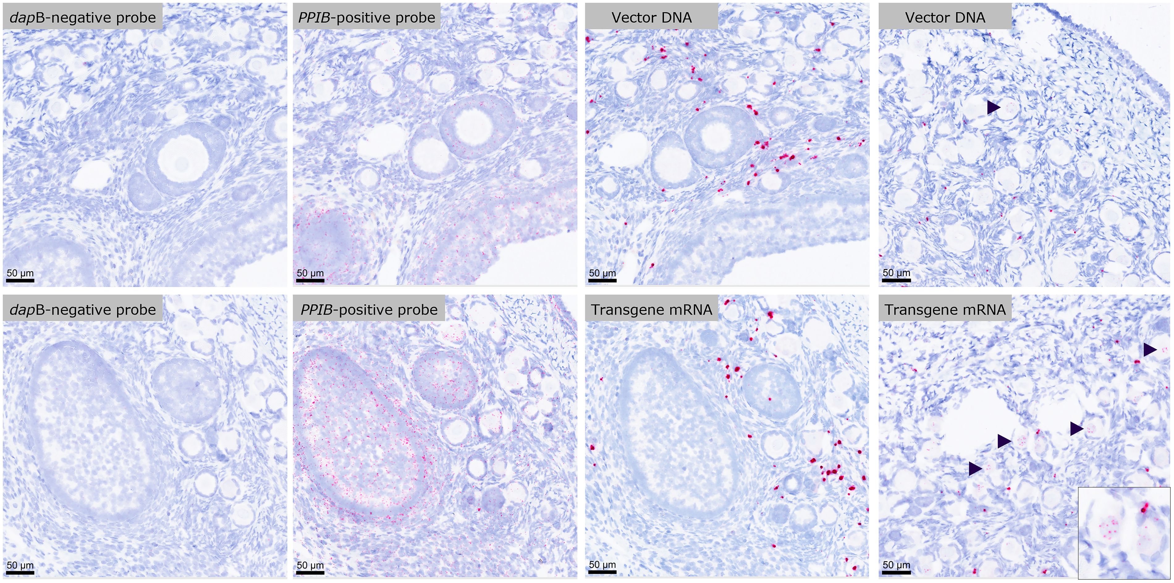
Vector DNA and transgene mRNA positive signal (red dots) evident in rare oocytes (germ cells; arrowheads; inset) within primordial and/or primary ovarian follicles in the left ovary obtained from a female animal administered adeno-associated virus (AAV) vector. Most of the signal was predominantly located in inter-follicular stromal cells and in a few follicular granulosa cells in the ovary samples of all female animals. Tissue evaluation was performed in relation to the in situ hybridization (ISH) staining outcomes with the dapB (negative control) and PPIB (positive control) probes for DNA and RNA in corresponding sections of the same tissue. All images are captured from the same female animal, with the Vector DNA and Transgene mRNA images representing two different regions within the same ovary.
ISH analysis performed on bilateral testes samples from all male animals terminated at 4- or 13-weeks post-vector administration demonstrated positive vector DNA and transgene mRNA in all but one low dose male, with positive vector DNA and transgene mRNA signal evident bilaterally in the testes and located in interstitial Leydig cells, interstitial vascular endothelial cells, peritubular myoid cells, and peritubular fibroblasts (Fig. 2). No evidence of male germ cell transduction was observed in any of the testes examined (32 testes—left and right sides—from a total of 16 male animals).
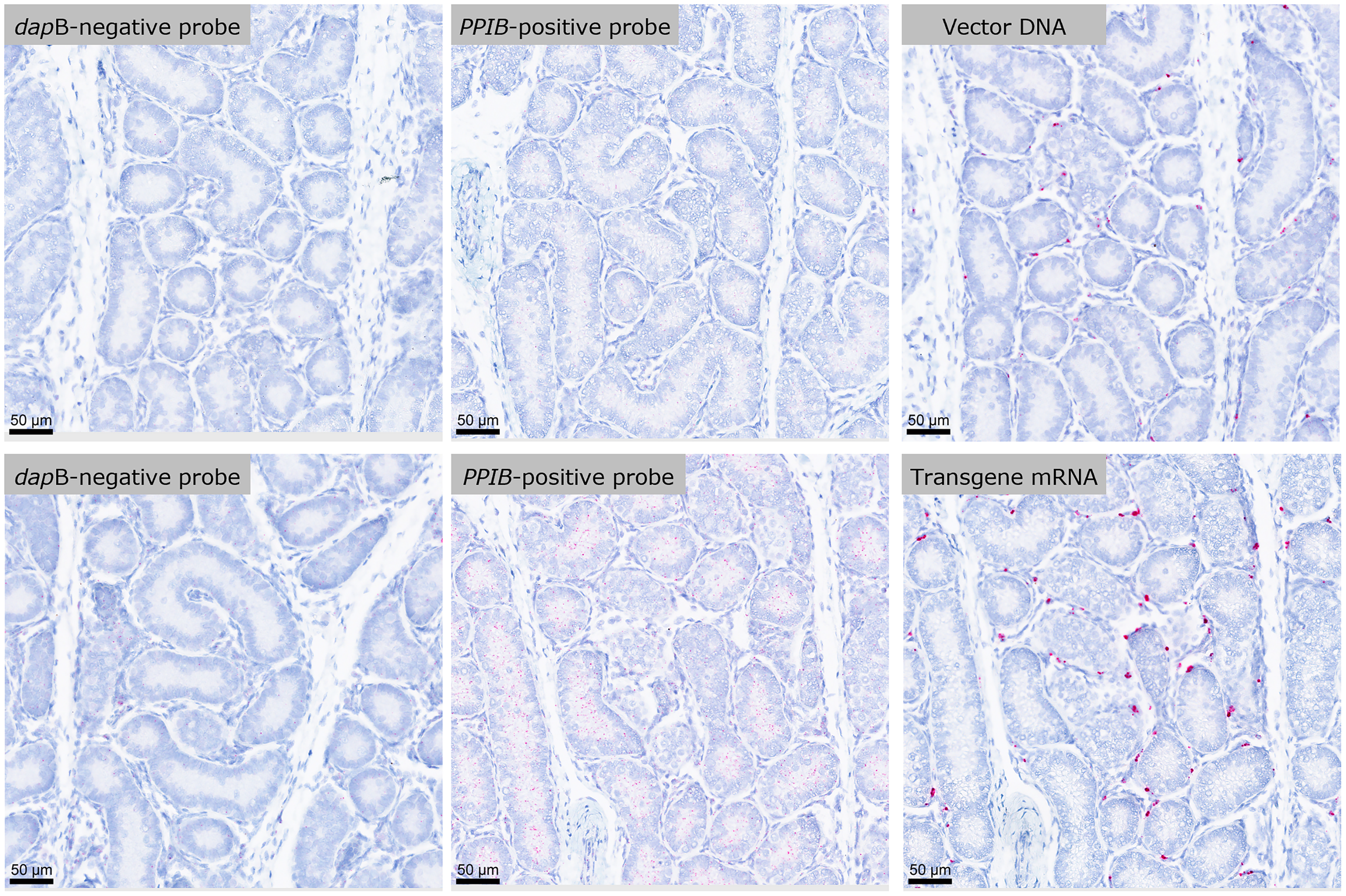
in situ hybridization (ISH) on bilateral testes sample from a male receiving dose level equivalent to the female in Figure 1 demonstrated positive vector DNA and transgene mRNA signal in interstitial Leydig cells, interstitial vascular endothelial cells, peritubular myoid cells, and peritubular fibroblasts. No evidence of male germ cell transduction was observed in any of the testes examined.
There was a dose-dependent pattern in the presence and distribution of vector DNA and transgene-positive signal in the gonads of male and female animals. The positive dots were sparse in the gonads of animals that received the low dose than in the gonads of animals that received the highest dose. The pattern and distribution of positive signal were comparable in the testicular and ovarian somatic and stromal (non-germ) cells in animals. Gonads from one male and one female control animals administered the formulation buffer were used as a negative control for the ISH assay, and as expected, there was no evidence of a positive signal in the germ cells or non-germ cells in these two animals (Supplementary Fig. S1).
DISCUSSION
In this brief report, we share the first case of AAV transduction of oocytes in cynomolgus monkeys 1 month following systemic intravenous administration. The vector DNA genome was likely transcriptionally active, as demonstrated by the more prominent signal detected by ISH mRNA probe compared with that of DNA probe, and consistent with PCR data that showed higher transgene mRNA compared with vector DNA copy numbers in the middle dose group animals (data not shown). The detection of AAV genome and transgene mRNA was unique to this single female administered the highest dose in the study and euthanized at 1 month post dose, while the vector DNA genome and transgene mRNA ISH signals in the remaining animals that received the same dose level or the 2 lower dose levels and euthanized at 1- or 3-months post vector administration was restricted to non-germ cells with no evidence of germ cell (oocyte) transduction. Vector DNA and transgene mRNA ISH signals in all male animals, at all dose levels and 2 euthanasia timepoints, were also equally restricted to non-germ cells with no evidence of germ cell (sperm) positivity. In our literature search, we were only able to identify a single report that documented the observation of oocyte transduction by ISH; the report was in mice 15 and 180 days following AAV2/8 systemic administration at the dose level of 2 × 10¹³ vg/kg (3 of 3 on day 15 and 2 of 3 on day 180). 9 However, no images were shared in the report to review or confirm the appearance or morphological localization of such finding within the ovaries. A recent study, among many others discussed below, utilized the sensitive RNAScope ISH technology and failed to detect sperm or oocyte transduction following systemic intravenous AAV9 administration at the high dose level of 6 × 10¹² vg/mouse in adult dystrophin null mice (equivalent to 1.7–2.5 × 10¹⁴ vg/kg). 10
Several observations are noteworthy in this brief report. Among all 16 females explored (at 3 dose levels and 2 euthanasia timepoints), oocyte transduction was uniquely detected in (1) a single female, that was (2) intravenously administered high dose of AAV at 1 × 10¹⁴ vg/kg, (3) examined at 1 month post vector administration, and (4) uniquely detected in early-stage primordial/primary follicles, but not in late-stage antral/Graafian follicles. The limited presence of germ cell transduction to one high-dose female may reflect the biological low incidence of AAV-mediated germ cell transduction even at high-dose systemic AAV administration and is consistent with the body of literature obtained from mouse studies.7,8 Germ cells are generally protected by the specialized blood-testis and blood-follicle barriers in testes and ovaries, respectively, that protect the developing sperm and oocyte from blood circulation and systemic exposures, and rAAV likely fails to cross or disrupt such barriers in order to transduce germ cells.11–13 The detection of oocyte transduction only in early-stage primordial or primary follicles and not in late-stage developed antral or Graafian follicles may reflect the limited susceptibility of early-stage follicles. Additionally, the observation was only at 1 month, but not 3 months, post-vector administration, which may also reflect that the transduced AAV DNA genome is transiently present and is expelled out over time or as the oocytes develop into late-stage follicles, similar to the loss of vector genome observed in hepatocytes.14,15
It should be emphasized that the low incidence of germ cell transduction reported herein is a separate risk from the potential for germline transmission, as a prerequisite for vector genome integration into the oocyte genome may be required in order to sustain the metabolic activity and many rounds of cell proliferation from the fertilized eggs into the newborn offsprings. This is clearly demonstrated in the mouse study that detected oocyte transduction following systemic administration, 9 or in several mouse breeding studies including those that actively transduced germ cells in male and female mice using several rAAV serotypes, yet failed to demonstrate any evidence of germline transmission to F1 progeny, even following the microinsemination of confirmed transduced germ cells.16,17
The observations and conclusions made in this report are limited by the study design and low number of animals. Nonetheless, this brief report highlights the potential of oocyte transduction, particularly in female non-human primates following systemic intravenous administration of high-dose rAAV and provides visual representation of such findings for future references. Additional work will be necessary to confirm the true incidence of findings, species restrictions, and to evaluate the potential for germline transmission.
AUTHORS’ CONTRIBUTIONS
B.T.A. contributed to the conception, organization, and drafting of the article. J.G.D. contributed to the PCR data and review of the article.
Footnotes
ACKNOWLEDGMENT
The authors would like to acknowledge all Sanofi team members and Charles River Laboratories scientists who contributed to this study.
FUNDING INFORMATION
Funding was provided by Sanofi.
AUTHOR DISCLOSURE
All authors are paid employees and have stock interests in Sanofi at the time of writing.
Supplemental Material
References
Supplementary Material
Please find the following supplemental material available below.
For Open Access articles published under a Creative Commons License, all supplemental material carries the same license as the article it is associated with.
For non-Open Access articles published, all supplemental material carries a non-exclusive license, and permission requests for re-use of supplemental material or any part of supplemental material shall be sent directly to the copyright owner as specified in the copyright notice associated with the article.
