
Abstract
This study investigated the influence of homeopathic medicines (HMs) on the in vitro behavior of stem cells from human exfoliated deciduous teeth (SHED). The HMs Symphytum officinale (SO), Calcarea carbonica (CC), and Calcarea phosphorica (CP) were evaluated, along with a vehicle control (V), using cells from a single donor in passages 4 (P4) and 5 (P5). Cellular responses were assessed using Neutral Red (viability-related activity), Crystal Violet (adherent cell density), MTT (metabolic activity), and scanning electron microscopy (SEM). Across assays, consistent modulation of cellular behavior was observed, particularly in P5 at 72 h. Neutral Red uptake increased over time in P4 and showed treatment-dependent responses in P5. Crystal Violet indicated increased adherent cell density, with higher values in P5 compared to P4. MTT demonstrated increased metabolic activity at 72 h, especially in P5. SEM analysis confirmed these findings, showing that cells treated with SO, CC, and CP remained well adhered and spread, with increased cell density and more compact organization. In contrast, the vehicle group exhibited reduced cell density and altered morphology. Overall, the tested HMs were associated with measurable changes in SHED behavior without evident adverse effects. These findings should be interpreted as exploratory and require further validation.
Introduction
Modern lifestyles characterized by physical inactivity and industrialized diets have increased the prevalence of bone diseases (Tamme et al., 2019). Among the pharmacological treatments, bisphosphonates are widely used to enhance bone mineral density, alleviate pain, and reduce fracture risk. However, prolonged administration may lead to Bisphosphonate-related Osteonecrosis of the Jaws (BRONJ).
Given these limitations, less toxic therapeutic alternatives have emerged, including molecular approaches such as microRNAs (Vázquez et al., 2021), macrophage exosomes (Hu et al., 2021), RUNX1 modulation (Tang et al., 2020), genetic therapies (Formosa et al., 2021), and integrative strategies involving homeopathic medicines (HMs).
Within homeopathy, Calcarea phosphorica (CP), Calcarea carbonica (CC), and Symphytum officinale (SO) are traditionally employed for bone and mineralization disorders. CP (calcium phosphate) supports bone formation and fracture healing, being indicated during growth, convalescence, and delayed consolidation (Tripathi and Choudhary, 2025). CC (oyster shell) contributes to structural balance and exhibits anti-inflammatory effects via COX modulation (Kumar et al., 2020). SO (“comfrey”) displays osteoregenerative and wound-healing properties, stimulating osteoblastic differentiation and tissue regeneration (Dey et al., 2020; Le et al., 2021).
At the cellular level, these medicines may modulate signaling pathways, proliferation, and the microenvironment, effects studied in mesenchymal stem cells (MSCs) (Valle et al., 2023). MSCs exhibit self-renewal and multipotency toward osteogenic, chondrogenic, and adipogenic lineages, making them valuable for regenerative medicine (Seigner et al., 2019). Stem cells from exfoliated deciduous teeth (SHED) represent an ethical and potent MSC source, with high proliferative and osteogenic capacity (Takebe et al., 2017; Shoushrah et al., 2021).
Integrative approaches combining homeopathy and cell therapy may enhance tissue engineering outcomes, providing safer and more accessible regenerative alternatives. Comparative studies have reported favorable outcomes for homeopathic over allopathic treatments in bone neoformation and with fewer adverse effects (Werkman et al., 2006). Ultradiluted preparations (e.g., 30CH) may influence cellular signaling through nanostructures or water clusters, affecting receptor–ligand interactions (Bell et al., 2013).
Clinical evidence supports homeopathic efficacy in conditions such as heterotopic ossification and knee bone marrow edema, with reduced pain, regression of calcifications, and improved joint function (Tsintzas et al., 2020; Tsintzas et al., 2019). In preclinical models, CP promoted bone repair in osteoporotic rats (Werkman et al., 2006), while CC reduced inflammation via COX modulation (Kumar et al., 2020). SO enhanced bone formation around titanium implants and demonstrated anti-inflammatory and reparative effects (Spin-Neto et al., 2010; Seigner et al., 2019; Staiger, 2012).
Therefore, this study aimed to investigate the influence of CP, CC, and SO in the culture of stem cells from human exfoliated deciduous teeth cells (SHED) in the first two passages after cryopreservation (P4 and P5).
Material and Methods
Ethical approval
The study was approved by the Research Ethics Committee of University Positivo (C.A.A.E.: 14634819.6.0000.0093). A single exfoliated deciduous tooth from a child treated at the Pediatric Dentistry Clinic was used, following parental consent. The tooth, which would otherwise be discarded, was donated for research purposes.
Due to ethical and practical constraints, only one donor was included, and the study was designed as a proof-of-concept exploratory model.
Isolation and culture of SHED
Stem cells from human exfoliated deciduous teeth (SHED) were isolated using the explant technique, which does not involve enzymatic digestion. Dental pulp was extracted, minced into small fragments, and washed with phosphate-buffered saline (PBS, GIBCO, Carlsbad, USA). Tissue fragments were placed in T25 flasks and incubated (InCuSafe, MCO-18AC-PA, Panasonic, Osaka, Japan) at 37°C and 5% CO2 for 20 min. Subsequently, 5 mL of Dulbecco’s Modified Eagle’s Medium (DMEM, Cytiva, Marlborough, USA), supplemented with 10% fetal bovine serum (FBS, Hyclone, Marlborough, USA) and 1% antibiotic–antifungal solution (GIBCO, Carlsbad, USA), was added.
The medium was replaced every 72 h until cell confluence. Cells were expanded to passage 3 (P3) and cryopreserved at 1 × 10⁶ cells per tube in liquid nitrogen. Cryopreserved cells were rapidly thawed at 37°C and cultured under standard conditions prior to experimental use in passages 4 (P4) and 5 (P5).
Preparation of HMs
According to the Brazilian Pharmacopeia (NatioBrazil, 2011), the HMs were prepared with homeopathic derivative dosage forms carried out in purified water, obtained using reverse osmosis (Quimis, Q842-210, Diadema, Brazil), in an amber 15 mL glass container, and dynamized in 30c potency through mechanical agitation (100 succussions for each energized potency) (Denise 10–50 Mechanical Arm Dynamizer, Citua, Campinas, Brazil), then filtered through a 0.022 µm membrane (Merck Millipore, Barueri, Brazil). In a previous and nonreported herein pilot, 5c, 6c, and 12c were also used but quickly demonstrated cell impairment.
Experimental design
All in vitro experiments were performed at 37°C in a humidified atmosphere containing 5% CO2 for 24, 48, and 72 h. Cells were maintained in DMEM (GIBCO, Carlsbad, USA) supplemented with 10% FBS (GIBCO, Carlsbad, USA).
SHEDs (P3), upon reaching 70%–80% confluence, were detached using trypsin/EDTA for 5 min, resuspended in DMEM supplemented with 10% FBS, and washed twice with PBS (pH 7.4). Cells previously characterized according to the Dominici et al. (2006) criteria were seeded in 96-well plates (Kasvi, São José dos Pinhais, Brazil) at a density of 4 × 10³ cells per well.
HMs—CC, CP, and SO—all at 30c potency, were administered daily. Treatments consisted of an initial addition of 50 µL of the respective HM, vehicle (ultrapure water), or culture medium (DMEM) to 250 µL of medium per well. Additional 50 µL doses were added every 24 h without medium replacement.
This approach resulted in cumulative exposure over time, with progressive changes in the proportion of administered solutions relative to the culture medium (∼16.7% at 0 h, 28.6% at 24 h, 37.5% at 48 h, and 44.4% at 72 h).
All solutions were vigorously shaken prior to application. Absorbance readings were performed in a blinded manner.
Experiments were conducted using cells derived from a single donor, expanded into different passages (P4 and P5). For each passage, assays were performed in duplicate cultures and in technical replicates (n = 6). Therefore, the data reflect technical variability within the same biological source, and comparisons between passages were considered descriptively.
Experimental groups were organized as follows: CC, CP, SO, vehicle control, and DMEM, which served as baseline control.
Cell viability and cell density assays (Neutral Red and Crystal Violet)
Cell viability was assessed using the Neutral Red assay as described by Borenfreund and Puerner (1985), and cell density was evaluated using the Crystal Violet method (Gillies et al., 1986).
For Neutral Red, SHEDs were seeded at 4 × 10³ cells per well. Treatments were applied at 24, 48, and 72 h. Cells were incubated with Neutral Red dye (10 µg/mL) for 2 h at 37°C and 5% CO2. After incubation, Neutral Red solution was removed, cells were washed with water then PBS, and the dye was solubilized using an ethanolic solution under gentle agitation for 10 min. Absorbance was measured at 550 nm using a microplate reader (ELx 800, Meridian Diagnostic, Inc.).
Subsequently, cell density was assessed on the same plate using the Crystal Violet method. Following removal of the ethanolic solution, cells were washed with PBS and fixed with methanol for 10 min at room temperature. The methanol was removed, and 75 μL of Crystal Violet solution (0.2% in 5% ethanol) was added for 10 min, followed by washing with PBS. The dye was solubilized using 0.1 M sodium citrate in 50% ethanol, and absorbance was measured at 550 nm (Epoch Biosystem, Biotek, USA). This sequential approach was used to obtain complementary measurements from the same cell population.
MTT (thiazolyl blue tetrazolium bromide) assay
Cell metabolic activity was evaluated using the MTT assay as described by Mosmann (1983). SHED cells were seeded at 4 × 10³ cells per well in 96-well plates. Treatments (50 µL) were administered daily for 24, 48, and 72 h. Prior to the assay, wells were washed with PBS, and 200 µL of MTT solution (0.5 mg/mL in PBS) was added to each well. Plates were incubated for 3 h at 37°C and 5% CO2. The MTT solution was then removed, and 200 µL of dimethyl sulfoxide (DMSO; Sigma-Aldrich, Vienna, Austria) was added to dissolve the formazan crystals. Absorbance was measured at 550 nm.
Scanning electron microscopy
Scanning electron microscopy (SEM) was used to evaluate cell morphology. The procedure was performed according to the protocol described by Biscaia et al. (2017).
Cells in P4 and P5 (2 × 10⁴) were cultured in 24-well plates on glass coverslips (Corning) and exposed to the experimental groups: CC, CP, SO, vehicle (ultrapure water), and control (DMEM only). Cells were maintained at 37°C and 5% CO2 for 72 h.
Cells were fixed for 1 h in Karnovsky solution (2% glutaraldehyde, 4% paraformaldehyde, 1 mM CaCl2 in 0.1 M sodium cacodylate buffer), washed, and postfixed in 1% osmium tetroxide. Samples were then washed in sodium cacodylate buffer for 1 h (in the dark at room temperature), dehydrated through graded ethanol series, and dried to the critical point. Subsequently, samples were sputter-coated with gold.
Images were obtained using a scanning electron microscope (TESCAN VEGA3 LMU, Brno, Czech Republic), and analysis was performed in a blinded manner.
Results
Cell viability (Neutral Red) assay
Figure 1 shows the Neutral Red assay results. Overall, Neutral Red uptake was higher in passage 5 (P5) than in passage 4 (P4) across all groups.

Neutral Red assay showing cell viability of cells exposed to homeopathic medicines and control groups at 24, 48, and 72 h in passages 4 (P4) and 5 (P5). Data are presented as mean ± standard deviation. Different uppercase letters indicate statistically significant differences among time points within the same treatment and passage (p < 0.05). All experiments were performed using cells derived from a single donor; therefore, results are based on technical replicates.
In P4, all treatments exhibited a consistent temporal pattern, with significantly higher values at 72 h compared to 24 h and 48 h (p < 0.05), while no significant differences were detected between 24 h and 48 h. In P5, the temporal profile was treatment-dependent: Calcarea carbonica and Calcarea phosphorica showed no significant differences over time, whereas SO and the vehicle group exhibited higher values at 24 h (p < 0.05). The control group showed increased values at 72 h compared to 48 h (p < 0.05).
Overall, P4 showed a uniform increase over time, while P5 presented variable responses, with consistently higher values than P4. Detailed numerical values are provided in Table 1.
Mean (±SD) Neutral Red Absorbance Values Indicating Cell Viability in Contact with Homeopathic Medicines and Control Groups at Different Time Points in Passages 4 and 5
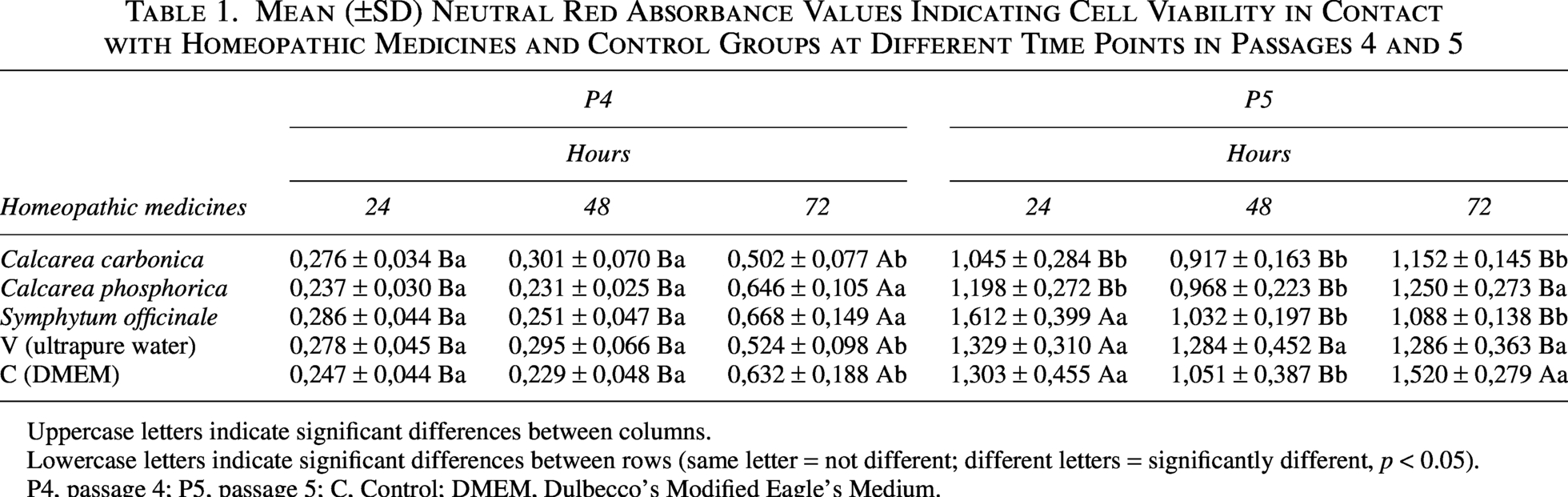
Uppercase letters indicate significant differences between columns.
Lowercase letters indicate significant differences between rows (same letter = not different; different letters = significantly different, p < 0.05).
P4, passage 4; P5, passage 5; C, Control; DMEM, Dulbecco’s Modified Eagle’s Medium.
Cell density (Crystal Violet) assay
Figure 2 shows the Crystal Violet assay results. As this assay was performed sequentially after Neutral Red, results reflect relative adherent cell density rather than direct proliferation.
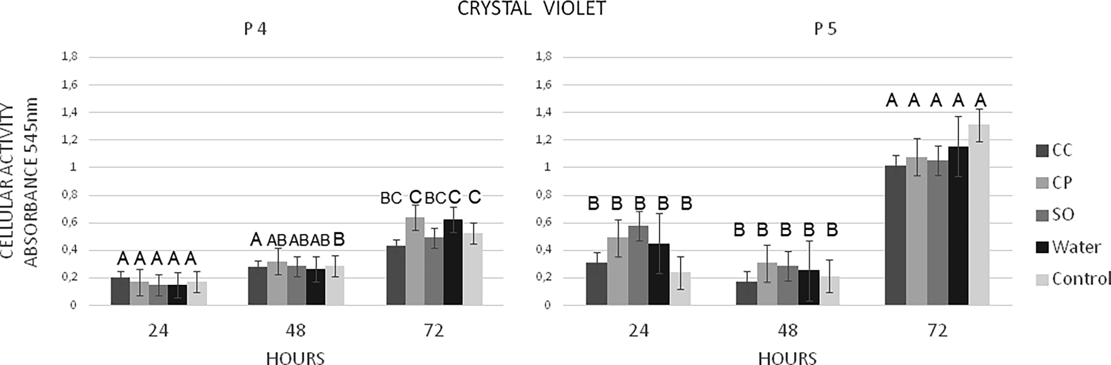
Crystal Violet assay showing relative adherent cell density of cells exposed to homeopathic medicines and control groups at 24, 48, and 72 h in passages 4 (P4) and 5 (P5). The assay was performed sequentially using the same cells previously subjected to the Neutral Red assay. Data are presented as mean ± standard deviation. Different uppercase letters indicate statistically significant differences among time points within the same treatment and passage (p < 0.05). All experiments were performed using cells derived from a single donor; therefore, results are based on technical replicates.
In P4, a gradual increase over time was observed, with intermediate values at 48 h varying among treatments. In contrast, P5 showed a consistent pattern across all groups, with significantly higher values at 72 h compared to earlier time points (p < 0.05), and no significant differences between 24 h and 48 h.
Overall, values were higher in P5 than in P4, particularly at 72 h, indicating a passage-related effect. Detailed data are shown in Table 2.
Mean (±SD) Crystal Violet Absorbance Values Indicating Cell Proliferation in Contact with Homeopathic Medicines and Control Groups at Different Time Points in Passages 4 and 5
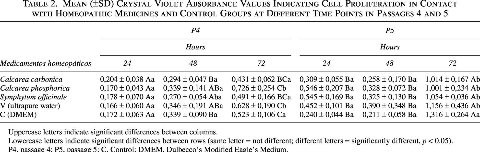
Uppercase letters indicate significant differences between columns.
Lowercase letters indicate significant differences between rows (same letter = not different; different letters = significantly different, p < 0.05).
P4, passage 4; P5, passage 5; C, Control; DMEM, Dulbecco’s Modified Eagle’s Medium.
Cellular metabolic activity (MTT) assay
Figure 3 shows the MTT assay results. As this assay was performed sequentially after Neutral Red and Crystal Violet, results reflect cellular metabolic activity rather than direct cytotoxicity.
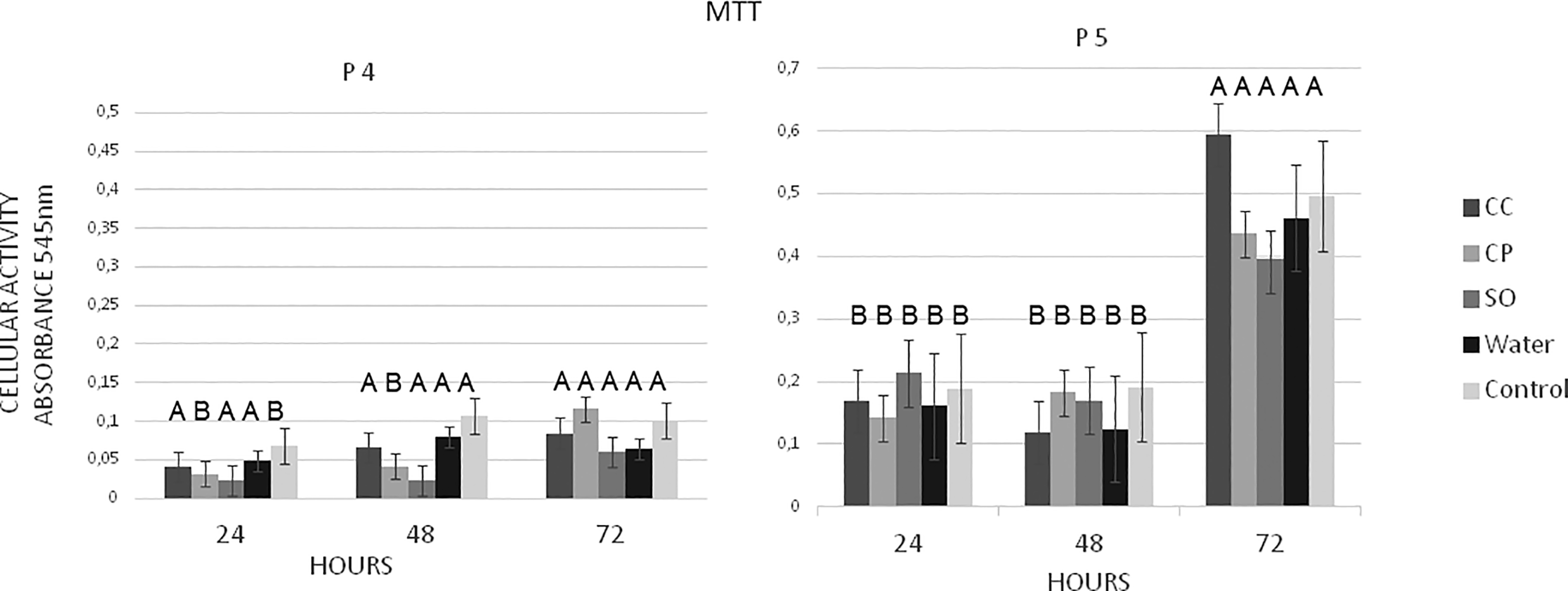
MTT assay showing cellular metabolic activity of cells exposed to homeopathic medicines and control groups at 24, 48, and 72 h in passages 4 (P4) and 5 (P5). The assay was performed sequentially using the same cells previously subjected to Neutral Red and Crystal Violet assays. Data are presented as mean ± standard deviation. Different uppercase letters indicate statistically significant differences among time points within the same treatment and passage (p < 0.05). All experiments were performed using cells derived from a single donor; therefore, results are based on technical replicates.
Overall, absorbance values were higher in P5 than in P4 across all groups. In P4, metabolic activity remained low and showed no consistent temporal pattern. In contrast, P5 exhibited significantly higher values at 72 h compared to 24 and 48 h (p < 0.05), with no significant differences between earlier time points, indicating a marked late-stage increase in metabolic activity, particularly in P5. Detailed numerical values are presented in Table 3.
Mean (±SD) MTT Absorbance Values Indicating Cytotoxicity in Contact with Homeopathic Medicines and Control Groups at Different Time Points in Passages 4 and 5
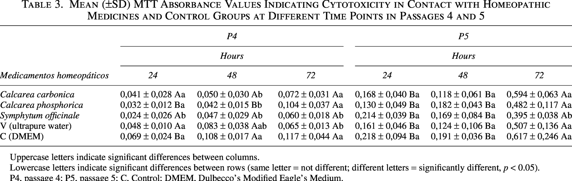
Uppercase letters indicate significant differences between columns.
Lowercase letters indicate significant differences between rows (same letter = not different; different letters = significantly different, p < 0.05).
P4, passage 4; P5, passage 5; C, Control; DMEM, Dulbecco’s Modified Eagle’s Medium.
Scanning electron microscopy
Figure 4 shows the ultrastructural features of cells under different experimental conditions.
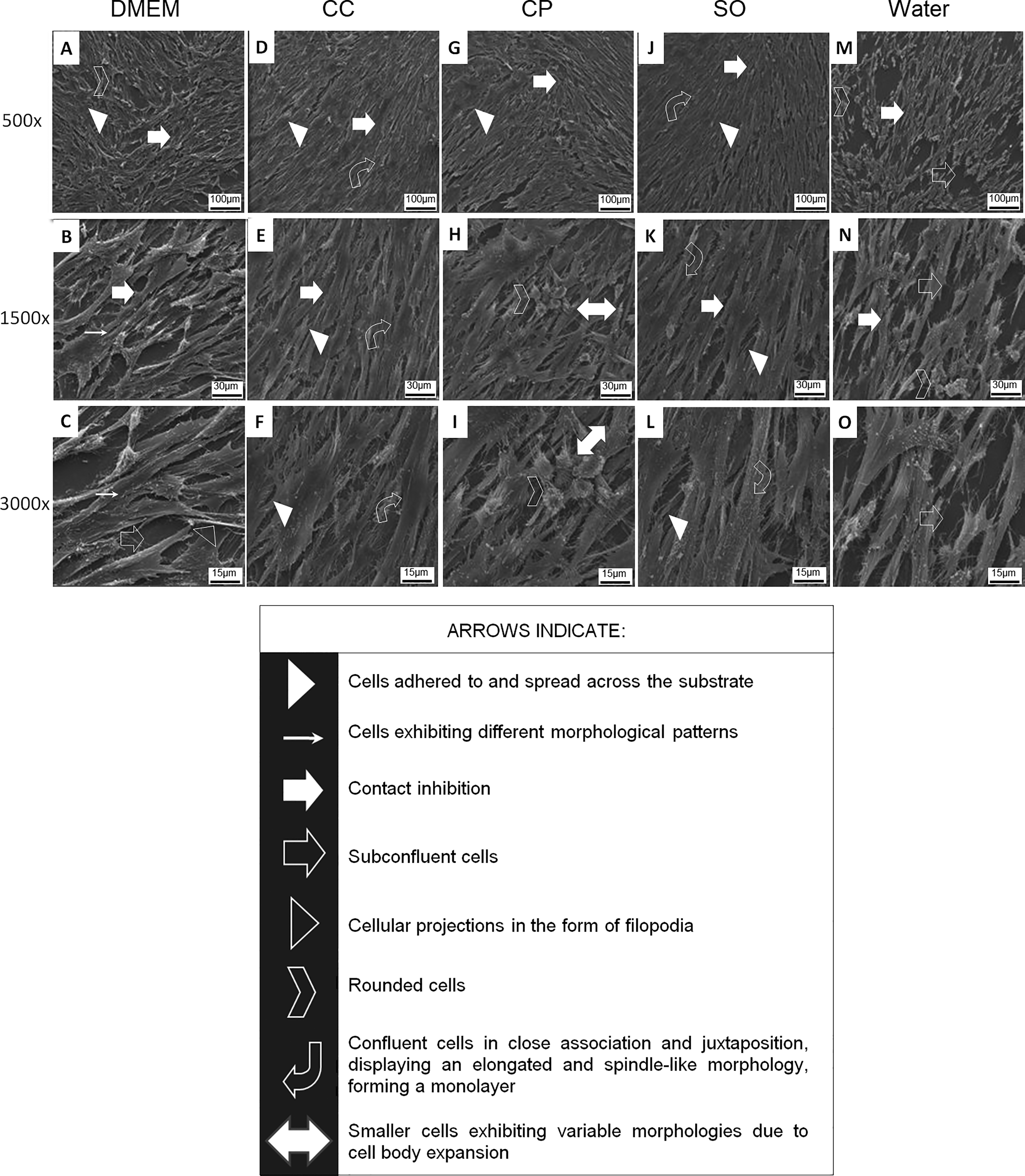
Ultrastructural analysis by scanning electron microscopy (SEM) of cells under different experimental conditions (in P5). Images
Control cells (DMEM only; Figure 4A–C) were well adhered and spread on the substrate, exhibiting typical morphology with contact inhibition and side-by-side organization. Subconfluent areas were observed, with cytoplasmic projections and sparse filopodia. Occasional small, rounded cells were also detected, suggestive of dividing or nonviable cells.
Cells treated with CC, CP, and SO (Figure 4D–L) remained adherent and spread, showing morphology similar to control cells. However, increased cell density and more compact organization were observed, suggesting higher confluence. CP and SO induced morphological changes characterized by elongated cells, reduced spreading, and closer cell–cell association, forming a more uniform monolayer. In contrast, CC treatment resulted in greater morphological heterogeneity.
Treatment with water (Figure 4M–O) induced the most pronounced changes, with reduced cell number, decreased spreading, and a higher proportion of rounded cells, suggesting impaired cell adhesion and/or altered cellular integrity.
Discussion
In vitro studies combining HMs and cell culture models remain limited and highly specific. Previous studies have explored different approaches, such as the use of low-diluted Viscum album in canine MSCs (Valle et al., 2023), CP-doped nanobioglass particles in murine stem cells (Kumar, 2018), and SO in osteogenic differentiation models (Dey et al., 2020). However, data involving human stem cells, particularly those derived from deciduous teeth (SHED), remain scarce.
In this context, the present study provides a proof-of-concept model demonstrating that SHED cells can be used to investigate cellular responses to HMs intended for bone-related applications. The results highlight SHED as a promising experimental model for studies in the homeopathic field.
A key aspect of this study was the comparison between closely related passages (P4 and P5). These passages are generally considered to maintain phenotypic stability following initial expansion and cryopreservation, while still allowing sufficient cell numbers for experimental assays (Perry et al., 2008; Takebe et al., 2017; Wang et al., 2018). Nevertheless, our findings indicate that even small differences in passage number may influence cellular behavior, as consistently higher activity, density, and metabolic responses were observed in P5 compared to P4. This suggests that passage-related effects should be carefully considered when interpreting in vitro data using stem cells.
In the Neutral Red assay, which reflects lysosomal activity associated with cell viability, a clear temporal increase was observed in P4, while P5 exhibited treatment-dependent variations. Although some groups showed comparable values, overall results suggest increased cellular activity at later time points and in higher passages, consistent with previous findings using MSCs (Kumar et al., 2008).
The Crystal Violet assay, performed sequentially after Neutral Red, provided complementary information on adherent cell density. A gradual increase was observed in P4, whereas P5 showed a more uniform and pronounced increase at 72 h across all groups. This pattern suggests enhanced cell accumulation or retention over time, particularly in later passages, although it does not directly indicate proliferation. Some HMs, as SO have demonstrated a trend for increased proliferation (Dey et al., 2020).
Similarly, the MTT assay, which reflects mitochondrial metabolic activity, showed a marked increase at 72 h in P5. These findings indicate a late-stage increase in cellular metabolic activity, reinforcing the passage-dependent pattern observed across assays. Previous studies have reported biological effects of SO in MSCs (Le et al., 2021), hepatotoxic effects have also been described (Sakakura et al., 2008), and associating SO with bone regeneration-related effects (Seigner et al., 2019). However, in the present study, metabolic activity changes should be interpreted cautiously, as they do not necessarily reflect cell number or viability. Therefore, further studies using molecular markers such as alkaline phosphatase, osteocalcin, and osteopontin are required to clarify potential differentiation pathways.
Ultrastructural analysis by SEM further supported these findings. Cells exposed to CC, CP, and SO maintained adhesion and spreading patterns similar to controls, while exhibiting increased cell density and closer cell–cell organization. In particular, CP and SO induced a more elongated and spindle-like morphology, with reduced spreading and tighter cell alignment. These features are consistent with typical mesenchymal stem cell morphology, which is characterized by fibroblast-like, elongated cells capable of forming monolayers and adapting their shape according to microenvironmental cues (Dominici et al., 2006). Additionally, SEM studies have shown that MSC morphology, including elongation and filopodia formation, is closely related to adhesion, cytoskeletal organization, and cell–substrate interactions, which may influence proliferation and differentiation processes (de Peppo et al., 2014).
In contrast, exposure to water resulted in reduced cell density, decreased spreading, and increased numbers of rounded cells, suggesting impaired adhesion and altered cellular integrity. Even though the evidence base for HMs in cell culture systems is growing rapidly across various cell types, basic cell experiments using stem cells with HMs still need to be made available (Sybil et al., 2020).
Despite these observations, several limitations must be considered. First, the use of SHED derived from a single donor limits generalizability and does not capture biological variability. Second, no evaluation of stemness, differentiation, or senescence markers was performed, preventing conclusions about functional cell status. Third, although early passages were used, passage-dependent effects were observed, indicating that even minimal expansion may alter cellular responses. Additionally, the absence of a succussed-only control limits the ability to distinguish specific homeopathic effects from possible mechanical or physicochemical influences.
Taken together, the present findings should be interpreted as a proof-of-concept model rather than definitive biological evidence. Further studies using multiple donors, extended time points, and molecular and functional analyses are required to validate these observations and better understand the mechanisms underlying the effects of HMs on MSCs.
Conclusion
The 30C homeopathic preparations (SO, CC, and CP) were associated with measurable changes in SHED cellular behavior in vitro, including increased cellular activity, adherent cell density, metabolic response, and morphological alterations. These effects followed a consistent passage-dependent pattern, with more pronounced responses observed in P5, particularly at 72 h.
Within this context, the present study should be considered an exploratory proof-of-concept model. The findings must be interpreted with caution, as the assays reflect indirect measures of cellular behavior, and the study is limited by the use of a single donor, the absence of stemness evaluation, and variability between passages. Further studies including multiple donors, molecular analyses, and improved experimental controls are required to validate these observations and clarify their biological significance.
Footnotes
Disclosure Statement
The authors declare that they have no conflicts of interest related to the work presented in this article.
Funding Information
This research received no specific grant from any funding agency in the public, commercial, or not-for-profit sectors.