



Abstract
Background
The Slump Test evaluates neural tissue mechanosensitivity in individuals with lower extremity symptoms. However, there has been little research assessing the role of altering lumbar spine positioning on sensory response. Therefore, this study compared differences in subjective and objective findings observed in different lumbar spine positions during neurodynamic testing.
Methods
Healthy participants were assessed using the 90/90 knee extension (KEA) to determine available ROM, followed by lumbar spine flexed slump (FS), and lumbar spine extended slump (ES). Outcomes included knee extension angle, hamstring EMG activity (%MVIC), distal extent of sensory response, and qualitative mechanosensory response.
Results
Forty participants (23.95 ± 2.64 years; 14 male/26 female) completed the study. Significantly less knee extension ROM was found for ES vs. FS (p < .001) and ES vs. KEA (p = .037). A greater proportion of distal symptoms were observed during ES vs. FS (p < .05). FS and ES showed higher EMG values than KEA (p < .001), with no difference between the two slump conditions.
Conclusion
In this healthy population, ES produced the greatest distal sensory responses, highest EMG activity, and lowest knee extension ROM. Incorporating lumbar extension during slump testing may be useful to determine mechanosensitive behavior, but further research is needed in clinical populations.
The trial was prospectively registered at clinical trials.gov. NCT05313217
Keywords
Background
Leg pain of neuropathic origin 1 or somatic referral is present in up to 57% of individuals with low back pain. 2 The use of provocative tests such as the slump test are clinical staples which help clinicians understand the behavior of sensory responses related to mechanosensitive sources.1,3,4 The positioning of the slump test combines axial spinal load while also placing increased tension on the neural tissues both distally and cranially. Mechanosensitivity results in the provocation of a sensory response, a reduction of range of motion of the affected side, or both.5,6 However, it remains unclear whether mechanical factors such as increased axial load and lumbar spinal position influence the neurosensory response to these tests.
McDevitt and colleagues have suggested that a greater understanding of mechanisms is required as we seek to understand musculoskeletal examination and intervention techniques. 7 Specific to neuropathic symptoms, a recent IASP consensus framework suggests treating the “container” (e.g., neuroforaminal interface) rather than the nerve itself in cases where neural response is due to space limitations. 8 Though the slump test is reliable for assessing neural mechanosensitivity in spinal and lower limb pain,9,10 both tension and compression have been implicated as sources of peripheral neuropathic pain in clinical populations, 11 and it remains uncertain if slump testing differentiates between stretch or tension-related mechanosensitivity and compression-related mechanosensitivity. Flexion of the lumbar spine is generally accepted to produce elongation and tension on the nervous system,12,13 with accompanying enlargement of the neural foramen. 14 When the lumbar spine is extended compared to neutral posture a significant decrease in cross-sectional area in the neural foramen has been observed, accompanied by shortening of the nervous system, potentially increasing the likelihood of neural compression and related neurosensory response.14,15 However, this is not the only potential source of compression. In a series of cadaveric spines, neural compression within the foramina was demonstrated to occur in over 15% of spines in flexed postures and 33% of spines in extended positions, supporting a need for dynamic testing to better understand the interface between the nerve and foramen. 14 Anecdotally, observation and clinical experience suggest a subset of individuals who experience a greater neurosensory response to slump testing performed with lumbar extension rather than lumbar flexion. To date, little research has explored how differences in trunk position affect mechanosensory response during the slump test.
Protective hamstring contraction has been observed during provocative neural testing, especially in symptomatic individuals.16-18 Since higher levels of hamstring contraction are thought to be protective of the nervous system, they may serve as an objective measure of stretch tolerance and/or mechanosensitivity, particularly if compared to baseline measures. The passive variation of the knee extension angle (KEA) has been suggested as the most accurate measure of hamstring length, serving as a baseline for both expected available ROM and tolerance to muscular stretch provocation with minimal neural involvement in an unloaded position. 19 Subsequent comparisons of range of motion, neurosensory response, and hamstring activity between the baseline measures of KEA and slump test variants will help clarify the role of spine position on neurosensory response.
This study aimed to explore the potential impact of spinal position on neurosensory response as observed through participant reported intensity and location, knee extension range of motion, and concurrent hamstring electromyographic (EMG) activity. A deeper understanding of these effects may improve clinical decision-making regarding neurodynamic assessment and intervention.
Methodology
Sample
The sample size was determined a priori. For the primary outcome of differences in ROM between individuals with and without neuropathic findings (lumbosacral radiculopathy), using a between group difference of 9 ± 9°, 16 α=0.05, and a power of 0.80, a minimum sample size of 32 participants was established. To account for unexpected variability in data or participant drop out, the sample was enlarged to 40 participants.
A convenience sample was recruited through word-of-mouth on a university campus between March 28-June 30, 2022. The trial protocol was approved by the University of Hartford IRB (approval #020220124), and the trial protocol was registered at clinical trials.gov; NCT05313217.
Procedure
Participants eligible for the study had to report no prior history of low back pain (LBP), surgeries, injuries to the lumbar spine or lower extremities, neurological symptoms, conditions affecting the lower extremities, or any other comorbidities. These exclusion criteria were established based on previous studies.16,20
All participants completed a medical screening form and informed consent before undergoing a physical screen that included spinal range of motion, myotome, dermatome, and reflex testing to screen for radicular symptoms and ensure participants were physically able to participate in the study. The height and weight of each subject were collected and recorded. All procedures were standardized using the right leg to ensure consistency, and all participants were tested at the same time of day in the University of Hartford motion analysis lab. Research has indicated that various sequences of neurodynamic testing do not change the overall mechanical load applied to the neural tissue. However, clinically, different sequences might affect the strain on the nerve due to variations in total time under tension or changes in the range of joint motion achieved by these sequences. 21 Thus, the performance sequence for all tests was kept identical for reliable comparisons.
Preparation for the EMG electrode was done by vigorously cleaning the skin over the biceps femoris with an alcohol wipe. The patient was instructed to lie prone with thighs flat on the table. The electrode (Norotrode 20, Myotronics Inc., Kent, WA, USA) was placed halfway between the ischial tuberosity and the lateral epicondyle of the femur 22 using double sided tape and an elastic wrap to fixate onto the skin. To ensure accurate placement of the electrodes, the signal was monitored during a trial of resisted knee flexion.
Measurements for maximum voluntary isometric contraction (MVIC) were recorded. The participant was positioned in prone with the involved knee flexed to 45° with slight lateral rotation, and a strap anchored to the table was secured around their ankle. The participant was instructed to pull against the strap as hard as possible for nine seconds. (Figure 1) The MVIC was selected from the middle three seconds of the test data to minimize motion artifact. During the remainder of the tests, EMG activity was recorded with the EMG data segment corresponding to maximal ROM obtained during assessment and normalized to the MVIC.

MVIC lateral hamstring. MVIC: maximum voluntary isometric contraction.
Available knee extension ROM, with 0 degrees representing full extension and a negative measure indicating the number of degrees they were lacking full extension, was selected as the primary outcome of interest and has previously been demonstrated to be a reliable (ICC = 0.96) measure within and between sessions during slump testing. 23 Available knee extension ROM during neurodynamic testing may be influenced by the degree of dorsiflexion at the ankle. 5 To standardize this variable, a custom, rigid ankle foot orthosis (AFO) consisting of small, medium, and large sizes to accommodate a range of foot sizes was used to place the ankle in zero degrees of dorsiflexion during all trials of KEA and slump testing.
ROM was assessed with a digital Android inclinometer. Phone based inclinometers have been found to have strong test-retest reliability when measuring range of motion, 24 with SEM ranging from 0.60–2.00°.25,26 The same device was used by the same rater for all participants. For all measurements, the Android inclinometer was placed along the anterior distal tibia, with the inferior edge of the phone aligned with the superior aspect of the medial malleolus. 19 An 8° difference in knee flexion was considered clinically significant, reflecting the range of motion variance previously observed between stretches targeted towards the hamstring musculature and neural sturctures. 3
The following tests were performed sequentially for all participants: KEA, FS, and ES. For all tests, at the end of the available range of motion the participant was asked to qualitatively describe the location and quality, (Table 1) and to quantify intensity using the Numeric Pain Rating Scale (NPRS) 27 of neurosensory response. (Table 2) Knee extension ROM and biceps femoris EMG activity were recorded for all test conditions.
Qualitative questions.

KEA: 90/90 knee extension; FS: Flexed slump test, ES: Extended slump test
To measure the KEA, participants were positioned supine with their hips and knees fully extended. 19 The Android inclinometer was placed on the distal thigh immediately proximal to the patella and the hip was passively flexed to 90 degrees. The examiner stabilized the thigh at 90° of hip flexion, then performed passive knee extension to a point where either firm resistance was elicited 28 or the participant reported that they had reached the limit of their tolerance to stretch/mechanosensory response. Knee extension ROM was recorded by a second examiner. The passive KEA demonstrates high reliability (ICC3,1 0.97–0.98), 28 an acceptable SEM of 7.60°, and MDD95 of 21°. 29
Two variations of the seated slump test were performed: (1) with the lumbar spine flexed and (2) with the lumbar spine extended. The participant was instructed to sit upright in a short sitting position at the edge of the table. To standardize a 90° trunk angle for this test, a backboard braced against the table was utilized. 10 To maintain a neutral pelvis, a seatbelt was secured across the participant's anterior superior iliac spines and fastened around the backboard. (Figure 2) For the FS, participants were instructed to sequentially slouch their trunk and neck until all segments of the spine were in flexion. A researcher maintained the position of cervical flexion while the participant actively extended their right knee to either their limit of ROM or symptom tolerance, whichever came first. Knee extension ROM was measured at this point. After the FS was performed, the participant returned to the upright position. A half foam roller was placed just above the participants PSIS to achieve and maintain lumbar extension. The participant was then instructed to extend their spine until their scapulae contacted the backboard. An additional belt was added across the sternum and under the axillae to maintain this extended positioning. The participant was then asked to flex their neck; data collection was then performed using the same protocol as described with the FS test. (Figure 2).

From the left: 90/90 KEA, flexion slump, and extension slump.
Data analysis
All data were analyzed quantitatively in aggregate form using SPSS v26.0 (IBM, Chicago, IL). The level of significance was established a priori with α set to 0.05 (type I error) and β set at 0.20 (type 2 error). All data were assessed for extreme outliers using SPSS defined as points ±3 standard deviations from the mean, as well as for normality using the Shapiro-Wilk test, visual inspection of the Q-Q plot and the Levene statistics for homogeneity of variance.
Assessments of between group differences for the primary outcome of knee extension ROM and the secondary outcome of EMG activity were performed using one-way repeated measures analysis of variance (ANOVA), followed by Bonferroni adjusted post hoc testing. In cases where the assumption of sphericity was not met, a Greenhouse-Geisser correction was applied. The magnitude of the effect was calculated using partial ή2 for ANOVA, interpreted as Negligible:
Results
A total of 40 asymptomatic individuals (age 24 ± 3 yrs.; 14 male/26 female) were enrolled in this study. (Table 2)
Demographics and baseline measures.
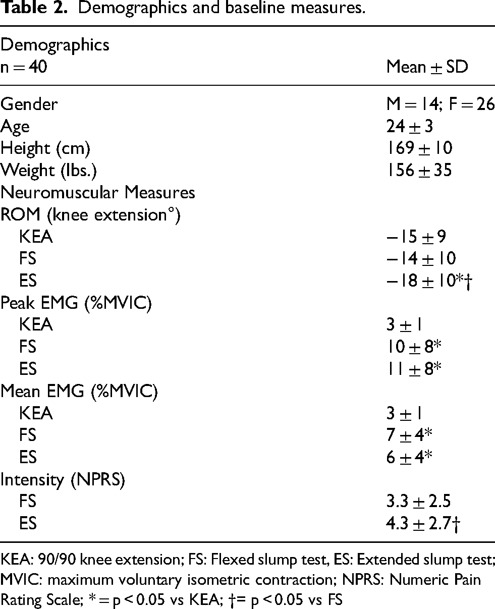
KEA: 90/90 knee extension; FS: Flexed slump test, ES: Extended slump test; MVIC: maximum voluntary isometric contraction; NPRS: Numeric Pain Rating Scale; * = p < 0.05 vs KEA; †= p < 0.05 vs FS
ROM
There was a significant overall difference in knee extension ROM between test conditions, F1.59, 61.85 = 6.04, p = .007,

KEA: 90/90 knee extension angle, FS: flexion slump, ES: extension slump * = p<0.05 vs KEA; † = p < 0.05 vs FS.
EMG
There were significant overall differences between tests for peak EMG values (%MVIC), F1.67,64.96 = 32.70, p < .001,
There were significant overall differences between tests for mean EMG values (%MVIC), F1.61,62.75 = 34.29, p < .001,
Mean and peak EMG values for each test condition are detailed in Table 2. (Figure 4)

Peak EMG activity, % MVIC: KEA: 90/90 knee extension angle, FS: flexion slump, ES: extension slump; MVIC: maximum voluntary isometric contraction; * = p<0.05 vs KEA.
Distal extent of sensory response
There was a significantly greater proportion of neurosensory responses reported distal to the knee during the ES (30%) compared to the FS (12.5%), p = 0.039. (Figure 5)

Location of Mechanosensory Response. ES: Extended Slump, FS: Flexed Slump; * = p < .05, ES vs. FS, proportion of responses below the knee.
NPRS
The ES resulted in a greater intensity of response (4.3 ± 2.7) than the FS (3.3 ± 2.5). There was a significant difference between groups for symptom intensity, mean difference 1.0, p = .001, d = 0.38. (Figure 6)
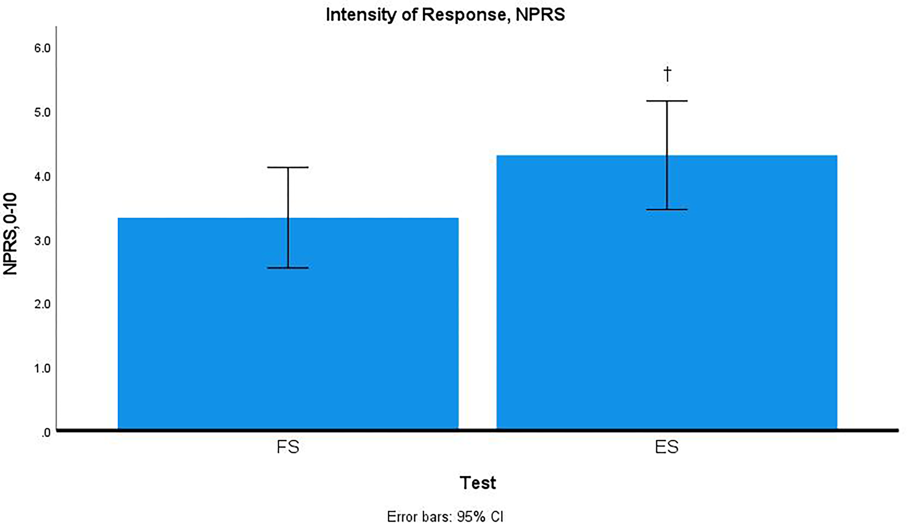
Intensity of response, NPRS. FS: flexion slump, ES: extension slump; NPRS: Numeric Pain Rating Scale; † = p<0.05 vs FS.
Qualitative
There were no statistically significant differences between groups for qualitative description of symptoms. The highest percentage described as anything other than “stretching” or “pulling” were observed during the ES (12.5%) followed by the FS (7.5%). There were no other responses observed during the KEA (0%). Findings are described in Table 1.
Discussion
The main findings of this study indicate that there is less knee extension ROM, greater hamstring activity, and a greater proportion of distal lower extremity mechanosensory responses during the extended slump compared to the flexed slump. The primary outcome of knee extension ROM was significantly less in the ES than in the FS or baseline measure of KEA. This finding is somewhat surprising, as knee extension is typically expected to be reduced in the flexed spine position, as the original slump test was intended to put maximal tension on the nervous system. 6 Theoretically, incorporating lumbar extension should reduce the overall tension on the nervous system and/or decrease the distance the nervous system must travel during knee extension, and therefore decrease the mechanosensory response. 33 However, the ES resulted in the greatest proportion of mechanosensory responses below the knee, aligned with accepted definitions of a peripheral neurogenic response. 11 The combination of a higher proportion of symptoms distal to the knee, decreased knee extension ROM, and increased hamstring EMG activity suggest that the nervous system experienced a provocative stimulus in the ES test position. While not the only potential source of decreased ROM and mechanosensory response (as increased hamstring length due to altered ischial tuberosity migration must be considered), the extended posture decreases the available space at the neuroforaminal interface, and this provocative stimulus may be the result of decreased foraminal space. Differentiating between compression and longitudinal strain in flexed and extended lumbar spine slump tests could be useful in clinical care, although further investigation is required in clinical populations.
The KEA was intended to serve as a baseline measure to evaluate hamstring flexibility while minimizing the role of neural tension. The KEA recorded the lowest hamstring EMG %MVIC, had no participants report responses distal to the knee, and 95% of participants reporting feeling only “stretching” during the test, supporting its effectiveness in assessing baseline ROM. The average lack of knee extension for the current sample was 15.13 ± 9.33°, indicating greater hamstring flexibility than the normative data (28° females, 38.6° males). 28 These ROM findings suggest that the mechanosensory responses observed during slump testing were less likely to have resulted from hamstring tightness. With baseline ROM established, response to mechanical stimulus was further analyzed through EMG and qualitative neurosensory reports. In this instance, the ES led to more distal responses. Overall, the observed differences between slump postures and baseline measures indicate that changes in spinal position during testing may allow the clinician to influence stresses on neural tissues and better understand dynamic influences on neural mechanosensitivity.
The influence of axial load on the mechanosensitivity of the nervous system is an important consideration for clinicians, as they must determine whether an issue originates from the nerve itself or from the neuroforaminal interface. This distinction directly impacts the decision to treat either the nerve or its “container."7,8,34 In the current study, both loaded tests (FS, ES) produced significantly higher hamstring EMG activity than unloaded test (KEA) despite similar ROM at the knee, highlighting the impact of axial load on neural responses. It remains unclear whether the increased EMG activity arises from proximal strain due to cervical flexion or from reduced space caused by the upright position during testing, although Ellis et al. found that isolated cervical flexion resulted in minimal excursion of the sciatic nerve, 33 perhaps lessening this likelihood. McHugh et al. previously observed a decrease in hamstring EMG activity when trunk flexion was added. 3 It is possible that in both the study by McHugh and the current study a posterior pelvic tilt during lumbar flexion may have alleviated hamstring tension, while foraminal opening is an additional possibility. Additionally, Hall et al. found a significant increase in hamstring stretch tolerance when a traction force was applied during a straight leg raise 35 They speculate that this improvement in tolerance may be due to the complex interplay of neurophysiologic factors associated with manual therapy, 35 proposing that “hamstring extensibility” is mediated by central neurophysiologic processing, 36 while foraminal opening remains an additional possibility. In view of the current findings, it is appealing to speculate that the interaction between axial load and lumbar joint positioning influence hamstring extensibility and the sensory response during testing. However, further work is required to fully elucidate the influence of axial load modulation on extensibility and sensory response during slump testing.
Several limitations should be considered when interpreting this research. Participants were exclusively healthy individuals without low back pain or radicular symptoms, and the majority were under 30 years of age. Accordingly, this study's findings may not be generalizable to a patient population with neuropathic symptoms, and further testing with a clinical population is needed. The KEA was assessed with the thigh manually stabilized, which could have introduced slight variability in accuracy. While previously reported to be a highly reliable test as performed, incorporating secure mechanical fixation of the thigh at 90° would have increased confidence in the KEA result. The trunk angle was not controlled between participants due to differences in tolerance and trunk flexibility, which could affect sagittal plane alignment. While efforts were made to control pelvic tilt, the FS naturally encourages a posterior pelvic tilt, thereby reducing resistance to hamstring stretch. In contrast, the ES encourages an anterior pelvic tilt, which increases the tension applied to the hamstrings. These factors likely contributed to the differences in knee extension range of motion observed in the study. It is possible that both ROM and sensory response may have increased following repeated testing. All tests were performed sequentially, and ordering effects were not controlled. In the current study, the ES was performed last and demonstrated the least knee extension ROM. In previous work investigating asymptomatic individuals, repeated slump testing resulted in increased knee extension ROM over successive trials.17,23 Thus, it is possible that the observed differences in ROM could have been more pronounced if the ES had been assessed earlier in the protocol. Fidel et al. observed a decrease in EMG activity with repeated slump testing in an asymptomatic population. 17 This does not, however, eliminate the possibility of altered mechanosensitivity following repeated testing in this trial. While the results demonstrated differences in ROM that did not meet the a priori clinical differences, the proposed meaningful difference was based on those observed in a) individuals with/without radiculopathy, and b) between hamstring and neural stretches. The MCID may be quite different for healthy individuals based on the onset of sensory response. Finally, there was an unequal distribution by gender within the sample, which may have influenced the overall ROM results.
Future studies with patient populations experiencing neuropathic symptoms are needed to generalize the results of this study prior to widespread implementation as a means to determine intrinsic vs extrinsic sources of mechanosensitivity.
Conclusion
The extended slump may be a useful test to assess neurodynamic behavior and this study serves as an initial proof of concept that alterations in lumbar spinal positioning can influence mechanosensory response. Potential clinical applications include the more accurate differentiation of load and positional sensitivity vs. tension sensitivity as a source of mechanosensory response, and thus the ability to better determine the associations between neurodynamics and lumbar directional preference. Further research is needed to validate these findings in clinical populations.
Clinical relevance
The extended slump test generated the greatest distal neurosensory response. The use of the extended slump may be helpful to determine the influence of lumbar position and axial load on mechanosensory response.
Footnotes
Acknowledgements
The authors thank Kaitlin Brosnihan and Chase Conway for their contributions during data collection for this study and Xin Ye, PhD, for his review and assistance with the data analysis plan.

Ethical approval
This study received ethical approval from the University of Hartford IRB (approval #020220124) on March 07, 2022.
Funding
The authors received no financial support for the research, authorship, and/or publication of this article.
Declaration of conflicting interests
The authors declared no potential conflicts of interest with respect to the research, authorship, and/or publication of this article.