



Abstract
Background
The effects of changes in cervicothoracic posture on respiratory and exercise capacity in healthy young adults are still debated.
Objective
This study aimed to investigate the effect of cervicothoracic posture on respiratory muscle strength and upper extremity function and to compare according to the activity levels in healthy young adults
Methods
Ninety-three healthy young adults were included in this study. Cervicothoracic posture, including the craniovertebral angle, cervical lordosis angle, forward shoulder angle (FSA), and thoracic kyphosis angle, was assessed using the photographic method. Chest wall mobility, respiratory muscle strength, upper extremity function, physical activity level, and severity of fatigue were assessed. Pearson's correlation analysis revealed significant associations between respiratory muscle strength and cervicothoracic posture, as well as anthropometric measures. Multiple linear regression identified independent predictors of MIP and MEP, adjusting for potential confounders such as smoking.
Results
The FSA demonstrated a weak but statistically significant correlation with both maximum inspiratory pressure (MIP) (r = −0.248, p = 0.021) and maximum expiratory pressure (MEP) (r = −0.219, p = 0.041). Axillary circumference measurements demonstrated a near-significant association with MEP (B = 4.93, p = 0.059). Upper extremity function did not correlate with cervicothoracic posture (p > 0.005). The MEP was significantly associated with fatigue severity (B = − 0.02, β = − 0.323, p = 0.022). Activity level was associated with MEP, chest mobility and fatigue (p < 0.05).
Conclusion
Cervicothoracic posture does not affect respiratory muscle strength or upper-extremity function in healthy young adults. The MEP is influenced by axillary chest mobility and affects fatigue. Activity is important for the respiratory health of healthy young adults.
Introduction
The increasing use of technological devices and decreasing age at the onset of their use have contributed to an increasing trend of physical inactivity among young individuals. 1 The spread of sedentary lifestyles negatively affects the physical health and posture of university students worldwide. Prolonged periods of sitting, coupled with inefficient biomechanics resulting from poor posture, are known contributors to a range of musculoskeletal and respiratory issues. These include, but are not limited to, rounded shoulders, thoracic kyphosis, forward head posture, and associated pain syndromes.2,3 Physical inactivity and sedentary behavior are associated with poor well-being in young people, with adverse effects extending into adulthood. 4 Specifically, students with low physical activity levels and more time spent sitting have imbalanced trunk muscles, worse respiratory function, and poorer quality of life and sleep. 4 These observations strongly suggest that sedentary behavior can precipitate significant musculoskeletal and respiratory challenges, even in young adult populations. Among these postural issues, forward head posture is particularly detrimental, as it has been shown to increase the activity of accessory respiratory muscles like the sternocleidomastoid and anterior scalene, thereby decreasing vital lung capacities such as forced vital capacity. 5
The chest wall, a critical structure supported by a complex network of respiratory and accessory muscles spanning the thoracic and shoulder regions, necessitates adequate mobility for optimal respiratory mechanics. In healthy individuals, typical chest wall alignment ranges from 4 to 7 cm; however, this mobility tends to diminish with age and in the presence of poor postural alignment.6,7 Existing research consistently demonstrates that altered posture significantly reduces chest wall mobility and impairs both respiratory muscle strength and overall lung function. 6 Beyond respiratory implications, individuals who maintain poor postural habits over extended periods, such as office workers and students, are highly susceptible to developing upper limb muscle imbalances and shoulder dysfunction.8,9 Poor posture has been associated with reduced trunk mobility, impaired shoulder kinematics, decreased upper-extremity muscle activation, and limitations in performing functional upper-extremity exercises.8,10 Rounded shoulder, kyphosis, and forward head posture reduce trunk mobility, negatively affect respiratory muscle strength and lung volume, 11 and impair shoulder kinematics, leading to decreased upper limb muscle activation. 9 This is particularly important because upper limb exercise capacity is closely related to cardiovascular endurance, fatigue levels, and perceived dyspnea, even in healthy populations. 12 Therefore, understanding the connection between exercise habits and physical activity levels, posture, respiratory muscle strength, and chest mobility in young, healthy individuals is crucial for proactively preventing potential respiratory difficulties, especially given the accelerating trend of inactivity driven by technological advancements.
A comprehensive examination of the relationship between cervicothoracic posture, chest wall mobility, and respiratory muscle strength is essential, particularly when evaluating the systemic effects of postural disorders on the respiratory system. The flexibility and mobility of the chest wall are paramount in maintaining respiratory capacity. Indeed, postural disorders in the cervicothoracic region are known to restrict chest wall mobility and subsequently reduce rib-cage movement. Research indicates a strong positive correlation between respiratory muscle strength and factors such as chest wall enlargement, diaphragm movement, and overall functional capacity in healthy participants.
13
This connection is particularly relevant in contemporary lifestyles: prolonged sitting positions, often adopted during office tasks or device use, increase neck flexion, thereby reducing lower cervical lordosis and causing an increase in thoracic kyphosis. This combination of increased tension in neck extensors and flexion of the cervical spine creates regions of stress in the neck.
14
Critically, this habitual sitting posture also leads to the contraction of chest muscles, limiting their mobility and consequently disturbing the humeroscapular rhythm.
14
This restriction of chest wall and shoulder girdle mechanics can directly interfere with the optimal functioning of the diaphragm and lead to a detrimental weakening of the primary respiratory muscles.
15
Furthermore, such cervicothoracic postural disorders often result in the overuse and eventual fatigue of accessory respiratory muscles
The primary objective of this study was to investigate the effects of cervicothoracic posture and chest mobility on respiratory muscle strength. Additionally, this study aimed to further assess the effects of respiratory muscle strength on upper extremity exercise capacity and fatigue. A crucial secondary objective was to conduct a comprehensive multivariate comparative analysis of cervicothoracic posture, chest wall mobility, respiratory muscle strength, upper extremity exercise function, and fatigue across distinct activity levels in a cohort of healthy young adults.
It is hypothesized that in healthy young adults, poorer cervicothoracic posture will be associated with reduced chest wall mobility and decreased respiratory muscle strength. Furthermore, it is hypothesized that lower respiratory muscle strength will correlate with reduced upper extremity exercise capacity and increased reported fatigue. Finally, it is anticipated that individuals with higher physical activity levels will demonstrate superior cervicothoracic posture, greater chest wall mobility, stronger respiratory muscles, better upper extremityfunctional exercises capacity, and lower fatigue levels compared to those with lower physical activity levels.
Methods
Study design and participants
This cross-sectional study was conducted at X University Department of Physical Therapy and Rehabilitation between March 2024 and March 2025. Healthy individuals aged 18–40 years with a body mass index (BMI) value between 18.5 and 30 kg/m2 were included in the study. Participants diagnosed with neurological, orthopedic (such as kyphoscoliosis), cardiac, and pulmonary diseases that would affect participation in physical activity and those who underwent thorax, upper extremity, and spine surgery were excluded from the study. This study was approved by the X University Non-Interventional Research Ethics Committee (Approval Number: 2024/34). The Informed Consent Form was explained to the patients one-on-one, and was filled in and recorded in accordance with the patient's request. This study was conducted in accordance with the principles of the Declaration of Helsinki. The study was conducted on healthy adults who agreed to participate in the study. Demographic information of the included individuals was recorded and posture, chest wall mobility, respiratory muscle strength, upper extremity mobility, physical activity level, and fatigue were evaluated.
Upper posture analysis
The craniovertebral angle (CVA), cervical lordosis angle (CLA), thoracic kyphosis angle (TKA), and forward shoulder angle (FSA) of the participants were measured using photogrammetry, which is a highly reliable intraclass correlation coefficient (ICC: 0.78–0.83) method that allows quantitative assessment of postural changes. 17 Although this method does not directly measure vertebral alignment, it offers a noninvasive and practical way to screen for postural deviations. 18 Photogrammetry consists of two stages, which are based on obtaining 2D images by a standard method and analyzing the images using a video analysis software.
The protocol developed by Belli et al. is preferred for photogrammetry. First, passive markers were attached to the right mastoid process; third (C3) and seventh cervical vertebrae (C7); sixth (T6) and twelfth thoracic vertebrae (T12); medial angle of the scapula; and acromion using double-sided adhesive tape by an experienced physiotherapist. 19 Participants were asked to look straight at the point at eye level and on the opposite wall to achieve natural head-body and shoulder alignment. Participants were asked to stand in their natural, relaxed posture with their arms resting at their sides and feet shoulder width apart. Thereafter, a 2D image (sagittal plane, lateral view) was captured using a camera (Galaxy S22 Ultra, 200 MP; Samsung, South Korea) positioned 2.5 meters from the participant.
In the second stage, digital images were analyzed using Kinovea software (version 0.8.15, Kinovea Open Source Project). All angle measurements were performed by the same and blinded rater to minimize inter-rater variability. A mark was placed on the ground to ensure that each participant was in the same position. The shooting was done with a height-adjustable tripod. The photographs were taken by an expert physiotherapist. The person taking the photographs and the one performing the measurements were different physiotherapists. The angles formed by the lines traced from the pre-labeled anatomical markers in the cervicothoracic posture were measured to identify possible asymmetries in cervicothoracic posture using 2D images. For CVA an angle was formed between a horizontal line passing through the C7 vertebra and a line drawn from C7 to the tragus of the ear.19,20 The CLA was formed by the lines between the occipital protuberance and C7, and the horizontal line between C4 and the lines intersecting the true vertical line. The FSA was measured as the angle between a vertical line passing through the spinous process of C7 and a line connecting C7 to the acromion process of the scapula. Three 2D images obtained from the participants were analyzed, and the average values were recorded. A CVA <50 ° indicates poor posture and an FSA <52° indicates rounded shoulder posture (RSP).21–24 For TKA, lines were drawn between C7, the inferior angle of the scapula (around T7), and the estimated T12. The angle between these lines reflected the curvature of the thoracic spine. A TKA angle greater than 40 °is considered hyperkyphosis.20,25
Statistical analysis
All statistical analyses were performed using IBM SPSS Statistics (version 27.0; IBM Corp., Armonk, NY, USA) and GraphPad Prism version 10.4.1 (GraphPad Software, LLC, San Diego, CA, USA). The distribution of the data was assessed using both visual (histograms and probability plots) and analytical methods (Kolmogorov–Smirnov test). Descriptive statistics were presented as the number of patients and percentages for categorical variables and as mean and standard deviation (SD) for continuous variables. Pearson's correlation analyses were performed to examine the relationship between participants’ cervicothoracic posture and anthropometric measurements with MIP and MEP. Power below 80 percent was considered weak. Power was calculated only for significant interaction. Variables that showed significant associations in the univariate analysis were entered into multiple linear regression analyses conducted in the two models. The purpose of using the two models was to determine the independent predictors of MIP and MEP, while adjusting for smoking as a potential confounding factor. A single-model multiple linear regression analysis was also conducted to determine the impact of respiratory muscle strength on the upper extremity functional exercise capacity and fatigue severity. Regression assumptions, including linearity, normality of residuals, homoscedasticity, and the absence of multicollinearity, were checked to ensure model validity.
One-way analysis of variance (ANOVA) followed by Tukey's post-hoc correction was used to perform multiple group comparisons. Statistical significance was set at P < 0.05.
The sample size calculation in the present study was originally based on the effect size (r² = 0.68) reported by Morais et al., 32 who examined the relationship between chest wall mobility and pulmonary function (FVC). However, since spirometry was not included in the present study, we acknowledge that this reference may not fully reflect the variables directly assessed herein (e.g., MIP, MEP, and upper extremity functional capacity). Although the initial estimated effect size was 1.0, yielding a minimum sample size of 46 (23 per group) with α = 0.005 and 80% power using PS-Power and SISA software, this estimation may not be sufficient for subgroup comparisons (e.g., by physical activity level). Therefore, we recognize this as a limitation of the study and acknowledge the potential risk of Type II error in subgroup analyses.
Results
Ninety-three healthy young individuals (mean age 21.84 ± 5.40, 40 (%43) male and 53 (%57) female) were included in the study. Among the healthy young people who participated in the study, 60.9% had an FSA <52, 41.4% had a CVA < 50, and 94.2% had TKA > 42. Of the participants, 52.7% were minimally active. While 49.50% of participants exercise habits, 35.50% were smokers (Table 1).
Demographic characteristics of the participants.
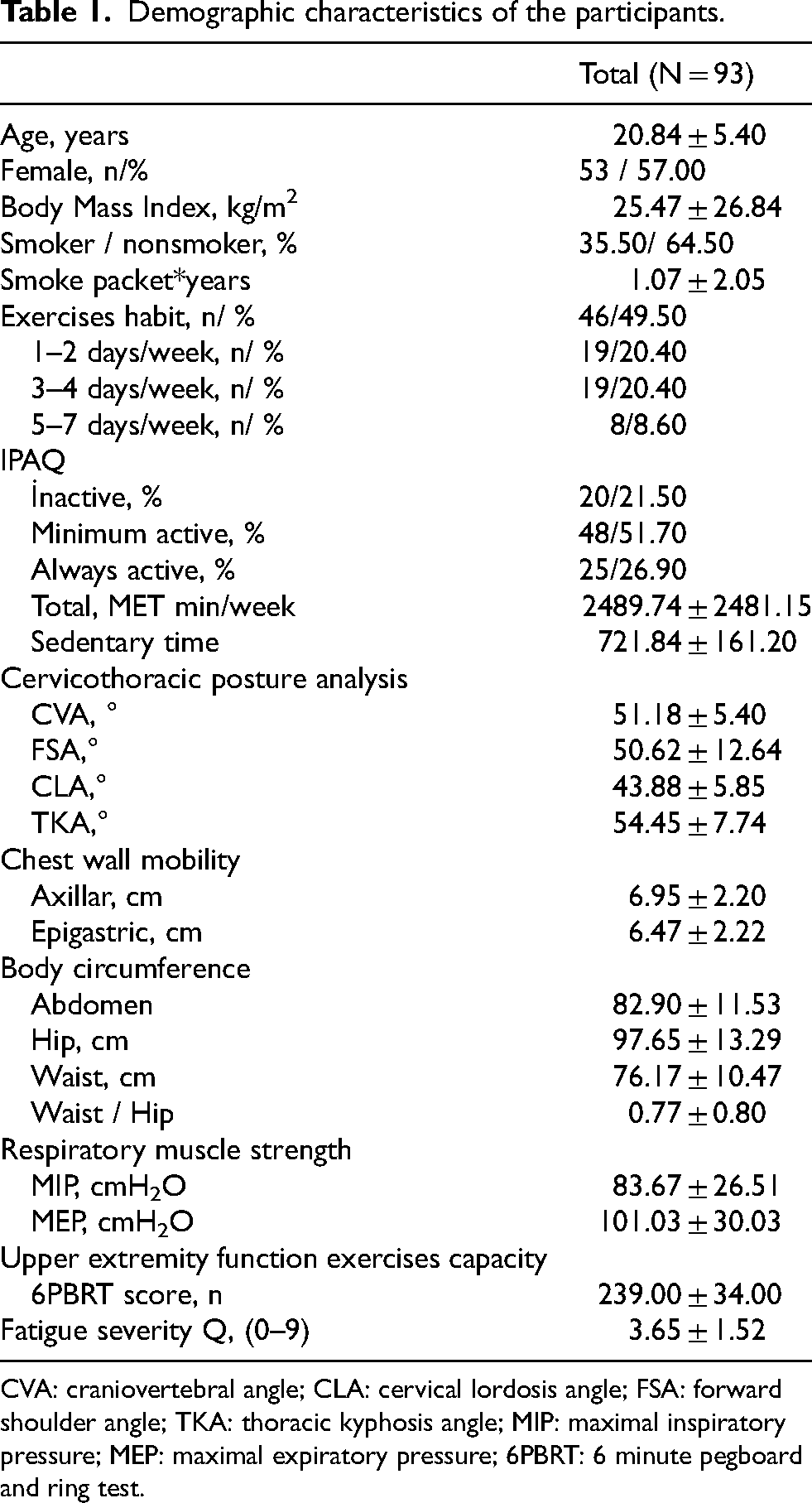
CVA: craniovertebral angle; CLA: cervical lordosis angle; FSA: forward shoulder angle; TKA: thoracic kyphosis angle; MIP: maximal inspiratory pressure; MEP: maximal expiratory pressure; 6PBRT: 6 minute pegboard and ring test.
FSA was significantly negatively correlated with CLA (p = 0.028) and TKA (p = 0.038). The CVA, FSA, CLA, and TKA values of participants with regular exercise habits (mean values: 51.10, 52.60, 44.29, and 54.18, respectively) were similar to those of participants without regular exercise habits (mean values: 51.27, 47.98, 43.44, and 54.74, respectively) (p > 0.05). The axillary circumference difference (p = 0.001), epigastric circumference difference (p = 0.001), MIP (p = 0.010), and MEP (p = 0.007) values were higher in patients who exercised regularly than those who did not.
There was no significant correlation between CVA, CLA, TKA, MIP, or MEP (p > 0.05). However, FSA demonstrated a weak but statistically significant negative correlation with both MIP (r = −0.248, p = 0.021) and MEP (r = −0.219, p = 0.041). Axillary and epigastric circumference measurements were moderately positive correlated with MIP (r = 0.455 and r = 0.438, respectively; p < 0.001) and MEP (r = 0.413 and r = 0.389, respectively; p < 0.001) (Table 2). Multiple linear regression analyses showed that in Model 1, which included FSA and axillary and epigastric circumference measurements, none of the predictors reached statistical significance for MIP or MEP. Axillary circumference measurements demonstrated a near-significant association with MEP (B = 4.93, p = 0.059). In the adjusted model (Model 2), which included smoking along with postural and anthropometric variables, the regression coefficients for FSA and circumference measurements remained relatively stable compared with those in Model 1. The FSA continued to show a negative association with both MIP (B = − 0.37, p = 0.079) and MEP (B = − 0.36, p = 0.135), with the association for MIP approaching statistical significance. Axillary circumference measurement retained its a near-significant positive association with MEP (B = 4.87, p = 0.064), approaching significance, whereas its relationship with MIP remained nonsignificant (B = 3.35, p = 0.989) (Table 3, Figure 1).

The association between participants’ cervicothoracic posture, chest mobility, respiratory muscle strength and fatigue severity RS: Rounded shoulder, ACM: Axillary circumference measurement, ECM: Epigastric circumference measurement, MIP: Maximal inspiratory pressure, MEP: Maximal expiratory pressure, UEFEC: Upper extremity functional exercise capacity.
Correlation of participants’ cervicothoracic posture, chest mobility, upper extremity function exercise capacity, respiratory muscle strength and fatigue severity.

CVA: craniovertebral angle; CLA: cervical lordosis angle; FSA: forward shoulder angle; TKA: thoracic kyphosis angle; MIP: maximal inspiratory pressure; MEP: Maximal expiratory pressure; 6PBRT: 6 min pegboard and ring test. Bold values indicate p < 0.05.
Multiple regression analysis for the association between participants’ cervicothoracic posture and anthropometric measurements and respiratory muscle strength.
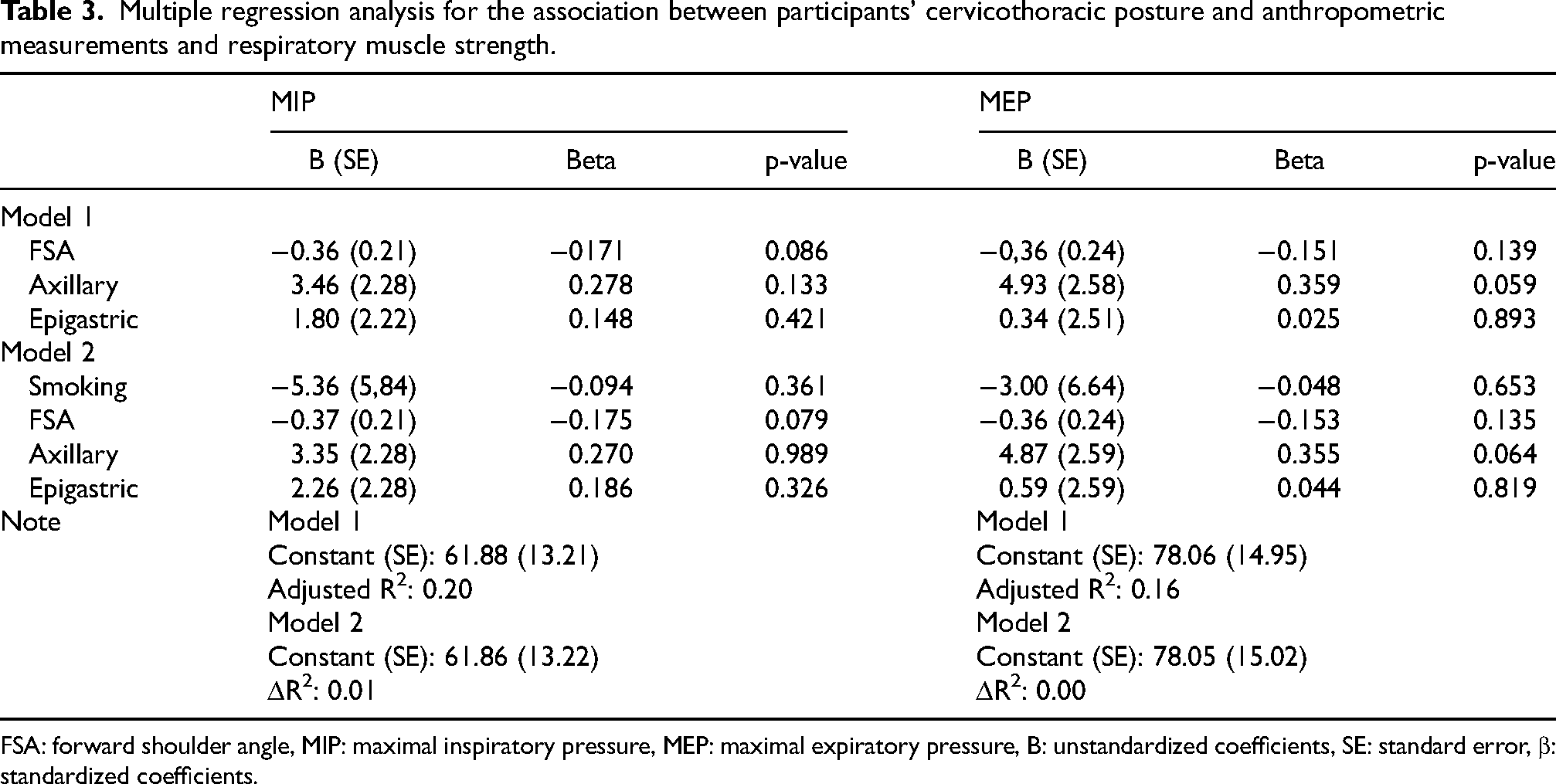
FSA: forward shoulder angle, MIP: maximal inspiratory pressure, MEP: maximal expiratory pressure, B: unstandardized coefficients, SE: standard error, β: standardized coefficients.
Regression analysis revealed no significant association between MIP and either fatigue severity or upper-extremity functional exercise capacity (p = 0.403 and p = 0.365, respectively). Although MEP was not significantly related to upper extremity function (p = 0.228), it showed a significant negative association with fatigue severity (B = − 0.02, β = − 0.323, p = 0.022), suggesting that greater expiratory muscle strength may be linked to reduced perceived fatigue (Table 4, Figure 1). There was no significant correlation between CVA, CLA, FSA, or TKA and 6PBRT (p > 0.05), and there was a statistically significant positive correlation between 6PBRT and both MIP (p = 0.013) and MEP (p = 0.009).
Multiple regression analysis for the association between participants’ respiratory muscle strength and upper extremity functional exercise capacity and fatigue severity.

MIP: Maximal inspiratory pressure; MEP: Maximal expiratory pressure; 6PBRT: 6 Minute Pegboard and Ring Test; B: Unstandardized coefficients; SE: Standard error; β, standardized coefficients. Bold values indicate p < 0.05.
Comparison analyses showed that MEP values differed significantly across the groups (p = 0.047), with the active group showing significantly greater MEP than the inactive group. No significant differences were observed in cervicothoracic postural parameters, including CVA, CLA, FSA, and TKA, across physical activity levels (p > 0.05). However, both axillary and epigastric circumference measurements were significantly greater in the active group than in the inactive and minimally active groups (p = 0.003 and p = 0.004, respectively). Although upper extremity function was similar across the groups (p > 0.05), fatigue severity was higher in the inactive group than in the active group (p = 0.004). (Table 5, Figure 2).
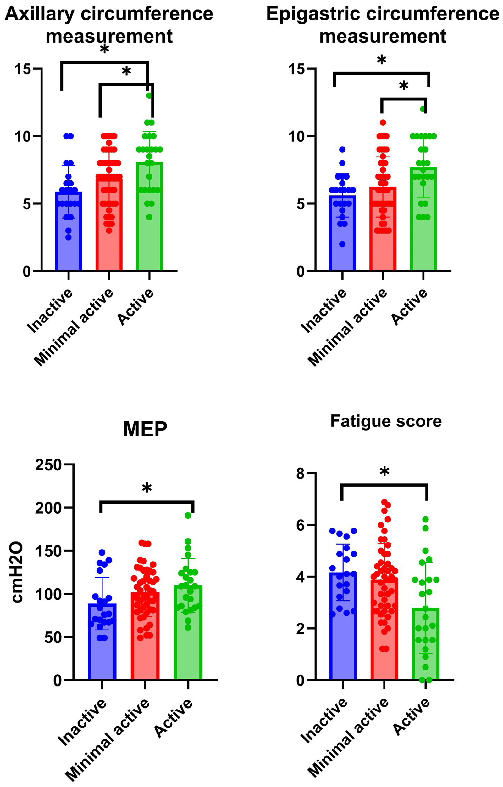
Variables that significantly differed across physical activity level groups MEP: Maximal expiratory pressure.
Comparison of respiratory muscle strength, cervicothoracic posture, upper extremity functional exercise capacity, chest wall mobility, and fatigue severity among individuals with different physical activity levels.
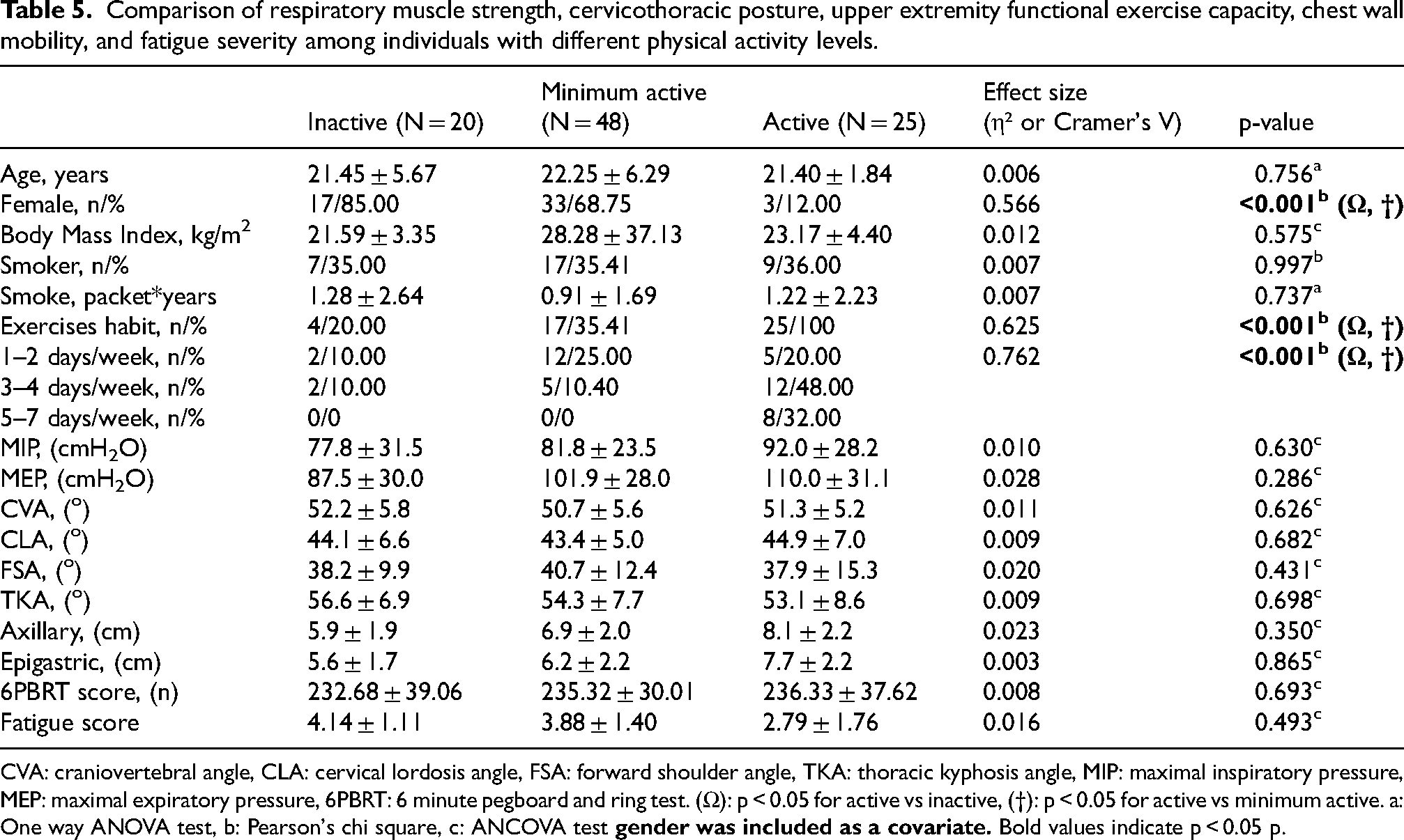
CVA: craniovertebral angle, CLA: cervical lordosis angle, FSA: forward shoulder angle, TKA: thoracic kyphosis angle, MIP: maximal inspiratory pressure, MEP: maximal expiratory pressure, 6PBRT: 6 minute pegboard and ring test. (Ω): p < 0.05 for active vs inactive, (†): p < 0.05 for active vs minimum active. a: One way ANOVA test, b: Pearson's chi square, c: ANCOVA test
Discussion
The aim of our study was to investigate the relationship and effects of cervicothoracic posture angles on chest mobility, respiratory muscle strength, upper extremity function, and fatigue and to compare these parameters according to the activity levels of healthy young adults. The findings showed that in healthy young adults, MEP was affected by axillary circumference measurement, but not cervicothoracic posture, and MEP affected fatigue severity.
In healthy young adults, changes in the biomechanics of the cervicothoracic spine and thorax may contribute to the short-term impairment of respiratory power.33,34 Forward head posture causes expansion of the upper thorax and contraction of the lower thorax; these morphological changes lead to decreased respiratory function. 16 In addition, rounded shoulders may restrict chest wall mobility by narrowing the anteroposterior diameter of the thorax, which may effectively prevent respiratory muscle contractions. Round shoulder posture causes abnormal breathing due to weakening of the respiratory muscles, and normal expansion of the lungs becomes difficult; therefore, vital capacity and gas partial pressure may decrease. 15 Similarly, the negative correlation of rounded shoulder posture with both MIP and MEP values in this study supports the notion that postural disorders may negatively affect thoracic expansion and, thus, the contractile power of respiratory muscles. However, the weak relationship suggests that postural change alone may not be a determining factor. These findings suggest that the relationship between chest wall mobility and respiratory muscle strength may be related to physical activity level and lifestyle habits rather than postural parameters. The FSA angles (37.9°, 40.7°, and 38.2°) decreased further when we categorized the participants as active, moderately active, and inactive according to their physical activity level. Those without regular exercise habits had an RSA <52° and kyphosis angles of 54°. Those with exercise habits had higher chest mobility, respiratory muscle strength, and fatigue perception than those without.
However, both chest wall mobility and respiratory muscle strength were higher in individuals who exercised regularly, indicating that physical activity may have a positive effect on both thoracic flexibility and respiratory muscle endurance. Changing the position of the head causes distortions in the three-dimensional shape of the chest and respiratory movements. 15 Indeed, an increase in the functional capacity of inspiratory and expiratory muscles with increasing levels of physical activity has been widely reported in literature. 13
FSA and axillary and epigastric chest mobility were associated with MIP and MEP; however, only axillary chest mobility tended to have an effect on expiratory muscle strength. Smoking did not affect the outcomes. The moderate positive correlation between chest wall mobility (axillary and epigastric circumferences) and respiratory muscle strength suggests that thoracic expansion capacity may be directly related to respiratory muscle strength. As the expansion capacity of the thorax increases, inspiratory muscles may work more efficiently, which may have a positive effect on respiratory muscle strength. 13 In this study, axillary and epigastric circumference differences were more pronounced, especially in active individuals, suggesting that physical activity may increase the flexibility of the thorax and thus the functionality of the respiratory muscles. Rodrigues et al. reported a correlation between upper extremity function and strength of the periscapular and trunk muscles. 10 In contrast, in our study, none of the cervicothoracic postures, including the round shoulder posture, were found to be associated with upper extremity function in healthy young subjects. The 6PBRT can be considered an assessment of functional arm exercise capacity in chronic diseases and healthy adults in clinical practice and research. Age was negatively associated with the 6PBRT score in healthy individuals who were mostly physically active. 35 Öncü et al. reported that younger age and higher PA levels were associated with better functional arm exercise performance in healthy young adults. 35 In our study, upper extremity function was similar between activity levels.
Padkao et al. reported a positive correlation between MIP and MEP and walking distance in their study. However, the significance values suggest that expiratory muscle strength may be more related to functional capacity than inspiratory muscle strength. 13 In our study, in parallel with this finding, MEP values of active individuals were higher than inactive individuals. Moreover, chest wall mobility was significantly higher in individuals who exercised regularly. This finding suggests that physical activity may positively affect chest wall flexibility and mobility.
Prolonged maintenance of poor posture may cause excessive pressure on the lumbar vertebrae, potentially leading to physical discomfort and the onset of myofascial pain syndrome. 36 Poor posture has a negative impact on the musculoskeletal system, causing localized muscle fatigue, and can affect physical function and skill levels. 37 Lu et al. showed that although poor posture is characterized by a forward head position, rounded shoulders, and increased thoracic kyphosis, maintaining this posture can affect other muscle groups. In the short term, poor posture can cause muscle fatigue in specific areas of the back and impair physical activity performance both immediately and over time. 38 In contrast, our findings show that fatigue severity in healthy young adults is not related to cervicothoracic posture but that fatigue is influenced by MEP. In addition, fatigue severity in inactive subjects was higher than that in active subjects. These findings suggest that factors beyond posture, such as MEP and physical activity level, may play a more significant role in fatigue severity in young adults. The discrepancy between our results and those of previous studies highlights the complexity of fatigue mechanisms and the need for a multifaceted approach to understand and address fatigue in this population.
The close to traditional significance of rounded shoulder posture on MIP and MEP suggests that changes in cervicothoracic posture may affect respiratory function in young individuals. Although these early postural changes have not yet significantly affected respiratory muscle strength, chest mobility, upper extremity function, or fatigue, they can potentially lead to more pronounced effects over time. This implies that early intervention and preventive measures such as promoting regular exercise and physical activity may be crucial for maintaining proper posture and preserving respiratory function as individuals age. Further research may be necessary to establish long-term relationships between cervicothoracic posture and respiratory health as well as to develop targeted interventions for young populations.
The strengths of our study are that it is the first to examine the effect of cervicothoracic posture angles on respiratory muscle strength, chest mobility, upper extremity function, and fatigue in healthy young subjects and the effect of smoking-adjusted data on these parameters. Our findings revealed that the negative effects of the round shoulder posture on MIP and MEP were of borderline significance. This finding suggests that larger sample sizes are needed to clarify the effects of rounded shoulder posture on respiratory muscle function. The limitations of this study are that pectoral muscle shortness was not measured in postural assessments, physical activity rate was not measured with objective measurements, and pulmonary function tests were not performed. Despite these limitations, this study provides valuable information on the relationship between cervicothoracic posture, respiratory muscle strength, upper-extremity function, and fatigue in young adults. The unequal group sizes and possible presence of confounding variables (e.g., age, BMI) represent limitations of the study. The unequal distribution of participants across subgroups and the limited sample size may have reduced the statistical power of the analyses, increasing the risk of a Type II error. This should be taken into account when interpreting the non-significant findings. Difficult to explain causality due to cross-sectional studies, correlation coefficients were not high. Further studies that comprehensively investigate the effects of unfavorable posture on long-term respiratory and exercise capacity in young adults are needed. Further research is needed to investigate the relationship between posture, MEP, physical activity, and fatigue severity in populations of various age groups under different health conditions.
Conclusion
In healthy young adults, cervicothoracic posture does not affect respiratory muscle strength or upper extremity function. The MEP is influenced by axillary chest mobility and affects fatigue. Activity level was related to MEP, chest mobility, and fatigue.
Footnotes
Acknowledgments
We thank the university students who voluntarily participated in this study.
Ethical approval
This study received prior ethical approval from the Tarsus University Ethics Committee (protocol number: 2024/34).
Informed consent
An informed consent form was included at the beginning of the survey, which presented the necessary information and recorded participants’ willingness to participate in the study. All the study participants provided written informed consent.
Author contributions
NE and FKÇ contributed to the design and implementation of the research, NE and VD analyzed the results, and wrote the manuscript. NE conceived the original and supervised the project.
Funding
The authors received no financial support for the research, authorship, and/or publication of this article.
Declaration of conflicting interests
The authors declared no potential conflicts of interest with respect to the research, authorship, and/or publication of this article.
