



Abstract
Background
Venous congestion is hypothesized to contribute to hand edema in patients with stroke; however, no studies have specifically investigated decreased venous return in the paretic hand.
Objective
This preliminary study explores this relationship by objectively measuring hand edema and venous return.
Methods
This cross-sectional study included patients who had experienced a first-ever stroke and presented with hand edema. Hand edema was measured bilaterally using a volumeter, and venous flow volume in the axillary vein was evaluated bilaterally using an ultrasound device. Hand edema and venous flow volumes were compared between the paretic and non-paretic limbs using a paired t-test.
Results
Thirteen male patients with stroke with hand edema were screened. Their median age was 69.3 years, and the median time since stroke onset was 117.5 days. Flow volume in the axillary vein was significantly lower on the paretic side (64.7 [95% confidence interval (CI): 41.8, 87.7] mL/min) than on the non-paretic side (115.9 [95% CI: 74.6, 157.2] mL/min).
Conclusion
The findings of this study suggest that venous return may be reduced in patients with stroke with hand edema. Future research should investigate the mechanisms underlying venous return impairment and other potential causes of hand edema.
Introduction
According to projections, by 2030, 3.4 million U.S. adults aged ≥18 years (3.9% of the adult population) will have experienced a stroke, representing a 20.5% increase in prevalence from 2012 (Martin et al., 2024). Therefore, treatment for hand edema, a secondary complication of stroke, will become increasingly important. Patients experiencing a stroke frequently experience edema in the paretic hand, with prevalence rates of 72.7% for hand swelling and 33.0% for edema (Boomkamp-Koppen et al., 2005). Persistent hand edema, accompanied by pain and tissue fibrosis, results in a decline in hand function (Boomkamp-Koppen et al., 2005) and negatively affects quality of life (Gustafsson et al., 2016).
Post-stroke hand edema is common; however, the exact etiology of upper limb edema after stroke remains unclear. Edema in patients with stroke is not classified as lymphedema (Geurts et al., 2000) but results from immobility-related physiological processes. Possible causes include not only autonomic nervous system dysfunction resulting from stroke (Bell & Muller, 2013; Giang et al., 2016) but also, as the most widely accepted hypothesis, increased venous congestion resulting from prolonged dependency and loss of muscle pump function in the paretic limbs (Gustafsson et al., 2016; Leibovitz et al., 2007).
Reduced venous return is likely a major contributing factor for hand edema. However, no studies have objectively investigated the relationship between hand edema and venous return. Traditional approaches, such as continuous passive motion and elevation (Giudice, 1990), compression therapy (Gustafsson et al., 2014), and intermittent compression (Roper et al., 1999), have been widely used to manage edema by enhancing venous flow. However, it remains unclear whether enhancing venous flow is actually desirable. Addressing the physiological basis of edema in patients with hemiparesis may improve management strategies and recovery outcomes. This preliminary study assesses the role of impaired venous return in hand edema formation after stroke, providing insights into its physiological mechanisms.
Methods
Participants
This study included eligible male patients who experienced their first unilateral stroke to examine differences in hand volume and upper extremity venous return between the paretic and non-paretic sides. All patients were admitted to the post-subacute rehabilitation units of three rehabilitation hospitals in Japan. The inclusion criteria were as follows: (1) diagnosis of stroke within the last 6 months; (2) presence of hand edema confirmed by a visible difference between the ipsilateral and contralateral hands, assessed by one occupational therapist at each hospital prior to the study recruitment; and (3) ability to tolerate assessments, such as maintaining a sitting position and following instructions. Patient recruitment was conducted by one occupational therapist from each participating hospital who was not involved in the study design.
All participants provided written informed consent before participation. This study was approved by the institutional ethics committee (Approval Number: 2021C0009) and adhered to the standards of the Declaration of Helsinki. Experiments were performed in a room maintained at a temperature of 24–27 °C.
Participants were assessed for the following demographic and clinical variables: age, days since stroke onset, height, weight, body mass index (BMI), systolic and diastolic blood pressure, heart rate, Fugl–Meyer Assessment Upper Extremity (FMA–UE) score, affected side, and stroke type.
Measures
Hand Edema Measurement
Hand edema was measured bilaterally using a portable volumeter based on the principle of water displacement. The participants were seated during the measurements and instructed to immerse their hands in the volumeter to determine the displaced water volume. The hand was immersed in the water-filled volumeter until the stop pin was positioned between the middle and ring fingers or between the index and middle fingers. The displaced water was collected, drained into a measuring cylinder, and weighed to calculate hand volume (Boomkamp-Koppen et al., 2005). The finger position in contact with the stop pin within the volumeter was standardized for both hands to minimize measurement error and ensure consistency.
Assessment of Venous Return
The diameter (mm) and time-averaged mean velocity (TAMV) (cm/s) of the axillary vein were measured bilaterally using an ultrasound device (Viamo C100, Canon Medical Systems, Tokyo, Japan). Measurements were performed in the supine position. The axillary vein was identified using a positive compressive pressure test and color Doppler imaging to confirm venous flow. The vein was visualized in the longitudinal plane, and the maximum diameter was assessed using B-mode imaging. TAMV was measured using pulse-wave Doppler imaging, with the angle of insonation maintained at <60°. The Doppler spectra were acquired in the longitudinal plane at the same site for 15 s. The venous flow volume (mL/min) on the paretic side was calculated using the following equation (Zamboli et al., 2014):

Data Presentation
Descriptive statistics were used to summarize the baseline characteristics of the study population, including age, days since onset, height, weight, BMI, systolic blood pressure, diastolic blood pressure, pulse rate, Fugl–Meyer Assessment of Upper Extremity, side of paresis, and type of stroke. Means and 95% confidence intervals (CIs) of hand edema volume and calculated venous flow volume were reported. We used the paired t-test to compare the paretic and non-paretic sides, opting for this parametric test despite the non-normality of our data. Effect sizes (d) were calculated and interpreted as follows: ∼0.20 indicates a small effect size, ∼0.50, a medium effect size, and ∼0.80, a large effect size (Maher et al., 2013). All statistical analyses were performed using HAD 18.0 software (Shimizu, 2016). A two-tailed p-value <0.05 was considered statistically significant.
Results
Fourteen patients who had experienced a stroke with hand edema (13 male and one female individuals) were initially enrolled in the study. However, during the research, one female participant withdrew due to personal reasons, resulting in the cessation of data collection for this individual. The final sample consisted of 13 participants (all male) who completed all phases of the study. Table 1 presents the demographic data. The mean age was 69.3 years, with a median of 102.2 days since stroke onset. The participants’ mean height and body mass were 165.6 cm and 61.0 kg, respectively, with a mean BMI of 22.3 kg/m2. The mean systolic and diastolic blood pressures were 134.2 mmHg and 76.3 mmHg, respectively, and the mean pulse rate was 66.4 bpm. The mean FMA–UE score was 25.7 points. Among the participants, 77.0% had right-sided paresis, and 61.5% experienced ischemic strokes.
Demographic Characteristics of the Sample (n = 13).
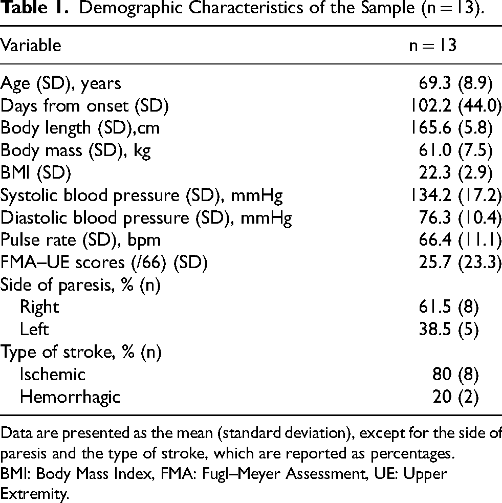
Data are presented as the mean (standard deviation), except for the side of paresis and the type of stroke, which are reported as percentages.
BMI: Body Mass Index, FMA: Fugl–Meyer Assessment, UE: Upper Extremity.
The measurement times for edema and venous return exhibited interpatient variability, with assessments conducted during two distinct time windows: between 10:00 and 12:00 or between 14:00 and 16:00. In all patients, hand volume was larger on the paretic side than on the non-paretic side. Furthermore, hand volume on the paretic side (448.3 mL [95% CI: 409.9, 486.8]) was significantly larger than that on the non-paretic side (408.2 mL [95% CI: 371.1, 445.2]; t = 5.479, p ≤ 0.001, d = 0.647, 95% CI: −0.198, 1.493). The axillary vein diameter was smaller on the paretic side (5.2 mm [95% CI: 4.4, 5.9]) than on the non-paretic side (6.8 mm [95% CI: 5.8, 7.7]; t = −3.710, p = 0.003, d = −1.130, 95% CI: −2.024, −0.237). No significant difference was observed in TAMV between the paretic (4.9 cm/s [95% CI: 4.0, 5.9]) and non-paretic sides (5.2 cm/s [95% CI: 4.1, 6.4]; t = −0.499, p = 0.627, d = −0.177, 95% CI: −1.000, 0.646). Flow volume in the axillary vein was significantly lower on the paretic side (64.7 mL/min [95% CI: 41.8, 87.7]) than on the non-paretic side (115.9 mL/min [95% CI: 74.6, 157.2]; t = −2.958, p = 0.022, d = −0.932, 95% CI: −1.803, −0.061) (Table 2).
Comparison of Hand Volumes and Axillary Vein Parameters between the Paretic and Non-paretic Sides.

95% CI: 95% confidence interval, TAMV: Time-averaged mean velocity.
The effect sizes were interpreted based on Cohen's criteria: small (0.2), medium (0.5), and large (0.8).
Discussion
We found a potential association between hand edema and reduced venous return in patients with stroke. This finding is supported by the observation of increased hand volume and decreased venous flow in the affected upper limbs.
Our results support the prevailing notion that impaired venous return contributes to the development of hand edema. In contrast, no difference in upper limb activity, measured using a uni-axial accelerometer constructed as a wristwatch, was observed between patients with stroke with and without edema, suggesting that reduced activity of the paretic hand alone may not cause upper limb edema after stroke (Gebruers et al., 2011). Reduced muscle activity is a contributing factor but not the sole cause; thus, other factors, such as microvascular damage, may play a more critical role. Previous studies have suggested that patients with stroke exhibit lower skin temperatures in the paretic limb, indicating autonomic dysfunction (Korpelainen et al., 1995). This implies that autonomic dysfunction may persist long-term in patients with stroke. A previous study demonstrated that patients with stroke exhibited decreased arterial inflow and impaired endothelium-dependent dilation in the skin vasculature of the paretic extremities. Furthermore, this dysfunction in the cutaneous microcirculation is more pronounced in the edematous limb, suggesting that impaired microvascular function may contribute to the development of hand edema in patients with stroke (Wang et al., 2002).
Patients with stroke exhibit severe vasodilatory dysfunction in the forearm, indicating significant impairment of vascular reactivity in the peripheral circulation (Stenborg et al., 2006). Thus, the function of the vessels and peripheral circulation on the paralyzed side may be impaired. The reduced venous return observed in this study may result from this reduced vascular function. The vascular diameter was slightly smaller on the paretic side than on the non-paretic side. Further research is required to clarify the cause of the reduced venous return.
Several factors, including diminished venous pump function resulting from reduced muscle activity in the hand and forearm, autonomic nervous system dysregulation potentially affecting vascular tone and venous capacitance (Giang et al., 2016), and post-stroke alterations in systemic and peripheral circulation (Wang et al., 2002) may contribute to reduced venous return in the affected hand after stroke. Therefore, further investigation is required to examine other potential factors and establish these mechanisms’ relative contributions.
It is imperative to gain a comprehensive understanding of the underlying pathophysiological mechanisms to develop optimal rehabilitation strategies for hand edema, which is frequently observed in patients with stroke. Our findings may provide a starting point for the development of new treatment strategies for hand edema after stroke. Clinical studies are warranted to investigate the efficacy of compression therapy and other interventions to improve venous return.
This study had some limitations. Given the small sample size of 13 participants, the results may be limited to a specific patient population. The 95% CI for the effect size (Cohen's d) was wide and included zero for all parameters except venous flow, indicating substantial uncertainty in the estimates. This suggests that the study may have been underpowered, likely due to a small sample size. However, the study confirmed that the paretic hand had a greater volume than the non-paretic hand in all patients. Notably, venous flow—the most critical parameter—demonstrated an effect size that did not include zero. This limitation underscores the need for more robust investigations. Increasing the sample size in future studies would help narrow the CI, yielding more precise effect size estimates and clearer conclusions regarding the magnitude and direction of the effect. Larger-scale studies with more diverse patient populations, particularly women, are necessary for additional validation. Future research should include a larger cohort to confirm these findings and apply statistical analyses, such as correlation coefficients, to better elucidate the relationship between venous return and hand volume. Additionally, factors such as impaired muscle pump function and autonomic dysfunction may contribute to edema, warranting further investigation into their interplay.
Conclusion
To our knowledge, this is the first study to objectively investigate the involvement of venous return in hand edema in patients with hemiplegic stroke. Our findings indicated that venous return may be reduced in patients with stroke with hand edema. These results suggest that future studies on hand edema in patients with stroke should consider strategies to enhance venous return as a potential treatment option. In the future, we will integrate our findings with other potentially relevant factors to elucidate the mechanisms associated with hand edema. Additionally, we will explore treatment approaches for hand edema in patients with stroke and examine their clinical implications.
Footnotes
Acknowledgments
We would like to express our sincere gratitude to the rehabilitation staff at Okazaki Higashi Hospital, Yamada Hospital, and Tokai Memorial Hospital for their invaluable contributions to this study.

Ethical Considerations and Informed Consent
The study was approved by the Ethics Committee of Seijoh University, Japan (Approval Number: 2021C0009). All participants provided written informed consent prior to participation.
Author Contributions/CRediT
Conceptualization: HH, AK, NT; Methodology: HH, AK, NT; Data Collection: HH, AK, NT, HO; Data Analysis: HH, AK, NT, HO; Writing – Original Draft: HH; Writing – Review & Editing: AK, NT, HO; Supervision: HH.
All authors have read and agreed to the final version of the manuscript.
Funding
The author(s) disclosed receipt of the following financial support for the research, authorship, and/or publication of this article: This research was supported by a grant from the Japan Society for the Promotion of Science (Grant Number: 19K19623).
Conflicting Interest
The author(s) declared no potential conflicts of interest with respect to the research, authorship, and/or publication of this article.
Data Availability
The data that support the findings of this study are available from the corresponding author upon reasonable request.