



Abstract
Study design
Prospective trial.
Objective
To evaluate the impact on muscle activation of different anatomical configurations of transcutaneous spinal cord stimulation electrodes amongst SCI and control patients.
Summary of background data
Spinal cord stimulation has garnered increased attention as a promising treatment for spinal cord injury (SCI). However, existing studies on transcutaneous spinal cord stimulation consist of highly variable methodologies and lack standard protocols.
Methods
Two adult SCI participants (PS01—T12 AIS D, and PS02—T2 AIS B) and two healthy adult controls (PS03, and PS04) were prospectively enrolled after IRB approval. Electromyography was used to record activity in the bilateral tibialis anterior (TA) and biceps femoris (BF). Four electrode configurations were trialed—cathode placement only at T11-T12 (montage T11-T12) versus combined configurations with cathodes at T11-T12 and at L1-L2, L3-L4, or S1-S2; anode placement was always at the bilateral iliac crests. Motor response was defined as EMG change >20 μV, as defined in the literature.
Results
SCI participants exhibited overall decreased motor responses compared to controls, other than PS01 at the T11-T12 montage. The majority of motor responses from SCI participants did not meet the 20 µV threshold, and did not show the same consistent cranial to caudal pattern. PS01’s highest response was at T11-T12/L1-L2 (BF: 265.52 µV), whereas PS02 was highest at T11-T12 (TA: 39.73 µV). Overall, the motor response from the motor complete SCI (PS02—T2 AIS B) was substantially less than the motor response from the motor and sensory incomplete SCI.
Conclusion
When performing transcutaneous spinal cord stimulation, the magnitude of motor response appears to vary with both the exact anatomical electrode configuration and the presence/severity of SCI. SCI participants demonstrated generally decreased motor responses, and a less predictable topographic pattern. Further work is needed to determine the ideal electrode configurations for different patients and spinal cord injury types.
Keywords
Introduction
Spinal cord injury (SCI) is unfortunately a prevalent neurologic condition, which can occur via traumatic or atraumatic mechanisms with an annual estimated incidence of 15–50 cases per million.1–4 SCI is associated with significant morbidity such as decreased functional ability, development of pressure injuries, and increased risk of medical complications such as deep vein thrombosis (DVT) and pneumonia.2,5 As a result, the management of SCI represents a significant source of healthcare expenditure with an estimated cost of more than $4 billion dollars annually in the United States. 1
Rehabilitation for patients with chronic SCI, and particularly patients with incomplete SCI, is focused on the implementation of assistive devices, functional training, and more recently muscle stimulation and neurostimulation techniques. 6 Spinal cord stimulation has garnered particular attention due to promising results of functional muscle activity generation through stimulation of spinal cord pathways in animal models.7,8 Moreover, epidural spinal cord stimulation has been demonstrated to elicit changes in the lower extremity musculature EMG activity in patients with SCI.9,10 Non-invasive means of spinal cord stimulation, such as transcutaneous spinal cord stimulation (tSCS), have also demonstrated the potential to activate spinal cord pathways in patients with SCI. 11
However, the efficacy of tSCS for SCI rehabilitation has not yet been conclusively demonstrated, partly because existing studies on tSCS consist of highly variable methodologies and lack standard protocols.12,13 As a result, studies focused on optimizing tSCS methodology are needed in order to more accurately evaluate the efficacy of tSCS for patients with SCI. This pilot study sought to evaluate the efficacy of different configurations of tSCS electrodes across a series of participants with and without SCI.
Methods
Participants
Four participants were included in this study: two control individuals without spinal cord injury (SCI) and two participants with chronic SCI. The SCI group included participant PS01 and PS02. PS01 is, a 52-year-old male with a traumatic International Standards for Neurological Classification of Spinal Cord Injury (ISNCSCI) injury level of T12 and American Spinal Injury Association Impairment Scale (AIS) D sustained 2 years prior. Surgical history for this participant included laminectomy and posterior fixation at C3-C6 consisting of paired rods and bilateral lateral mass screws (Figure 1). MRI for patient PS01.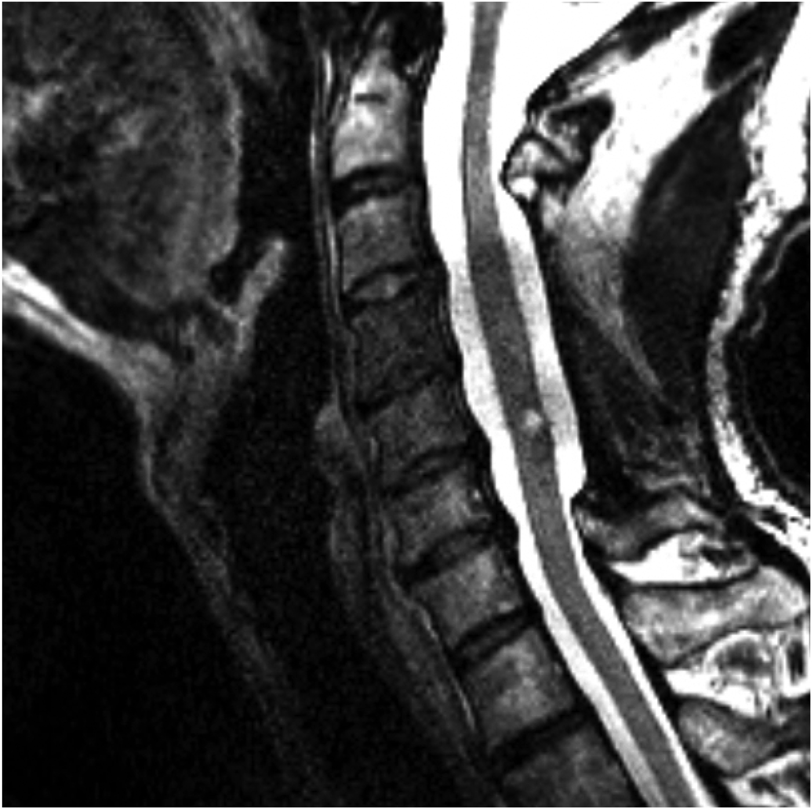
PS02 is a 41-year-old female with a spinal cord injury classified as T2 according to the ISNCSCI and graded as AIS B, sustained 2 years prior secondary to a spinal hemorrhage during labor. Her surgical history includes a posterior T1-T2 thoracic laminectomy and resection of an epidural hemorrhagic angiolipoma in the posterior right margin, no instrumentation was used (Figure 2). The control group consisted of participant PS03, a 27-year-old male, and participant PS04, a 50-year-old female, both with no history of neurological disorders or surgery. All participants provided informed consent and completed baseline assessments of lower extremity motor function. MRI for patient PS02.
Electromyography data collection
Participants were positioned in either a seated upright position or flat in supine position, such that one patient with an SCI and a control were seated and the other two were supine, as joint angles can contribute to changes in EMG amplitudes. Following positioning, electromyography (EMG) recording sites including the tibialis anterior (TA) and the biceps femoris (BF) bilaterally were prepared by removing hair and dead skin using a disposable razor. The skin was then thoroughly cleaned with alcohol swabs to enhance signal quality. After skin preparation, EMG sensors were centered and placed over the belly of each muscle. Following placement, transpore surgical tape was used to secure the EMG sensor in order to reduce movement artifact. EMG data were collected using Delsys wireless EMG sensors (Delsys, Natick, MA). Motor response was defined as an EMG change >20 μV, per prior literature. 14 This value represents the smallest detectable change required to distinguish from artifact.
Electrode configurations
Following EMG placement, spinal levels of interest were identified by palpating key anatomical landmarks, including the bilateral iliac crests and the vertebra prominence, to ensure accurate electrode positioning. Photographs were taken of each electrode configuration to assist with consistency in placement across visits. Electrode application and placement confirmation were performed by a trained physical or occupational therapist.
Two electrode configurations were utilized in this study. In the first configuration, a self-adhesive circular electrode (1.25-inch diameter) serving as the cathode was placed in the intervertebral space between the T11 and T12 spinal levels (montage T11-T12). A large rectangular self-adhesive electrode (3 × 4 inches), serving as the anode, was placed unilaterally on either the left or right anterior iliac crest. The second configuration involved two interconnected circular electrodes, both serving as cathodes. One of these cathodal electrodes was positioned in the T11-T12 intervertebral space, while the other was placed between the L1-L2 (montage T11-T12/L1-L2), L3-L4 (montage T11-T12/L3-L4), or S1-S2 spinal levels (montage T11-T12/S1-S2). Large interconnected rectangular electrodes, acting as anodes, were placed bilaterally on the anterior iliac crests (Figure 3). Depiction of electrode placement.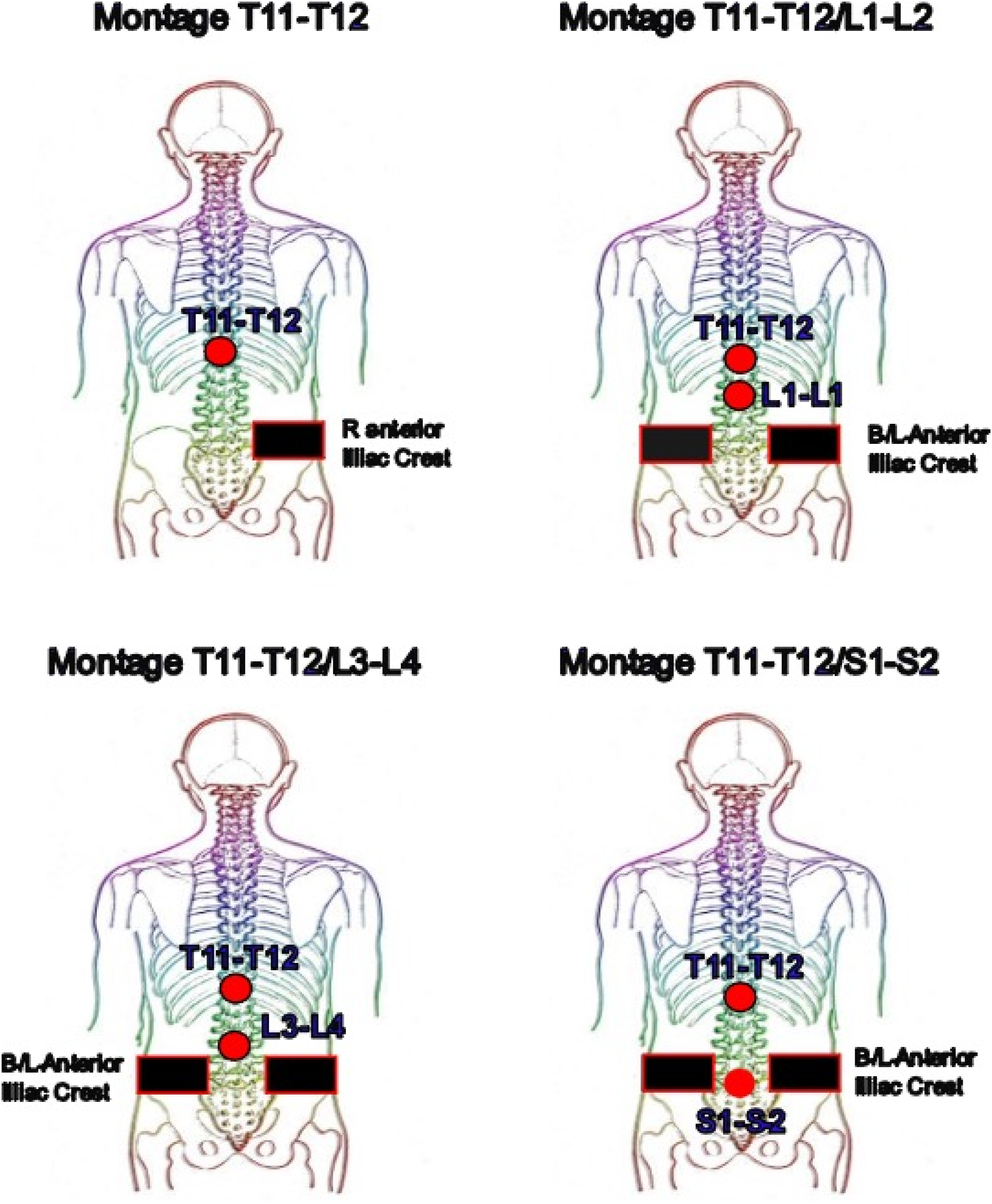
Stimulation procedure
Stimulation was delivered using a 1 millisecond (ms) pulse width biphasic waveform with an interstimulus interval of 50 ms, generated by the Xcite stimulator (Restorative Therapies Inc., 1434 Fleet St, Baltimore, MD 21231, USA), with participants either positioned in a seated up right position or lying flat in a supine position. Stimulation parameters and output data were controlled and monitored via a computer tablet using custom-built software. The stimulation amplitude was gradually increased, with at least 5 s between paired pulses, until each participant reported reaching their subjective maximum sensory tolerance level. This stimulation procedure was repeated for each electrode configuration and spinal level starting from T11-T12 and ending with T11-T12/S1-S2.
Motor response analysis
Electromyography (EMG) data were recorded at a sampling rate of 1259 Hz to 2148 Hz. to ensure high temporal resolution of the muscle activity signals. The raw EMG signals were processed offline using a 4th-order zero-phase Butterworth band-pass filter with cut-off frequencies set between 20 Hz and 500 Hz. This filtering removed movement artifacts and high-frequency noise while preserving the physiological signal of interest.
Motor responses were analyzed by measuring the peak-to-peak amplitude within a specific time window of 10 ms–50 ms post-stimulation. This window was selected based on the expected latency of motor-evoked potentials in the target muscles, ensuring that the analysis captured the relevant neural responses. Each motor response was visually inspected to confirm its presence and to differentiate true motor responses from potential artifacts or background noise.
Results
Montage T11/T12
Motor responses amongst different montages at the biceps femoris and tibialis anterior.
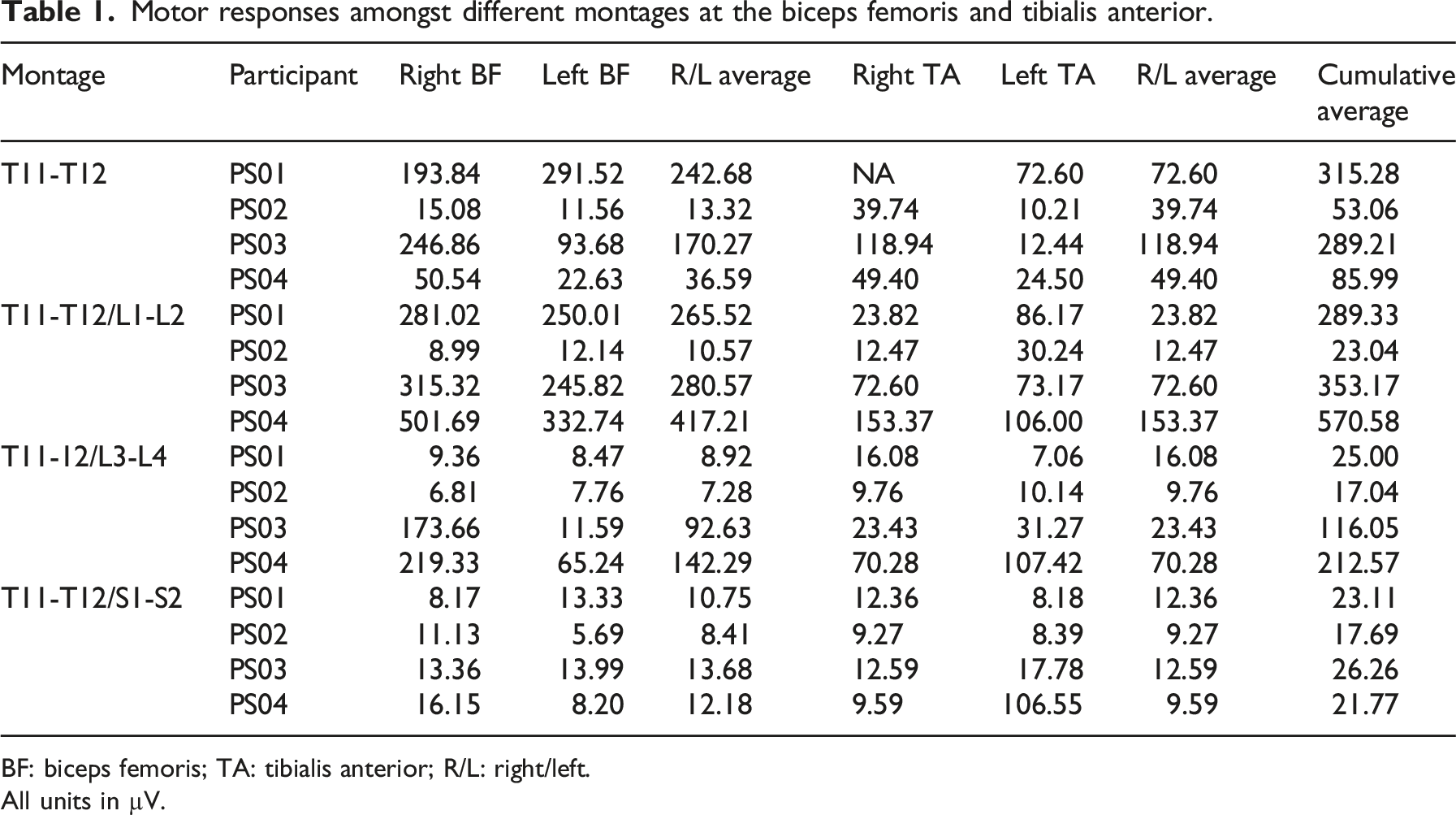
BF: biceps femoris; TA: tibialis anterior; R/L: right/left.
All units in µV.

(a–d): Motor response outputs at different montages.
Montage T11-T12/L1-L2
PS04 showed the highest average values for both the BF (417.21 μV) and TA (129.68 μV), and the highest cumulative motor response (546.9 μV) between the two averages. PS01, PS03, and PS04 all had their highest cumulative averages at this montage. In the SCI group, PS01 exhibited strong BF motor responses with peak-to-peak amplitudes of 250.0 μV on the left BF and 281.0 μV on the right BF. The left TA also showed a significant response of 86.1 μV, while the right TA amplitude was 23.82 μV. PS02 showed an elevated TA motor response on the left side, with a peak-to-peak amplitude of 30.24 μV, while the right TA amplitude was below the response threshold. PS02 did not demonstrate any significant BF responses (Table 1, Figure 4(b)).
In the control group, PS04 demonstrated significant responses in both BF and TA muscle groups. The BF responses had peak-to-peak amplitudes of 332.74 μV on the left and 501.69 μV on the right. The TA responses were also substantial, with amplitudes of 106 μV on the left and 153.37 μV on the right. PS04 demonstrated significant responses in both BF and TA muscle groups. The BF responses had peak-to-peak amplitudes of 245.82 μV on the left and 315.32 μV on the right. The TA responses were also substantial, with amplitudes of 73.17 μV on the left and 72.6 μV on the right (Table 1, Figure 4(b)).
Montage T11-T12/L3-L4
In the SCI group, there was a substantial decrease in motor responses across all muscle groups with all peak-to-peak amplitudes below the 20 μV threshold. In the control group, both PS03 and PS04 demonstrated substantial decreases in both the BF (PS03—92.63 μV; PS04—142.29 μV) and TA (PS03—27.35 μV; PS04—88.85 μV) average motor responses, and the cumulative averages. The PS03 left BF was below threshold, but all other motor responses were detectable (Table 1, Figure 4(c)).
Montage T11-T12/S1-S2
In the SCI group, both PS01 and PS02 exhibited further decreased muscle motor responses across all muscle groups, all below the response threshold. In the control group, the only detectable motor response was the left TA of PS04 (106.55 μV), all other responses were sub-threshold (Table 1, Figure 4(d)).
Peak-to-peak (p-p) EMG amplitudes compared to the T11-T12 baseline
Motor responses compared to the T11-T12 baseline amongst different montages at the biceps femoris and tibialis anterior.

BF: biceps femoris; TA: tibialis anterior; R/L: right/left.
All units in µV.
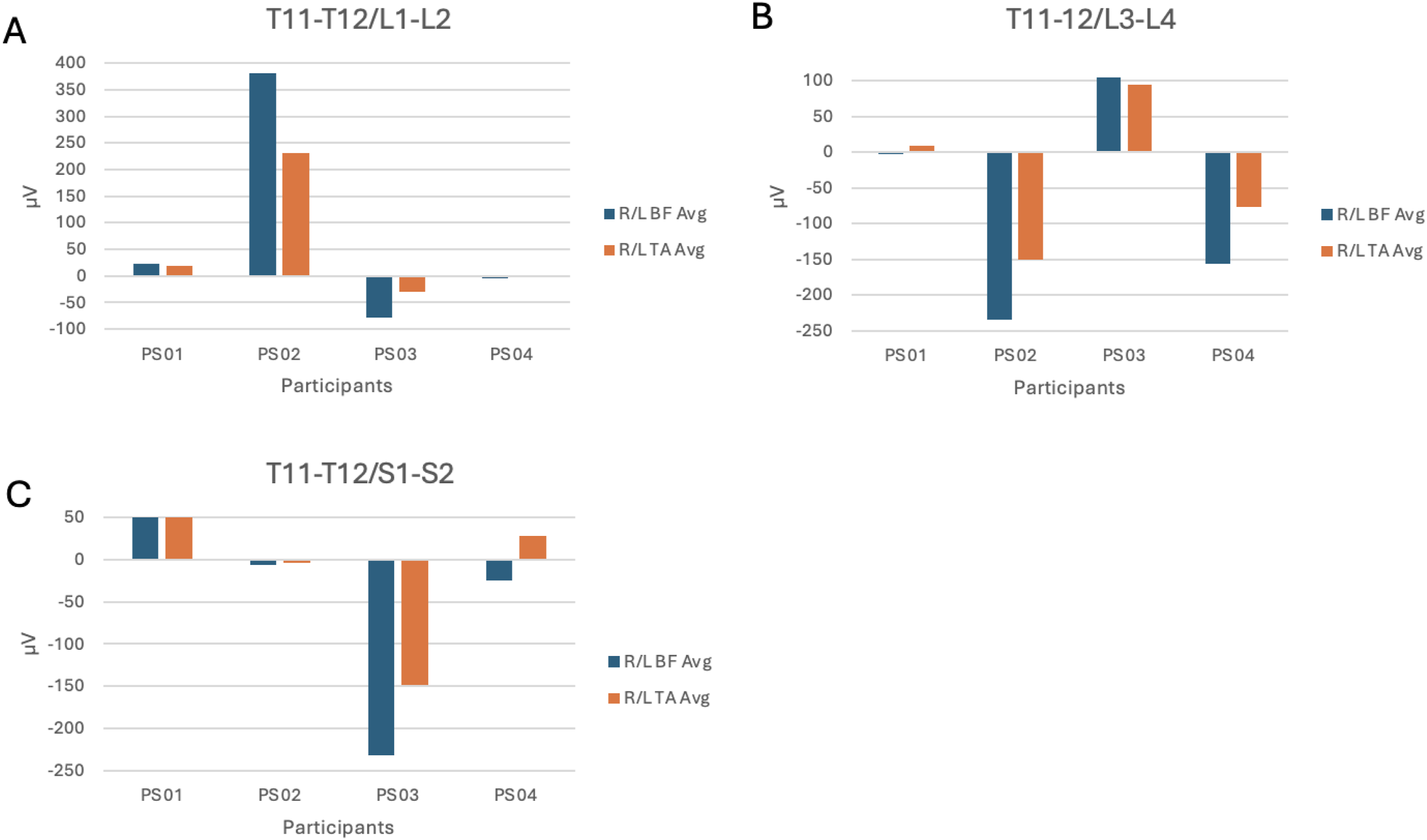
(a–c): Motor response outputs at different montages compared to the T11-T12 baseline.
Discussion
Most groups investigating the effects of tSCS are arbitrarily choosing anatomical montages of electrodes across participants. This pilot feasibility study sought to lay the groundwork for precise and personalized tSCS based upon the first principles understanding that even slight changes in the path of electrical energy can have dramatic effects on which excitable elements within the nervous system are affected. The literature on tSCS appears to imply that it provides a non-specific electroceutical plasticity-enhancement benefit independent of the anatomical pathway of the applied electrical energy. Rather than waiting to see if months of functional task practice and other therapies paired to tSCS could yield functional benefits beyond sham stimulation, we sought to first identify immediate activation effects to inform a precise, personalized approach that could increase the probability of inducing a more durable benefit when paired with therapies in the future.
In this pilot study, we demonstrated that transcutaneous stimulation elicited responses in the SCI population. In addition, the degree of muscle response from tSCS was impacted by topographic electrode configuration, and by the presence of spinal cord injury. Likewise, degree of muscle response in the participants with SCI may have also been influenced by severity (AIS classification) and etiology (traumatic vs vascular). Control participants demonstrated consistently more muscle activation at the T11-T12/L1-L2 montage, and then decreased relatively consistently as the electrode placement moved caudally. The participant with a motor and sensory incomplete SCI (PS01) functioned more similarly to the control participants in terms of the degree and electrode pattern of motor response. In contrast, the participant with a motor complete SCI (PS02) had globally decreased motor responses and had slightly more cumulative motor response at the T11-T12 montage rather than the T11-T12/L1-L2 montage. This may indicate that severity of injury as measured by AIS may influence motor response in terms of electrode placement. Interestingly, PS01 sustained a traumatic injury while PS02 sustained a vascular injury, which may indicate etiology of injury may also influence tSCS response. Further work is warranted.
Prior studies evaluating tSCS in patients with SCI have similarly demonstrated efficacy for lower extremity muscle activation.15,16 Gerasimenko et al. demonstrated that epidural spinal cord stimulation induced involuntary coordinated, cyclic, oscillatory, locomotor-like steps people with and without spinal cord injury when placed in a gravity-neutral position. 15 Interestingly, this study also showed that stimulation of lower extremity motor function was dependent on electrode configuration, with some SCI participants demonstrating more muscle activation with electrode placement over T11 as compared to electrodes placed along more distal segments. 15 This finding is generally consistent with the results of our study, which noted that electrode placement over the T11-T12 and L1-L2 interspaces generated the most lower extremity muscle activation for control SCI participants compared to more distal placement. The increased muscle activation found with additional electrode placement at the L1-L2 level in our study may be due to preferentially greater stimulation at the lumbosacral enlargement which is typically located from the T10-L1 vertebrae. 16 Notably, the present study observed that participant PS02, who had a T2 AIS B injury, demonstrated substantially lower global motor response and a less consistent cranial-caudal pattern of motor activation. In contrast, PS01, who had a T12 AIS D injury, was more similar to the control patients in terms of pattern and output of motor responses.
The current study found that tSCS resulted in variable activation of lower extremity muscles between different participants, even with similar electrode configurations. For example, although T11-T12/L1-L2 montage generally produced the most robust responses for all participants except for PS02, there was substantial side-to-side variation in motor responses amongst muscle groups for the same participants. Additionally, motor responses were markedly different between participants, even for the control participants. These results are similar to a systematic review by Garcia et al. which showed that tSCS resulted in variable lower extremity muscle activation across different participants. 17 This finding may imply that the ideal placement of electrodes may vary among individuals and that electrode configurations may have to be specifically optimized for participants in order to effectively stimulate spinal cord pathways for SCI rehabilitation. Further studies with a larger cohort of control and SCI participants are needed to more definitively establish the efficacy of tSCS with different electrode configurations.
This study is associated with several limitations, such as a small number of participants. Consequently, more granular questions such as clarifying if the severity and mechanism (e.g., vascular vs traumatic) of spinal cord injury could play a role in motor response and pattern were not able to be answered. In addition, this study specifically focused on the activation of the biceps femoris and tibialis anterior muscle groups and did not evaluate other lower extremity muscle groups. As a result, this study could have potentially missed differential activation of other lower extremity muscle groups with different configurations of electrodes. PS02 had a vascular injury, which sometimes results in a more diffuse mixed lesion (both upper motor neuron and lower motor neuron) injury, than a traumatic event. Spinal cord injury mechanism (vascular vs high impact mechanical trauma) could affect the way tSCS induced muscular activation, regardless of AIS level. Bony landmarks may not correlate to spinal cord anatomy given known variation across uninjured people (e.g., while the gastrocnemius is primarily innervated from the S1 spinal nerve, the L5 and S2 nerves may contribute to varying degrees). Additionally, the conus ends at different places in different individuals and this could also alter stimulation effects. Further studies with more control patients and SCI participants are needed to more definitively confirm the results of this study.
Conclusion
Within the limitations of this pilot study, tSCS appears to be able to stimulate lower extremity motor function through spinal cord pathways in participants with and without SCI. The efficacy of tSCS is impacted by electrode configuration, with combined electrode positioning at the T11-T12 and L1-L2 interspaces generally demonstrating the greatest lower extremity muscle activation amongst control patients, and in the participant with the less severe SCI. The effects of tSCS at a given set of anatomical landmarks exerts variable effects across individuals, and it is evident within individuals the degree to which tSCS results in muscle activation, varies considerably by varying the anatomical pathway of the electrical energy.
Footnotes
Acknowledgments
The authors would like to thank the following individuals for their assistance and input: Namrata Grampurohit, Jennifer Rexon, Erica Jones, Maclain Capron, Thomas Hulcher, Nicole Gerhardt, and Josiah Carberry. The authors are grateful for the altruism, engagement, and feedback from the participants.
ORCID iDs

Ethical considerations
This is an IRB-approved prospective study.
Consent to participate
All participant information was deidentified and participant data will not be shared with third parties.
Funding
The authors disclosed receipt of the following financial support for the research, authorship, and/or publication of this article: This work was supported by the Reynolds Foundation.
Declaration of conflicting interests
The authors declared no potential conflicts of interest with respect to the research, authorship, and/or publication of this article.
Data Availability Statement
Deidentified participant data available upon reasonable request.