



Abstract
Cancer -associated thrombosis (CAT), particularly venous thromboembolism (VTE), is a major contributor to mortality in cancer patients and is widely recognized as a leading cause of death after the direct effects of cancer progression. Cancer patients have significantly higher VTE risk than the general population, due to hypercoagulable states and anticancer therapies, with those with advanced malignancies carrying the highest risk. Primary thromboprophylaxis and anticoagulation are pivotal for CAT management. Despite advances, key challenges include different thrombotic and bleeding risks across cancers and how recurrent VTE affects anticoagulation duration. Low-molecular-weight heparin (LMWH) has largely replaced warfarin, and direct oral anticoagulants (DOACs) are challenging LMWH’s first-line role with proven efficacy. However, the key dilemma is balancing thromboprophylaxis and treatment against anticoagulant-induced bleeding, particularly in the context of recurrent VTE. Current CAT guidelines show discrepancies and gaps in clinical coverage; some conclusions derived from meta-analyses need validation via more randomized controlled trials. This review synthesizes recent CAT research (focused on VTE) across epidemiology, pathophysiology, laboratory assessments, and management. It analyzes how cancer type, patient conditions, and drug-drug interactions influence anticoagulant selection, supported by a review of the corresponding experimental evidence. Additionally, the article addresses key clinical scenarios (e.g., intracerebral hemorrhage, pregnancy, pediatric and adolescent patients, and COVID-19) to aid clinical decision-making, delineates unresolved clinical controversies, and integrates high-quality cohort/subgroup data to guide meta-analysis validation. By summarizing risk-benefit consideration, this article provides a framework for complex cases and informs future RCT design.
Keywords
1. Introduction
Cancer remains a major global health burden and one of the leading causes of mortality, with an estimated 20 million new cases and 9.7 million deaths reported worldwide in 2022. 1 Beyond direct invasion and metastasis, cancer can induce systemic complications, among which venous thromboembolism (VTE) carries a high mortality rate (second only to cancer progression) and profoundly influence prognosis and therapy. 2 Cancer patients are at a markedly increased risk of VTE, with incidence rates up to nine times higher than in the general population. 3 Moreover, approximately one-fifth of patients presenting with VTE have an underlying active malignancy, and in some cases, VTE may even serve as the first clinical manifestation of an occult cancer. 4 The close and complex bidirectional relationship between cancer and thromboembolic events is termed cancer-associated thrombosis (CAT), with venous thromboembolism (VTE) being the most common manifestation.5,6 Owing to its high incidence in cancer patients and its substantial contribution to mortality and adverse outcomes, most contemporary research and clinical guidelines on CAT have concentrated on VTE. Given the intricate interplay between malignancy and thrombosis, the pathogenesis and management of thrombosis in cancer patients are shaped by tumor-related factors, host characteristics, and treatment modalities, collectively defining a unique clinical entity that demands individualized therapeutic strategies. 7
In this review, we focus on VTE as the predominant manifestation of CAT, synthesizing current evidence and guidelines to address its growing clinical burden. We examine its epidemiology and pathophysiology, with a focus on cancer type, treatment phase, and individualized patient profiling to guide subsequent management. A core theme throughout is the critical balance between preventing recurrent thrombosis and mitigating bleeding risks inherent to anticancer therapy. Key clinical scenarios are also explored to offer practical insights. Ultimately, this review aims to equip clinicians with actionable, evidence-based strategies for optimizing VTE management in cancer patients.
2. Epidemiology
In absolute terms, the cumulative incidence of VTE in cancer patients ranges from 1% to 8%, depending on cancer type, stage, treatment modality, and patient characteristics. Advanced (especially metastatic) cancer is associated with significantly higher VTE risk. In terms of incidence density, Horsted et al reported a VTE incidence of 13 per 1000 person-years among average-risk cancer patients (representing the general cancer population), while high-risk patients (e.g., those with advanced/metastatic disease or undergoing high-risk treatments) showed a markedly elevated incidence of 68 per 1000 person-years. A large UK database study (involving >82,000 patients) further confirmed an overall VTE incidence of approximately 13.9 per 1000 person-years in all cancer patients. 8 The incidence of tumor-associated VTE correlates positively with the biological aggressiveness of the cancer, such as one-year mortality. 9
Unexplained deep vein thrombosis (DVT) often serves as the initial clinical manifestation of an underlying malignancy. 10 Up to 10% of patients with unprovoked VTE are diagnosed with cancer within one year following the VTE diagnosis. Over 60% of occult cancers are detected shortly after an unprovoked VTE event. At the population level, higher VTE incidence has been observed among older and obese cancer patients and those with multiple comorbid conditions, particularly renal or respiratory diseases, acute infections, and prolonged immobility.11,12 Elevated VTE incidence is also reported in relation to cancer treatment. Following cancer surgery, VTE incidence remains increased for approximately two to four months after the procedure and varies across tumor types. 13 Chemotherapy is associated with an approximately six-fold higher incidence of VTE. 14 Thrombosis related to central venous catheters (CVC) accounts for most cases of secondary upper-extremity DVT, comprising approximately 80 to 90% of such events. 15 Other treatments, including hormone therapy, erythropoiesis-stimulating agents, blood transfusions have also been linked with higher thrombotic incidence. 14 More recently, observational studies have reported an incidence of VTE among patients receiving immune checkpoint inhibitors (ICIs), including programmed cell death protein 1 (PD-1) inhibitors (e.g., pembrolizumab and nivolumab), cytotoxic T-lymphocyte–associated-4 (CTLA-4) targeting agents (e.g., ipilimumab), and programmed death-ligand 1 (PD-L1) inhibitors (e.g., atezolizumab and durvalumab), administered either as monotherapy or in combination regimens. The estimated cumulative incidence was 7.6% at 6 months and 11.1% at 12 months in a large cohort, which was higher than that observed among cancer patients not receiving ICIs, although substantial heterogeneity exists across tumor types and clinical settings.16,17
Representative Incidence of Venous Thromboembolism by Tumor Type
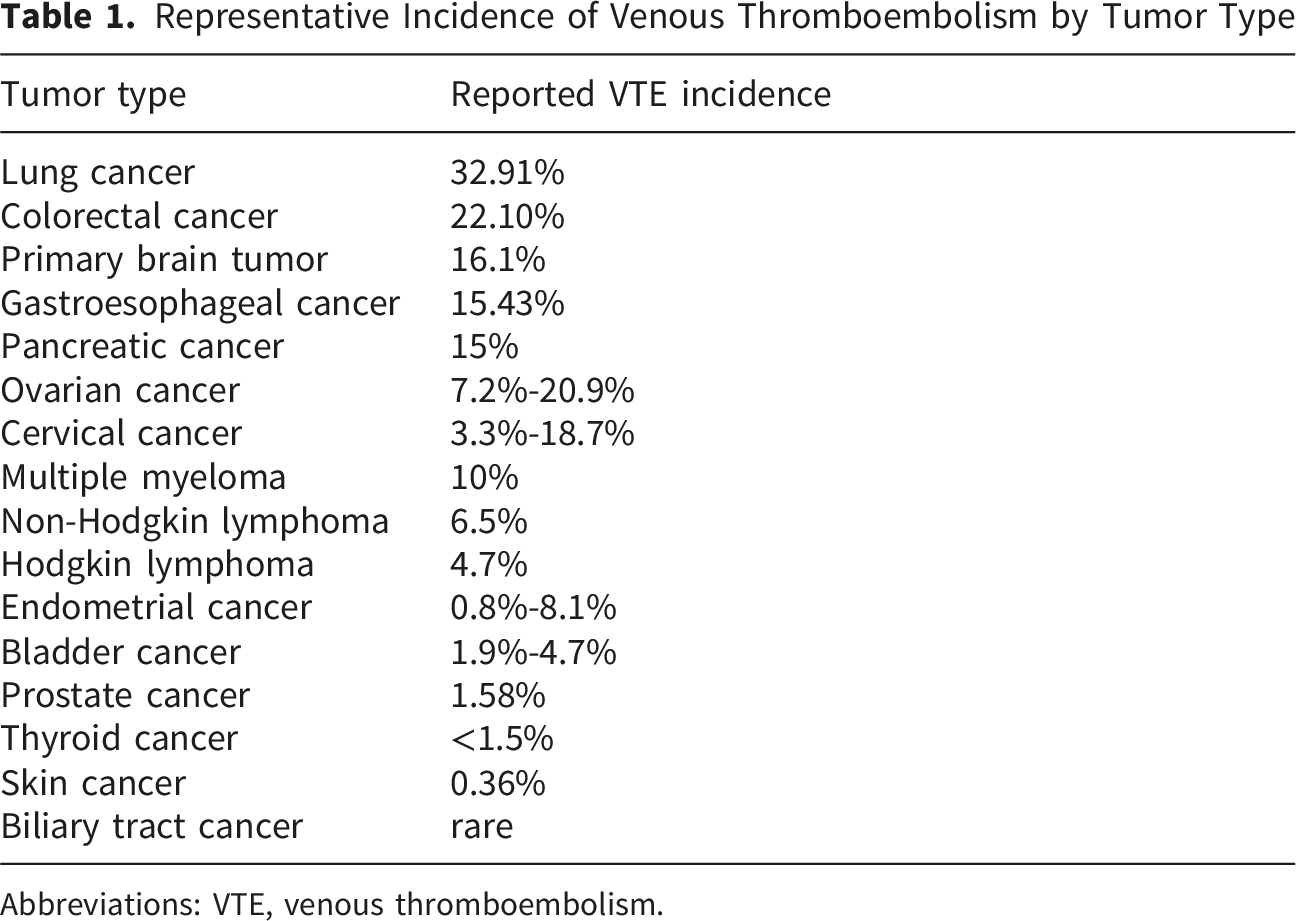
Abbreviations: VTE, venous thromboembolism.
Among hematological malignancies, VTE incidence varies by disease subtype. For lymphoma, pooled evidence suggests a higher incidence in non-Hodgkin lymphoma at approximately 6.5% than in Hodgkin lymphoma at about 4.7%. 29 Multiple myeloma, as another major hematological malignancy, is also strongly associated with thrombosis, with about 10% of patients developing thrombosis during the disease course. 30
3. Mechanisms of Tumor-Induced Thrombosis
3.1. Combined Effects of Virchow’s Triad
Virchow’s triad, comprising endothelial injury, venous stasis, and hypercoagulability, describes the core pathophysiological framework of thrombosis in cancer. 31 The thrombotic burden arises from the malignancy itself, as well as its treatments, and associated clinical complications.13-15
Endothelial injury and dysfunction reflect both tumor invasion and hypoxia-related activation, and are prominently augmented by iatrogenic influences such as surgical trauma and the placement of CVC. 32 Catheter-related infection may further amplify inflammatory activation and prothrombotic conditions. 15
Venous stasis results from tumor-related vascular compression and reduced mobility in advanced disease, and is frequently exacerbated by postoperative immobilization, prolonged hospitalization, and catheter-related flow disturbance. 33
Hypercoagulability is driven by tumor-derived procoagulant factors and a systemic inflammatory state, and is further shaped by systemic cancer therapies, particularly chemotherapy. 34
Thus, CAT reflects the convergence of disease-specific, treatment-related, and care-related factors within the framework of Virchow’s triad.
3.2. Tumor Cell Expression of Procoagulants Leading to Hypercoagulability
Tumor cells release large amounts of procoagulant substances, leading to a hypercoagulable state. Tumor cells can release extracellular vehicles (EVs), which are lipid bilayer-bound particles that cannot replicate and do not contain a functional nucleus. 35 In the resting state, phosphatidylserine (PS) is located on the inner leaflet of the EVs’ lipid membranes but translocates to the outer membranes upon stimulation. 36 The exposed PS provides a negatively charged membrane surface for coagulation and offers more accessible sites for the assembly of the prothrombinase complex and tenase complex, thereby promoting thrombin generation and blood clotting.37,38 Tumor-derived EVs carry a significant amount of intracellular procoagulant material upon release, which is closely related to systemic coagulation. 39 In clinical settings, PS-positive EVs have been detected in the circulation of patients with cancer, 40 and observational data indicate that EV profiles and inflammatory profiles are dynamic during anticoagulant treatment, supporting the clinical relevance of PS-positive EVs in CAT.40,41 Tissue factor (TF), a transmembrane receptor and primary initiator of coagulation, can be expressed by tumor cells or spontaneously released as TF-positive microparticles into the bloodstream, both of which contribute to thrombosis. 42 The expression of the TF gene is regulated through various signaling pathways, transcription factors, and microRNAs (miRNAs). 43 TF expression is often upregulated in tumors with specific oncogene (e.g., K-ras, c-Met) and tumor suppressor gene (e.g., p53, PTEN) mutations, as well as in tumor cells undergoing epithelial-mesenchymal transition (EMT) or hypoxia. 44 Mutations in oncogenes and tumor suppressor genes activate the mitogen-activated protein kinase/phosphatidylinositol-3 kinase signaling pathway, subsequently inducing full-length tissue factor (flTF) expression. 45 TF-mediated coagulation is inhibited by tissue factor pathway inhibitor (TFPI). 46 Under hypoxic conditions created by tumor tissues, hypoxia-inducible factor hypoxia-inducible factor-1α (HIF-1α) accumulates and binds to the hypoxia-responsive element (HRE) region of the TFPI promoter, suppressing TFPI transcription while simultaneously activating pro-survival genes such as vascular endothelial growth factor (VEGF), thereby upregulating TF.47,48 Epigenetically, abnormal downregulation or overexpression of miRNAs such as miR-19 and miR-520g can upregulate TF by suppressing flTF levels.49,50
Studies have shown elevated plasma levels of von Willebrand factor (vWF) in patients with various cancers. 51 This reflects endothelial activation, with increased vWF release and surface presentation that promotes platelet adhesion and immune cell recruitment. 52 Tumor-derived factors may further amplify this process by promoting vWF secretion and regulating its expression within the tumor microenvironment. High plasma vWF levels have been linked to VTE risk in cancer patients and tend to cluster with more advanced disease and higher clinical risk strata.53,54
Tumor cells can also secrete Cancer Procoagulant, which directly activates coagulation factor X independently of factor VII and is unaffected by TFPI. 55 Podoplanin, a transmembrane protein, is abnormally elevated in many tumors. It significantly induces platelet aggregation and promotes coagulation by activating platelets through the platelet receptor C-type lectin-like receptor 2 (CLEC-2).56,57
3.3. Induction of Host Immune and Inflammatory Responses
Tumor development is accompanied by recruitment and activation of neutrophils within the tumor microenvironment, with release of inflammatory mediators that further amplify local immune cell accumulation. 58 Activated neutrophils can express tissue factor on the endothelial surface and release neutrophil elastase, thereby promoting thrombin generation, impairing TFPI activity, and facilitating the formation of neutrophil extracellular traps (NETs). Tumor cells can further directly induce NETs formation by neutrophils.59,60
Experimental studies from the Wagner laboratory have demonstrated that cancer creates a systemic milieu that primes neutrophils for NETs formation and that NETs release directly contributes to CAT in murine models. 61 Consistent with this, inhibition of NETs formation in mouse thrombosis models reduces thrombus burden, supporting a causal role for NETs in tumor-associated hypercoagulability. 62 In the intravascular environment, NETs form extracellular chromatin networks that provide a structural scaffold facilitating platelet adhesion and retention of circulating coagulation factors, thereby promoting thrombus stabilization and local propagation. Beyond this physical scaffold function, NETs also serve as a biochemical platform that concentrates procoagulant molecules within the thrombus microenvironment. Exposure of extracellular DNA and polyphosphates on NETs enhances platelet activation and thrombin generation, while neutrophil-derived proteases can impair endogenous anticoagulant pathways, further shifting the hemostatic balance toward hypercoagulability.63,64 These effects of NETs occur within established platelet and coagulation pathways, in which vWF-mediated platelet tethering and adhesion contribute to platelet recruitment and thrombus propagation. Work from the Dubois laboratory has shown that tumor-derived microparticles bearing tissue factor can localize to thrombotic sites and interact with intravascular scaffolds, linking tumor-derived procoagulant activity with thrombus propagation associated with NETs. 65 Together, these findings establish NETs as a central structural and biochemical amplifier within the tumor-associated thrombotic microenvironment, integrating immune cell activation with thrombus growth dependent on platelet and coagulation pathways. 66 Consistent with these mechanisms, elevated NETs levels in CAT patients are associated with an increased risk of thrombosis. 67
Immune checkpoint blockade of the PD-1/PD-L1 and CTLA-4 pathways may amplify immune-driven inflammatory activation and thereby promote prothrombotic pathways relevant to CAT. 68 Consistent with this, ICI therapy has been linked to a systemic pro-inflammatory state with elevated cytokine levels; activated T cells may promote hypercoagulability by inducing tissue factor expression in monocytes/macrophages. Emerging translational data also suggest that patients who subsequently develop VTE during ICI treatment may exhibit higher baseline inflammatory signatures, including increased myeloid-derived suppressor cells and CXCL8, which may facilitate NETs formation and thereby promote a prothrombotic state.68,69
3.4. Platelet Activation
Tumor cells secrete platelet agonists such as adenosine diphosphate and thrombin to directly activate platelets. They can also activate platelets through interactions between membrane proteins (e.g., podoplanin) on the tumor cell surface and their specific receptors on platelets.
70
Mucinous adenocarcinomas secrete abnormally glycosylated mucins into the bloodstream, which interact with leukocyte L-selectin and platelet P-selectin, leading to platelet activation and aggregation.
71
Collectively, these interconnected mechanisms contributing to tumor-induced thrombosis are illustrated in Figure 1. Schematic overview of key mechanisms contributing to cancer-associated thrombosis.
4. Clinical Evaluation
4.1. Clinical History and Physical Examination
For cancer patients suspected of having thrombosis, rapid clinical assessment is critical. Medical history should clearly identify the onset time, location, and severity of symptoms, and identify potential precipitating factors such as hospitalization, surgery, chemotherapy, radiotherapy, or central venous catheter placement. Physical examination must check for signs of cancer-associated VTE and bleeding. A comprehensive assessment of comorbidities and medications that may affect bleeding risk or interact with anticoagulant drugs is equally important. According to guideline recommendations, attention should also be given to comorbidities and concomitant therapies that may influence thrombotic or bleeding risk, such as atrial fibrillation, hepatic or renal insufficiency, thrombocytopenia, and anticoagulant or anticancer drugs that may interact with anticoagulants. 72
4.2. Laboratory Assessment
In clinical laboratory assessments, conventional coagulation tests (PT/INR/APTT) are widely used to screen for coagulation disorders. However, these tests have limited clinical utility in detecting hypercoagulable states or predicting thrombotic risk in cancer patients. D-dimer, a fibrin degradation product, is the most widely used laboratory biomarker in routine clinical practice to aid the evaluation of suspected VTE in cancer patients. Due to its low specificity, it is primarily used as an exclusionary test, while persistent elevation during follow-up may indicate a higher risk of VTE recurrence. 73 Parameters such as leukocyte count, soluble P-selectin, and platelet count have been investigated in research settings and shown to be associated with VTE risk. These parameters may provide mechanistic insights or contribute to future risk models. However, it is important to note that these associations have not been validated for clinical application, therefore they are not currently recommended for inclusion in the clinical evaluation of VTE.4,74
4.3. Imaging Evaluation
Ultrasound is the first choice for imaging examinations of venous thrombosis. Different examination techniques have different degrees of accuracy. If the purpose is to diagnose proximal DVT, compression ultrasound (CUS) alone may be the most appropriate technique (with a sensitivity of over 90% and specificity of nearly 100%). 75 Conversely, if the aim is to identify distal DVT, duplex or triplex ultrasound may be more appropriate techniques. 76 Based on the latest literature, CUS has become the preferred diagnostic method for detecting DVT and has been applied in multiple departments, including hospital emergency rooms and outpatient clinics. 75 Study has suggested that emergency physicians can use portable ultrasound machines in the emergency room to perform 3-point CUS examinations of the lower limbs, which can effectively detect possible DVT in a short period of time. 77 For pregnant women, patients with severe renal failure, or patients with a history of severe reactions to iodine contrast agents, CTPA is typically contraindicated. In such cases, CUS can provide a bedside, simple, and non-invasive examination to determine the presence of DVT. When assessing for DVT in the abdomen and pelvis or planning preoperatively, CT venography and MR venography are more appropriate. However, the ionizing radiation from CTV and the cost-effectiveness of MRV, along with the specialized expertise required for each modality, must be considered. 78 For patients suspected of pulmonary embolism (PE), CT angiography remains the gold standard for diagnosis.
5. Management
Anticoagulation remains the cornerstone of VTE management in cancer patients. Clinical decision-making is complicated by significantly higher risks of both VTE recurrence and anticoagulant-related bleeding in cancer patients compared to the general population.
79
This complexity necessitates a multidisciplinary team approach. Accordingly, this section is structured to reflect a stepwise clinical management paradigm for CAT, progressing from primary prevention to anticoagulant therapy and special clinical scenarios. A clinical management algorithm (Figure 2) is provided to assist in the translation of these concepts into practice, with the caveat that individualized expert judgment remains essential in complex cases. Algorithm for the management of CAT.
5.1. Primary Thromboprophylaxis
Cancer patients are at a significantly elevated risk of VTE compared with the general population. Predictors of VTE include patient-related factors such as a prior history of VTE, advanced age, obesity, and comorbidities; tumor-related factors, particularly cancer site (pancreatic, gastric, and biliary tract regions); anticancer treatment such as chemotherapy, and biomarkers, among which D-dimer is a promising candidate for baseline risk stratification.80-84 The Khorana score is the most widely validated and applied tool to address this heterogeneous risk, stratifying patients into low (0 points), intermediate (1–2 points), and high (≥3 points) VTE risk categories prior to the initiation of systemic anticancer therapy.82,85 However, external validation studies indicate that the Khorana score may have a limited ability to reliably distinguish patients at higher and lower risk of VTE across diverse cancer populations. In particular, analyses from the Van Es group have shown reduced sensitivity for capturing patients who subsequently develop VTE, with variable performance across major cancer subgroups.85,86 In addition to the Khorana score, other risk assessment models, including the Vienna CATS and ONKOTEV scores, have been proposed for VTE risk stratification in cancer patients.87,88 More recently, the COMPASS-CAT score, prospectively developed in common solid tumors and incorporating broader clinical and comorbidity-related factors, has been proposed as an alternative tool that, unlike the Khorana score, can be applied after initiation of anticancer treatment. Subsequent studies suggest moderate discriminatory performance and potential advantages in selected populations, including lung cancer and immunotherapy-treated cohorts, although substantial heterogeneity and limited prospective external validation remain.89,90 Accumulating evidence suggests that exposure to ICIs is independently associated with an increased risk of venous thromboembolism, even after adjustment for traditional clinical and tumor-related factors. However, existing risk assessment models, including the Khorana score, were developed prior to the widespread use of ICIs and primarily in chemotherapy-treated populations, and their performance in ICI-treated patients appears limited and inconsistent, highlighting the need for dedicated, prospectively validated risk stratification tools and more individualized approaches to thromboprophylaxis in this population.91,92 Routine primary thromboprophylaxis is not advised for all ambulatory cancer patients owing to variable absolute VTE risk and possible bleeding complications.82,93 Current clinical guidelines therefore recommend applying validated risk stratification tools to identify ambulatory cancer patients who have an intermediate to high risk of VTE (e.g., Khorana score≥2) and a low bleeding risk, and to consider initiating primary pharmacological thromboprophylaxis in this subgroup.82,93,94 In this population, prophylaxis with low-molecular-weight heparin (LMWH) or direct oral anticoagulants (DOACs) is recommended and should generally be continued for at least 12 weeks.94,95 For example, in ambulatory patients with pancreatic cancer receiving systemic chemotherapy, primary pharmacological thromboprophylaxis is recommended with LMWH (grade 1A) or DOACs (rivaroxaban or apixaban; grade 1B). 94 In hospitalized cancer patients without VTE, pharmacological thromboprophylaxis is generally recommended in the absence of contraindications, with LMWH preferred.94,95 However, extended thromboprophylaxis is not routinely recommended; a study by Osataphan et al showed no significant reduction in VTE incidence between the extended (28-42 days) and standard (14 days) prophylaxis group, while the extended group exhibited a higher risk of clinically relevant bleeding. 96 For cancer patients undergoing surgery, thromboprophylaxis should be administered both preoperatively and postoperatively according to guidelines, and mechanical methods such as intermittent pneumatic compression (IPC) may be used as adjuncts.93-95 Overall, personalized risk assessment remains essential, incorporating not only thrombotic and bleeding risks but also factors such as treatment-related thrombocytopenia, renal and hepatic function, and potential drug interactions. 97 These prophylactic strategies form an integral component of a clinical management paradigm for VTE in cancer patients, laying the groundwork for subsequent treatment options and ongoing follow-up and reassessment throughout the disease course.
5.2. Anticoagulant Therapy
5.2.1. Comparison of Anticoagulants
More than two decades ago, vitamin K antagonists (VKAs), particularly warfarin, were the mainstay for CAT until the landmark CLOT trial in 2003 showed that LMWH significantly reduced recurrence (9% vs. 17%; HR 0.48; P = 0.002), with no significant difference in major bleeding (6% vs. 4%; P=0.27).98,99 Warfarin has largely lost patient favor due to the frequent and challenging INR monitoring it requires, as well as its associated complications. 100 However, daily injections, cost, and injection-related discomfort (e.g., hematoma) diminished patient adherence,101,102 prompting demand for effective and convenient oral anticoagulants. 103
Comparison Between DOACs and LMWH in CAT From Key RCTs

Abbreviations: VTE, venous thromboembolism; LMWH, low-molecular-weight heparin; DOACs, Direct oral anticoagulants; HR, hazard ratio; SHR, Subdistribution Hazard Ratio.
Notably, apixaban demonstrated a major GI bleeding rate similar to dalteparin in the Caravaggio trial. 104 Furthermore, it showed a significantly lower rate of pulmonary embolism recurrence and severe bleeding in another comparison with LMWH. 111 An observational study suggests that apixaban may be associated with lower bleeding and fewer hospitalization events than rivaroxaban. 112 A network meta-analysis also suggests that in patients with CrCl >80 mL/min, apixaban is associated with a lower risk of major bleeding than warfarin, rivaroxaban, or dabigatran. 113 These results highlights the potential advantages of apixaban among the numerous DOACs, though no head-to-head RCTs of DOACs exist, and dabigatran is not included in current recommendations. Currently, international guidelines all generally consider LMWH and DOACs (apixaban, rivaroxaban, and edoxaban) as the preferred treatment options for CAT. In many eligible patients without contraindications, DOACs are preferred owing to their inherent advantages, especially during the short-term (3 to 6 months) maintenance phase after initial therapy, with careful assessment for the patients’ suitability required. In contrast, warfarin is classified as a backup or secondary option in guidelines, to be considered when LMWH or DOACs are not feasible, with decisions based on individual patient circumstances.94,95,114,115
5.2.2. Drug-Drug Interactions
Anticoagulant therapy in cancer patients requires careful attention to drug-drug interactions (DDIs), which can significantly alter drug levels or bleeding risk, particularly with concomitant chemotherapy, targeted agents, or immunotherapy. Although DOACs exhibit fewer DDIs than VKAs, their interaction potential remains clinically relevant in polypharmacy settings. 116 DOACs are P-glycoprotein (P-GP) substrates; apixaban and rivaroxaban are mainly metabolized via CYP3A4, whereas edoxaban and dabigatran involve minimal CYP metabolism. 117 Concomitant use with strong inhibitors or inducers of these pathways may elevate bleeding or thrombotic risk. Tyrosine kinase inhibitors (TKIs), commonly used in oncology, frequently modulate P-GP and CYP3A4 activity and independently increase bleeding risk through effects on endothelium and platelet function.101,116 Bleeding rates vary among TKI classes, with Bruton’s TKIs associated with the highest reported bleeding risk, whereas VEGFR-TKIs and other VEGF-pathway inhibitors show elevated but heterogeneous bleeding and thrombotic risk across agents and settings. 118 Agent-specific data are therefore important: in patients with CAT receiving therapeutic anticoagulation, continuation of bevacizumab was not associated with increased risks of recurrent VTE or major or CRNMB compared with discontinuation. 119 These findings underscore the importance of individualized assessment when combining DOACs with targeted anticancer therapies. When combining DOACs with interacting anticancer drugs, consider dose adjustment, such as reduction with inhibitors or escalation with inducers; alternatively, substitution with LMWH may be employed.120,121 Further studies are needed to establish evidence-based dosing guidelines for specific TKIs-DOAC combinations.
5.2.3. Anticoagulation Duration - Consideration for VTE Recurrence
Cancer patients exhibit a substantially higher risk of current VTE recurrence than non-cancer patients, contributing to poorer clinical outcomes and increased mortality. 122 Independent predictors of recurrence include specific cancer type (e.g., brain, gynecological, urogenital, gastrointestinal, hepatobiliary, pancreatic, and lung), poor performance status (ECOG ≥1), ongoing anticancer therapy, and higher creatinine clearance.123,124 The presence of residual venous thrombosis (RVT) after 6 months of treatment also signifies elevated recurrence risk, 125 whereas recent surgery prior to VTE is associated with lower recurrence. 126 Meta-analyses of RCTs demonstrated that DOACs carry a similar or potentially reduced risk of recurrence compared to LMWH and VKAs.127,128 However, due to increased bleeding risks with DOACs, particularly in gastrointestinal and genitourinary cancers, LMWH is typically the first-line option in these populations. 129 Treatment duration significantly influences recurrence rates. Anticoagulation for less than 3 months markedly increases risk, whereas extending therapy to 3 to 6 months or beyond 6 months substantially reduces recurrence compared to shorter courses. 122 However, prolonging anticoagulant after 6 months does not confer additional recurrence benefit but increases bleeding risk. 130 Recent evidence from the API-CAT trial suggests that extended anticoagulation beyond 6 months with reduced-dose apixaban (2.5 mg bid), compared with the full dose (5 mg bid) therapy, can maintain noninferior protection against recurrent VTE while reducing clinically relevant bleeding, thereby supporting a novel dose-reduction strategy for extended therapy in patients with CAT. 131 In addition to clinical efficacy and bleeding reduction, a recent environmental impact analysis suggests that reduced-dose apixaban strategies for extended VTE treatment may also be associated with a lower carbon footprint, introducing sustainability as a potential ancillary consideration in selected long-term treatment contexts. 132 Current guidelines recommend at least 6 months of therapy, with extended treatment reserved for high-risk subgroups, such as those with active malignancies, ongoing antitumor therapy, a history of RVT, specific cancer types, or metastatic and progressive disease, following regular reassessment of thrombotic and hemorrhagic risk.122,123 Long-term anticoagulation management remains a dynamic and challenging process, and accordingly, validated prognostic models are urgently needed to optimize indefinite strategies. 126
5.2.4. High-Risk Bleeding Factors and Therapeutic Considerations
Assessing bleeding risk is a critical component of developing an appropriate anticoagulation strategy. Despite the availability of traditional bleeding risk assessment models used in VTE, such as VTE-BLEED and RIETE, 133 as well as cancer-specific models derived from secondary analyses of the Hokusai VTE Cancer trial, these models show limited predictive performance and remain insufficient to reliably guide current clinical decision-making. 134 In practice, clinicians must consider established bleeding risk factors and adopt individualized therapeutic strategies that balance thrombotic control with bleeding risk to maximize treatment safety and clinical benefit.
5.2.4.1. Cancer Site
Bleeding risk varies substantially by tumor site, which has important implications for anticoagulant selection. Gastrointestinal (GI) cancer is associated with a heightened bleeding risk, particularly evident in some DOACs. The subgroup analysis in the SELECT-D and Hokusai VTE trials showed more frequent major bleeding with DOACs (rivaroxaban: 36%, edoxaban: 12.7%) than LMWH (11%, 3.6%) in patients with GI cancer.104,135 An early meta-analysis of RCTs also reported higher major bleeding rates with DOACs than LMWH in GI cancer patients (RR 2.55; 95% CI 1.24-5.27, P = 0.01). 136 In contrast, subsequent meta-analyses and systematic review indicated that, among patients with GI tumors treated with DOACs (apixaban, edoxaban, and rivaroxaban), rate of CRNMB was significantly elevated in the DOAC-treated group compared with patients receiving LMWHs, while there was no significant difference in the incidence of major bleeding. 110 The Caravaggio trial similarly found no bleeding difference between apixaban and LMWH for GI cancer, though it had insufficient data to reliably detect differences in major bleeding. 107 Head-to-head comparisons revealed that apixaban was associated with lower incidences of both major bleeding (3.4% vs. 8.5%) and CRNMB (2.2% vs. 7.6%) in GI cancer patients than rivaroxaban. 137
Emerging evidence suggests bleeding risk varies by GI cancer subtype and resection status, with unresected upper GI cancers carrying the highest risk, 138 upper GI cancer and non-resected luminal GI cancer have been identified as predictive factors for major bleeding. The PROLAPS-II randomized trial has demonstrated rivaroxaban’s efficacy and safety in colorectal cancer resection patients, 139 and some data support DOACs may be eligible for resected lower GI cancer patients. 140 Current guidelines universally classify GI cancer as high-risk and typically favoring LMWH as the first choice, or apixaban if an oral agent is preferred. Specific guidance for DOACs use in lower GI or resected tumors remains undefined, underscoring the need for subtype-specific randomized trials. 140
Genitourinary (GU) cancer is also associated with an increased risk for bleeding, with rivaroxaban showing a significantly higher incidence of major bleeding in GU patients than in non-GU patients1 141 ; accordingly, current guidelines list GU cancer as a high-risk factor of bleeding and recommend LMWH over DOACs, particularly in patients with unresected tumors or active lesions. 7
Brain cancer presents unique challenges due to the inherent risk of spontaneous intracerebral hemorrhage (ICH), which is elevated in both primary and metastatic tumors. 142 A meta-analysis showed that metastatic disease confers higher ICH rates than primary brain cancer patients and anticoagulation increase ICH risk in primary cancer but not in metastatic cases. In contrast to GI cancer or GU cancer, patients with brain cancer receiving DOACs experience lower rates of ICH and mortality compared to those taking VKAs or LMWH.143,144 Guidelines recommendations vary considerably. The ASH 2021 guidelines recommend DOACs (apixaban, edoxaban, and rivaroxaban) as an alternative to LMWH, 145 while the ESMO 2023 guidelines advise LMWH for brain metastases. 146 Other guidelines offer no specific preference, reflecting exclusion of these patients from major RCTs and reliance on retrospective data.94,114 Therefore, anticoagulation choice must be individualized by balancing thrombotic and bleeding risks, including thrombocytopenia and other patient-specific factors.
5.2.4.2. Hepatic and Renal Dysfunction
Impaired hepatic function significantly increases bleeding risk in cancer patients due to reduced synthesis of clotting factors and altered anticoagulant metabolism. Approximately 66% of rivaroxaban and 25% of apixaban is metabolized via the CYP3A4 pathway, underscoring their susceptibility to hepatic dysfunction. Hepatic dysfunction and coagulopathy influence anticoagulant choice, with LMWH often favored and DOAC use constrained by Child-Pugh class.147,148
Based on the latest ISTH guidance, anticoagulation with either a DOAC or LMWH may be considered in patients with Child-Pugh A or B hepatic dysfunction. However, rivaroxaban should be avoided in patients with Child-Pugh B because of pharmacokinetic concerns. In patients with Child-Pugh C hepatic dysfunction, DOAC therapy is generally discouraged due to limited safety and efficacy data, and LMWH-based regimens, with transition to VKAs in selected cases, are more commonly used.149,150
Renal impairment is associated with an increased risk of major bleeding in patients with CAT. In the CATCH study, patients with renal impairment had higher rates of major bleeding compared with those without renal dysfunction (6.1% vs. 2.0%; RR 2.98; 95% CI 1.29 to 6.90). 151 Accordingly, reduced renal clearance necessitates careful consideration of anticoagulant selection and dosing. Guidelines generally favor DOACs over VKAs in mild-to-moderate renal impairment (CrCl 30 to 59 mL/min), provided there are no contraindications or significant DDI. 94 Apixaban appears to be a reasonable option in patients with VTE and concomitant renal impairment, with observational and pharmacokinetic data suggesting acceptable efficacy and bleeding risk relative to VKAs. 152 In patients with moderate renal impairment receiving standard-dose apixaban, trough plasma concentrations are approximately twofold higher than in those with normal renal function. 153 However, the clinical implications of this increased exposure for bleeding risk remain uncertain. Because different DOACs vary in renal clearance, both rivaroxaban and edoxaban generally require dose reduction according to labeling in patients with renal dysfunction, highlighting important inter-drug differences relevant to individualized anticoagulant selection.154,155 Patients with CAT and severe renal impairment (CrCl ≤ 30 mL/min) were excluded from major DOAC trials. Management therefore generally relies on UFH with transition to VKAs, or LMWH with laboratory-guided dose adjustment. Given the absence of CAT-specific evidence, DOACs are not routinely recommended in this setting. 146
5.2.4.3. Thrombocytopenia
Cancer patients with thrombocytopenia face an elevated risk of bleeding during anticoagulation therapy. 156 Evidence remains limited, however, particularly in hematologic malignancies which exhibit higher rates of thrombocytopenia than solid tumors(5.6% vs. 0.7%).157,158 Prior major trials often excluded patients with significant thrombocytopenia (platelet counts < 75,000/µL).104,107 Observational data suggests that dose-adjusted anticoagulation may reduce major bleeding compared with full-dose regiments (5.6% vs. 2.6%). 156 Similar findings were reported in a cohort where LMWH was used predominantly in hematologic malignancies with thrombocytopenia (platelet count < 100,000/µL). 159 The ongoing START randomized trial aims to compare LMWH dose adjustment versus platelet transfusion in patients with platelet counts <50,000/µL, which may inform future practice. 160 Current guidelines recommend standard anticoagulants for platelet counts 50,000 to 100,000/µL, with LMWH preferred in cases of acute VTE with unstable thrombocytopenia. For counts between 25,000 and 50,000/µL, LMWH may be used cautiously, combined with platelet-activating factor receptor agonists or direct platelet transfusion, to maintain platelets at safe concentrations. Anticoagulants are generally discontinued when platelet fall below 25,000/µL. 161
Although pivotal clinical trials evaluating anticoagulant strategies in CAT have been predominantly conducted in Western populations, the applicability of these findings to Chinese and other Asian patients warrants further validation. Given potential differences in baseline thrombotic risk, bleeding susceptibility, body composition and clinical practice patterns, future multicenter cohort studies and real-world data analyses in Asian populations are needed to clarify population-specific considerations regarding anticoagulant dosing, effectiveness, and safety, thereby improving the regional implementation of evidence-based CAT management.
5.3. Clinical Scenarios With Special Considerations
5.3.1. Intracerebral Hemorrhage
ICH represents a frequent complication in cancer patients, often associated with hematologic malignancies, primary brain tumors, hemorrhagic metastases, or treatment-related coagulopathies, such as chemotherapy-induced thrombocytopenia and the use of anticoagulant for VTE. 162 In acute ICH, according to guidelines from the American Heart Association/American Stroke Association, therapeutic anticoagulation should be immediately discontinued. Mechanical thromboprophylaxis with IPC is recommended to reduce the risk of DVT, 163 and insertion of a retrievable inferior vena cava filters should be considered for patients with established DVT or PE. The optimal timing for resuming anticoagulation is complex and there is no precise or stable time for the management. Early studies have shown that initiating oral anticoagulants within 7 days or 2 weeks is safe and reasonable, does not increase the risk of ICH, and can reduce thromboembolic events.164,165 Therefore, researchers suggest that when the cause of ICH is corrected and there is a high risk of thromboembolism, anticoagulant therapy should be initiated at 4 weeks. 166 Kareem also considered 30 days as the average time for restarting after the hemorrhage event. 167 However the timing must be individualized based on hematoma stability, thrombotic risk, and the underlying prothrombotic state of cancer. Earlier resumption (2 weeks after ICH) in patients with artificial mechanical heart valves and a postponement (8 to 10 weeks after ICH) for patients with bleeding in the cerebellum and brainstem may be necessary. 168 For patients with atrial fibrillation who experience ICH, the restart period is 4 to 8 weeks post event. Furthermore, multidisciplinary discussions involving neurology, oncology, hematology, and cardiology is strongly recommended before restarting therapy. 167
5.3.2. Pregnant and Postpartum Women
Cancer during pregnancy significantly elevates thrombotic risk, with a meta-analysis reporting an approximately 6.5-foldhigher risk compared with pregnancy alone. 169 This risk stems from synergistic hypercoagulability due to both pregnancy-related physiological changes and cancer-specific prothrombotic mechanisms, 169 the postpartum period, particularly the first 6 weeks, represents the highest risk phase. LMWH constitutes the anticoagulant of choice throughout pregnancy and the postpartum period, owing to its established safety profile characterized by minimal placental transfer and no known teratogenic risk. 170 DOACs are contraindicated due to placental passage and potential fetal toxicity, and VKAs are generally avoided except in exceptional circumstances with no alternative options (e.g., mechanical heart valves).170,171 Peripartum management requires meticulous timing. LMWH should be withheld at least 24 hours (therapeutic dose) and 12 hours (prophylactic dose) prior to delivery, and resumed 6 to 12 hours after vaginal delivery or 12 to 24 hours after cesarean section, provided hemostasis is confirmed adequate. 172 Treatment should be continued for at least 3 months, extending prophylactic anticoagulation for a minimum of 6 weeks postpartum. 170
5.3.3. Pediatric and Adolescent
Evidence on CAT in pediatric and adolescent populations remains limited and is largely derived from studies in children with acute lymphoblastic leukemia during induction therapy. 173 Recent randomized and pooled analyses indicate that LMWH is the only pharmacological strategy consistently associated with a reduction in VTE risk in this setting, without an apparent increase in major bleeding, and it is therefore recommended for thromboprophylaxis.94,174 Catheter-related thrombosis represents an important clinical consideration in children with cancer. Prospective data suggest that peripherally inserted central catheters are associated with a substantially higher risk of catheter-related VTE compared with centrally inserted catheters. 175 These findings underscore the heterogeneity of thrombotic risk in pediatric cancer patients, supporting an individualized anticoagulation approach with dosing typically adjusted according to body weight and clinical context.
5.3.4. Patients With COVID-19
Evidence regarding VTE in cancer patients and COVID-19 also remains limited. 176 International guidelines indicate that strategies for the treatment and prophylaxis of VTE in cancer patients should not differ solely on the basis of COVID-19 status, and all patients should undergo standard VTE risk assessment. During hospitalization, pharmacological thromboprophylaxis is recommended using the same anticoagulant type and dosing as in cancer patients without COVID-19, whereas post-discharge thromboprophylaxis and primary pharmacological prophylaxis in ambulatory cancer patients are not recommended, emphasizing the need for individualized benefit–risk evaluation. 94
6. Conclusion
CAT represents a severe and complex complication in oncology, presenting significant challenges in prevention and treatment. Substantial gaps persist in predicting thrombotic and hemorrhagic risks, determining optimal anticoagulation duration, and guiding management in high-risk populations such as patients with brain tumors, GI or GU malignancies, thrombocytopenia, pregnancy, pediatric cancers, and those receiving ICIs. Due to the lack of large-scale RCTs in high-risk subgroups, current guidelines largely rely on subgroup analyses, observational studies, and expert consensus, leaving many aspects of CAT management undefined. Anticoagulation therapy requires balancing the risks of thrombotic recurrence and bleeding, with decisions needing to be individualized based on cancer type, treatment stage, organ function, and patient-specific factors. Multidisciplinary collaboration involving oncologists, hematologists, pharmacists, and related specialist is crucial for optimizing CAT anticoagulation strategies. Future prospective studies and collaborative research efforts are needed to address clinical questions, refine risk assessment tools, and evaluate novel anticoagulants. Ultimately, personalized, evidence-based management is essential for improving thrombosis-related outcomes in cancer patients.
Footnotes

Author Contributions
Funding
The authors received no financial support for the research, authorship, and/or publication of this article.
Declaration of Conflicting Interests
The authors declared no potential conflicts of interest with respect to the research, authorship, and/or publication of this article.