
Abstract
Background
Acute coronary syndrome (ACS), a cardiovascular disease with high mortality, is closely associated with microRNA (miRNA) dysregulation, making miRNAs promising targets for diagnostic and therapeutic development.
Aim
This study evaluated the clinical value of miR-3615 in ACS and its pathological mechanism.
Methods
This study included 121 ACS patients and 72 controls; miR-3615 and DPF3 expression were quantified by qRT-PCR. The clinical diagnostic and prognostic value of miR-3615 was analyzed through the ROC curve, correlation analysis, multivariate regression analysis, and K-M survival analysis. In vitro, a cell model of human coronary artery smooth muscle cells (HCASMCs) was established by inducing oxidized low-density lipoprotein (ox-LDL). Inflammatory markers were analyzed using ELISA kits. CCK-8 and Transwell assays were used to evaluate cell proliferation and migration abilities. The targeting effect of miR-3615 on DPF3 was confirmed by dual luciferase assay.
Results
MiR-3615 expression was elevated in ACS patients, especially among those who experienced major adverse cardiovascular events (MACE). Furthermore, its levels showed a positive correlation with both the Gensini score (indicating coronary lesion severity) and cardiac troponin I (cTnI, a marker of myocardial injury). MiR-3615 has a high degree of accuracy in diagnosing ACS and predicting poor prognosis. In an ox-LDL-induced HCASMCs model, inhibition of miR-3615 effectively attenuated pathological cell proliferation, migration, and inflammatory responses. DPF3 was identified as a target of miR-3615.
Conclusions
MiR-3615 is expected to become a potential biomarker for the diagnosis and prognosis of ACS. Moreover, the upregulation of miR-3615 may induce damage to HCASMCs, thereby participating in the occurrence and development of ACS.
Keywords
Introduction
Acute coronary syndrome (ACS), recognized as the most severe clinical phenotype of coronary atherosclerotic heart disease, is featured by abrupt onset, rapid progression, and substantial mortality risk. 1 It is categorized by electrocardiogram (ECG) into ST-segment elevation ACS (STE-ACS, equivalent to STEMI), typically caused by complete coronary occlusion, and non-ST-segment elevation ACS (NSTE-ACS), encompassing NSTEMI and unstable angina (UA), often due to incomplete occlusion or plaque erosion.2,3 Plaque rupture or erosion is the central pathological process that activates secondary platelet activation, thrombosis, and acute myocardial ischemia. 4 Despite the remarkable efficacy of contemporary revascularization strategies in restoring coronary blood flow, a subset of patients still suffer from adverse outcomes, including in-stent restenosis, recurrent myocardial infarction, and other severe cardiovascular complications, which ultimately compromise long-term clinical prognosis. 5 Therefore, screening for specific biomarkers associated with the initiation, progression, and prognosis of ACS is pivotal to improving diagnostic precision, refining risk stratification strategies, and individualizing clinical treatment regimens.
MicroRNAs (miRNAs) are endogenous, short non-coding RNAs with well-conserved sequences. 6 By binding complementarily to the 3′ untranslated regions (3′UTRs) of target mRNAs, thereby exerting precise post-transcriptional regulation of gene expression. 7 This regulates core cellular functions, like proliferation, differentiation, apoptosis, and inflammation, and thereby broadly impacts many physiological and pathological conditions.8,9 Benefiting from their pivotal regulatory roles in ACS pathogenesis, excellent stability in peripheral circulation, and facile detection, miRNAs have emerged as a category of highly promising diagnostic and prognostic biomarkers. In recent years, numerous miRNAs have been demonstrated to possess diagnostic and prognostic value for ACS. 10 For example, miR-9 was reported to inhibit the progression of atherosclerosis through SDC2-FAK/ERK signaling pathways, which could provide a new theoretical basis for the treatment of such diseases. 11 Serum miR-145 levels are significantly downregulated in ACS patients, identifying it as a potential diagnostic biomarker. 12 Additionally, the miR-101/CHD5 pathway has been shown to modulate endothelial cell function during ACS progression, underscoring its dual potential as a therapeutic target and a novel diagnostic indicator. 13
MiR-3615 has been identified as playing roles in various diseases. It shows differential expression in retinal pigment epithelial cells under oxidative stress conditions. 14 Moreover, miR-3615 has prognostic value for inflammatory bowel disease. 15 In cardiovascular diseases, miR-3615 has been reported to be upregulated in patients with acute heart failure (AHF) and shows diagnostic potential. 16 And in the GEO dataset GSE202937, miR-3615 was observed to be differentially expressed in patients with ACS, demonstrating its great potential in the diagnosis of ACS. However, the exact function of miR-3615 in the pathogenesis of ACS, its value in early diagnosis and prognosis assessment, as well as its downstream regulatory pathways, have not been systematically elucidated.
Based on these, this study proposes the following hypothesis: The expression level of miR-3615 has certain clinical significance in patients with ACS and is involved in the occurrence and progression of ACS. The main research approach is to verify the diagnostic and prognostic value of miR-3615 in clinical patients’ serum, and to validate the function and mechanism of miR-3615 in ACS through cell models that simulate the pathological environment of ACS.
Materials and Methods
Bioinformatic Analyses
Differentially expressed genes (DEGs) of patients with ACS were downloaded from the GEO database (https://www.ncbi.nlm.nih.gov/geo/), using the dataset GSE202937, with screening criteria of P<0.05 and ∣log2 (fold change) ∣>1.
Using the miRWalk (https://mirwalk.umm.uni-heidelberg.de/), miRDB (https://mirdb.org/), and miRTarBase (https://mirtarbase.cuhk.edu.cn/∼miRTarBase/miRTarBase_2025/php/index.php) databases, the potential downstream target genes of miR-3615 were predicted.
Patient Enrollment and Sample Collection
This study consecutively enrolled 121 ACS patients admitted to Xinxiang Central Hospital from February 2024 to March 2025, alongside 72 healthy controls identified through concurrent routine exams. The Ethics Committee of the affiliated institution approved this study. Key inclusion criteria were: (1) age 18–80 years; (2) chest pain duration < 24 hours; (3) availability of complete clinical data; (4) ACS diagnosis confirmed by standard evaluations including medical history, physical examination, laboratory tests, echocardiography, coronary computed tomography angiography(CCTA), or coronary angiography; and (5) provision of signed informed consent by the patient or guardian, with willingness to comply with follow-up. Key exclusion criteria comprised: (1) comorbidities including malignant arrhythmias, infective endocarditis, aortic coarctation, valvular heart disease, or pulmonary embolism; (2) History of cardiac surgery or antithrombotic therapy; (3) Presence of infectious diseases, hemorrhagic disorders, or severe anemia; (4) Diagnosis of any malignancy; (5) Accompanied by severe hepatic or renal dysfunction; (6) Pregnancy or lactation; (7) Presence of immunodeficiency diseases.
Baseline Information and Blood Collection
Baseline demographic and clinical characteristics were retrieved from all participants, including age, gender, body mass index (BMI), smoking, and alcohol history. After hospital admission, subjects were placed on an overnight fast, and blood samples were drawn from their peripheral veins. Following centrifugation at 3700 rpm for 20 minutes, serum aliquots were prepared and cryopreserved at -80°C for subsequent laboratory assays. Serum biochemical markers, including total cholesterol (TC), triglycerides (TG), high-density lipoprotein cholesterol (HDL-C), low-density lipoprotein cholesterol (LDL-C), creatine kinase-MB (CK-MB), and cardiac troponin I (cTnI), were measured with an automated biochemical analyzer (ARCHITECT c8000; Abbott Diagnostics, USA). The severity of coronary lesions among the ACS patients was assessed via the Gensini scoring system.
Prognostic Analysis in ACS Subjects
During the 6-month follow-up period through outpatient and telephone visits, major adverse cardiovascular events (MACE; defined as death, recurrent MI, angina, heart failure, or malignant arrhythmia) and survival status were documented for all ACS patients. Based on this, the patients were divided into MACE and non-MACE groups.
Cell Culture and Transfection
The in vitro studies utilized human coronary artery smooth muscle cells (HCASMCs, ATCC, USA). These cells were propagated in Dulbecco’s Modified Eagle Medium (DMEM, Cytiva, USA) containing 10% FBS (Cytiva, USA) and 1% penicillin/streptomycin (Sigma-Aldrich, USA), under standard incubation conditions of 37°C and 5% CO2.
To establish an induced model, cells were challenged with 100 μg/mL oxidized low-density lipoprotein (ox-LDL, Kalen Biomedical, USA) for 24 hours. 3 Following ox-LDL induction, cells were transfected using X-tremeGENE 360 (Roche, Switzerland) with either an miR-3615 inhibitor or an miR-3615 mimic (RiboBio, Guangzhou) to downregulate or upregulate miR-3615 expression, respectively. The cells after transfection were cultured for 24 hours before being used for subsequent verification. Control groups were established by transfecting cells with a corresponding inhibitor negative control (NC) or mimic NC under identical conditions.
RT-qPCR Analysis
RNA from serum and cells was extracted with TRIzol reagent (Sigma-Aldrich, USA). cDNA was reverse-transcribed using the GoScript™ system (Promega, USA) and the Mir-X™ kit (Takara Bio). MiR-3615 and DPF3 expression was quantified via SYBR Green Master Mix (Invitrogen, USA) on a Rotor-Gene Q system. U6 and GAPDH served as internal references. Relative expression was determined by the 2−ΔΔCt method. The sequences of the primers used are shown in Supplementary Table 1.
Enzyme-Linked Immunosorbent Assay (ELISA)
Quantification of inflammatory cytokines (TNF-α, IL-6, IL-1β) was performed on cell culture supernatants from treated HCASMCs using corresponding commercial ELISA kits (Abcam, Shanghai). Before assay, the supernatant samples were centrifuged (3000 rpm, 10 min) for clarification. Cytokine levels were assayed according to the manufacturer’s instructions.
CCK-8 Assays
Cell proliferation was assessed at 24, 48, and 72-hour intervals using a CCK-8 assay (Beyotime, Shanghai, China). Following the designated treatments, HCASMCs plated in 96-well plates (5,000 cells/well) were incubated with CCK-8 solution for 2 hours under standard conditions. The absorbance at 450 nm, indicative of proliferative capacity, was subsequently recorded with a microplate reader.
Transwell Migration Assay
Treated cells (2.5×10⁴) were seeded in serum-free medium into the upper compartment of a 24-well Transwell insert, with 10% FBS medium in the lower chamber as a chemoattractant. After incubation, migrated cells on the lower membrane were fixed with 20% methanol and subsequently stained with a 0.2% crystal violet solution. Subsequently, multiple fields of view were randomly selected under the optical microscope, and counted under a microscope from randomly selected fields.
Dual-Luciferase Reporter Assays
The wild-type (DPF3-WT) and a mutated (DPF3-MUT) version of the DPF3 3′UTR sequences were cloned into a pmirGLO vector (Promega, USA). After seeding in 6-well plates, cells were co-transfected with either the DPF3-WT or DPF3-MUT plasmid, in combination with miR-3615 mimic, a mimic NC, miR-3615 inhibitor, or an inhibitor NC. Luciferase activity was measured with a commercial kit 48 hours post-transfection, and Firefly luciferase activity was expressed relative to Renilla luciferase values.
Statistical Analysis
Data analysis was performed using SPSS and GraphPad Prism, and charts were drawn. Continuous data are reported as mean ± SD. Group comparisons were made with the t-test or Chi-square test, as appropriate. The comparisons among multiple groups were conducted using one-way analysis of variance. Correlations were analyzed by Pearson’s test. Diagnostic value was assessed by ROC analysis. Multivariate logistic and Cox regressions identified ACS risk factors and MACE predictors, respectively. Kaplan-Meier(K-M) survival analysis was conducted, applying a significance threshold of P < 0.05. All in vitro experiments were independently repeated at least three times.
Results
MiR-3615 Expression Was Increased in the Serum of ACS
Volcano plot analysis of the GSE202937 dataset identified miR-3615 as significantly upregulated in ACS patients (Figure 1A). RT-qPCR assays confirmed that miR-3615 expression was upregulated in ACS patients (P < 0.001, Figure 1B). Correlation analyses revealed positive correlations between miR-3615 expression and coronary artery disease severity (Gensini score: r = 0.751, Figure 1C), and myocardial injury marker cTnI levels (r = 0.782, Figure 1D). Expression of miR-3615 in ACS. (A) Volcano plot of differentially expressed miRNAs. (B) The expression level of miR-3615 in the ACS and the control group. (C) Correlation analysis of miR-3615 and Gensini score. (D) Correlation analysis of miR-3615 and cTnI. (***P< 0.001)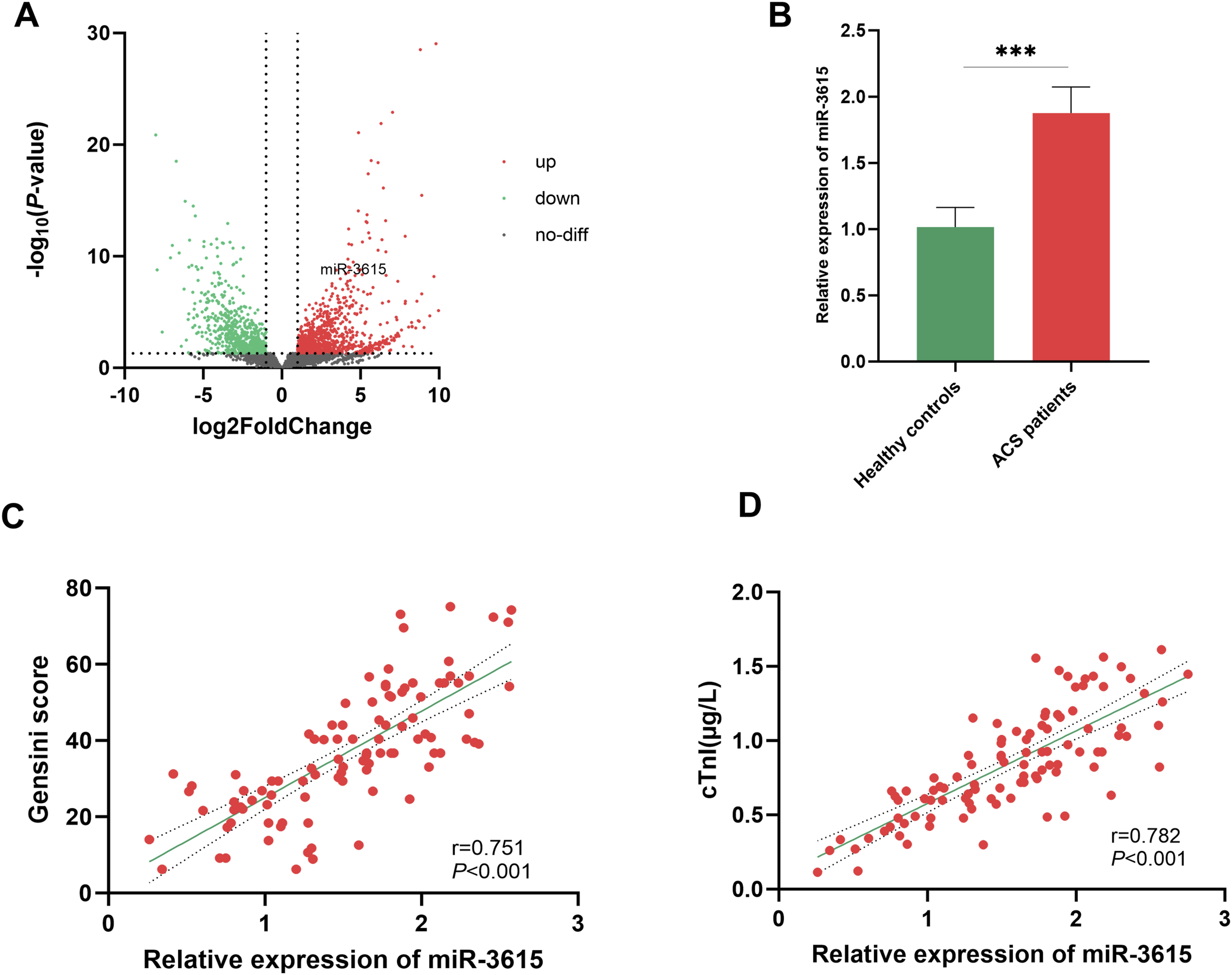
Evaluation of Risk Factors for the Progression of ACS
Comparison of Baseline Data Between ACS Patients and Healthy Controls
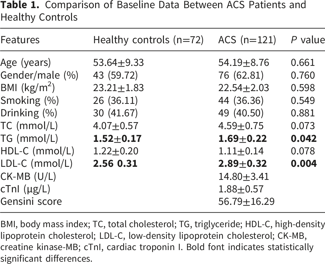
BMI, body mass index; TC, total cholesterol; TG, triglyceride; HDL-C, high-density lipoprotein cholesterol; LDL-C, low-density lipoprotein cholesterol; CK-MB, creatine kinase-MB; cTnI, cardiac troponin I. Bold font indicates statistically significant differences.
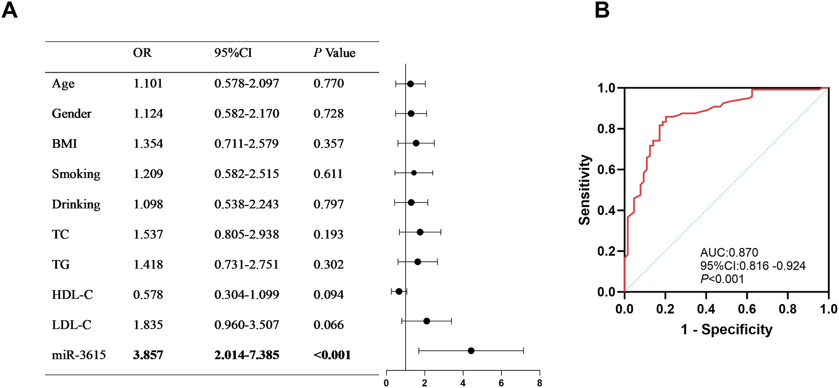
Evaluation of the diagnostic value of miR-3615 for ACS. (A) Logistic regression analysis of risk factors associated with ACS. (B) ROC curve assessing the diagnostic efficacy of miR-3615
ROC curve analysis demonstrated that miR-3615 has a strong discriminatory ability for ACS, with an AUC of 0.870 (95% CI: 0.816-0.924, P < 0.001, Figure 2B), a sensitivity of 0.933, and a specificity of 0.875.
Association Between miR-3615 and ACS Prognosis
Comparison of Clinical Characteristics Between the MACE and Non-MACE Groups
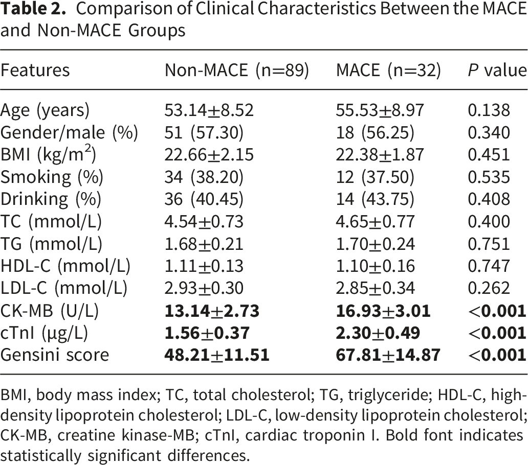
BMI, body mass index; TC, total cholesterol; TG, triglyceride; HDL-C, high-density lipoprotein cholesterol; LDL-C, low-density lipoprotein cholesterol; CK-MB, creatine kinase-MB; cTnI, cardiac troponin I. Bold font indicates statistically significant differences.
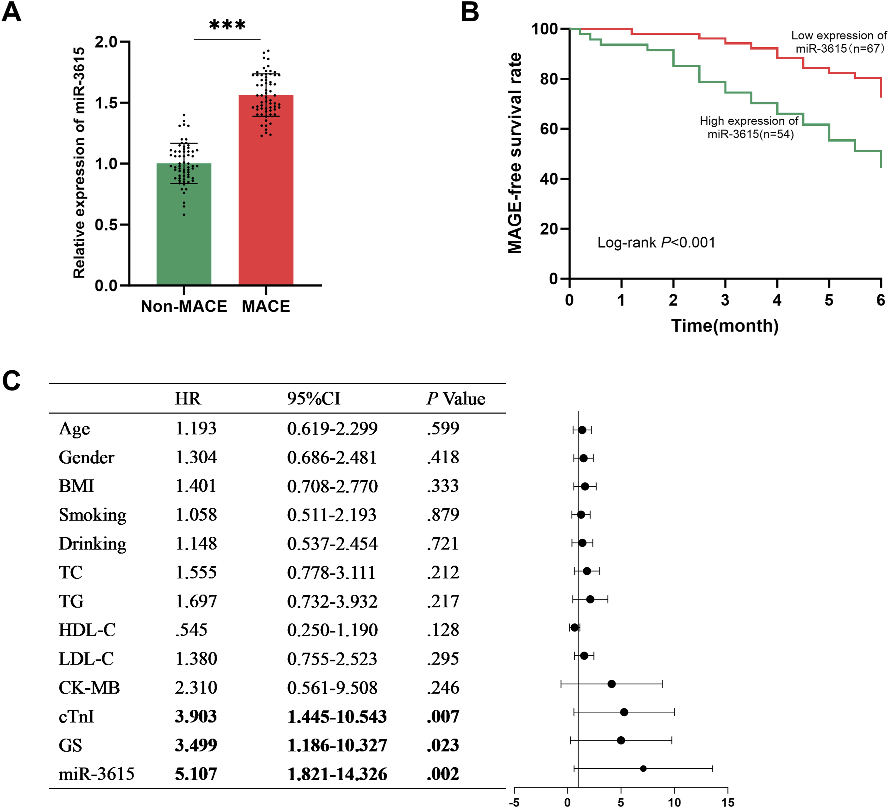
Prognostic prediction of miR-3615 in ACS. (A) Expression of miR-3615 in the non-MACE group and the MACE group. (B) Prognosis of patients with different miR-3615 levels. (C) Independent risk factors for MACE in ACS patients. (***P< 0.001)
The Effect of miR-3615 Expression on HCASMCs Induced by Ox-LDL
After treatment with ox-LDL, the level of miR-3615 in HCASMCs was significantly increased (P < 0.001, Figure 4A), accompanied by a significant enhancement in cell proliferation and migration ability (P < 0.001, Figure 4B,C), and the secretion of inflammatory cytokines (TNF-α, IL-6, and IL-1β) also increased (P < 0.001, Figure 4D). However, after inhibiting the expression of miR-3615, the above effects could be reversed. The main finding was a decrease in the level of miR-3615 (P < 0.001, Figure 4A), and the cell proliferation, migration ability (P < 0.001, Figure 4B,C), as well as the levels of inflammatory cytokines were also significantly reduced (P < 0.001, Figure 4D). Investigating the functional role of miR-3615 in ox-LDL-stimulated HCASMCs. (A) RT-qPCR was used to verify the changes in the level of miR-3615. (B) CCK-8 assay to confirm the changes in cell proliferation levels. (C) Transwell assay to verify the changes in cell migration levels. (D) ELISA verification of changes in the levels of related inflammatory factors. (***P< 0.001)
MiR-3615 Targets DPF3
By integrating the miRWalk, miRDB, and miRTarBase databases, DPF3 was demonstrated as a potential target of miR-3615 by prediction algorithms (Figure 5A). In ACS patients, the expression level of DPF3 was lower than that in healthy controls (P < 0.001, Figure 5B), and it was also lower in MACE patients compared to non-MACE patients (P < 0.001, Figure 5C). The targeting relationship was confirmed by a dual-luciferase assay: the miR-3615 mimic suppressed and its inhibitor enhanced luciferase activity specifically in the DPF3-WT group (P < 0.001), but not in the DPF3-MUT group (Figure 5D). In ox-LDL-induced HCASMCs, the level of DPF3 was significantly decreased; however, after inhibiting miR-3615, the level of DPF3 significantly increased (P < 0.001, Figure 5E), demonstrating direct regulation of DPF3 by miR-3615. DPF3 is a target gene of miR-3615. (A) Downstream target gene prediction Venn diagram. (B) The expression level of DPF3 in the serum of ACS patients. (C) The expression levels of DPF3 in the serum of MACE patients and non-MACE patients. (D) The dual-luciferase assay confirmed the interaction between miR-3615 and DPF3. (E) RT-qPCR was used to verify the changes in the expression level of DPF3. (***P< 0.001)
Discussion
ACS is the most severe type of coronary heart disease, accounting for about half of the deaths related to cardiovascular diseases. 17 Elderly people, women, and patients with chronic kidney disease are important high-risk groups for ACS. Finding accurate and clinically applicable biomarkers is the key to improving the early diagnosis and prognosis assessment of ACS. 18 MiR-3615 is involved in the occurrence process of various cancers and diseases, and has been proven to have diagnostic potential in acute heart failure. 16 This study confirmed that miR-3615 was significantly upregulated in ACS, and the trend of the analysis results in the GSE202937 dataset was consistent with this. Moreover, the level of miR-3615 was positively correlated with the Gensini score and cTnI. The Gensini score quantitatively assesses the degree and extent of vascular stenosis through coronary angiography, and can be used to explain the cause and predict the long-term prognosis of patients. 19 cTnI is a core indicator for diagnosing myocardial injury and infarction, and its elevation provides a key basis for acute myocardial infarction. 20 These indicate that miR-3615 may play an important role in ACS. Furthermore, the ROC analysis demonstrated that miR-3615 has a good diagnostic efficacy for ACS. The Logistic regression analysis further confirmed that miR-3615 is an independent risk factor for the occurrence of ACS, suggesting that miR-3615 has potential application value in the clinical diagnosis of ACS.
It is worth noting that some ACS patients remain at risk for MACE after discharge. Therefore, it is crucial to conduct effective monitoring for these patients to prevent MACE. 21 Existing studies have confirmed that miR-497-5p has predictive value for the clinical outcomes of patients with acute coronary heart disease after treatment. 11 This study shows that the expression level of miR-3615 in the serum of patients who experienced MACE was significantly increased. The K-M curve also proved that patients in the high-expression group of miR-3615 had a higher risk of MACE, suggesting that it is related to the occurrence of MACE. COX regression analysis further proved that miR-3615 is an independent prognostic factor for MACE. At the same time, the Gensini score, cTnI, and CK-MB are also independent prognostic factors for the occurrence of MACE. Among them, CK-MB, as a traditional marker for judging the time of myocardial infarction and monitoring reinfarction, 22 together with cTnI and Gensini score, are all commonly used clinical prognostic evaluation indicators.23,24 The above results suggest that miR-3615 may serve as a potential new biomarker for assessing the occurrence and prognosis of ACS, providing new theoretical references for clinical early diagnosis and risk stratification.
ACS pathological basis entails the erosion or rupture of coronary atherosclerotic plaques, leading to secondary incomplete or complete occlusive thrombosis, impaired myocardial perfusion, and decreased cardiac function, often complicated by heart failure.25,26 The treatment of HCASMCs with ox-LDL can simulate the local pathological microenvironment of atherosclerosis at the cellular level, and is used to study the biological behavior changes of vascular core cells (smooth muscle cells) under pathological stimuli related to ACS.27,28 Previous studies have investigated the effects of ox-LDL on HCASMCs, revealing the significance and biological impacts of miR-139-5p in the context of ACS. 3
The phenotypic switching of HCASMCs triggered by ox-LDL serves as a pivotal pathogenic event in atherosclerosis development. The characteristics of this transformation are accelerated proliferation, enhanced migration ability, and secretion of various inflammatory mediators. 29 These phenotypic alterations foster the formation and destabilization of atherosclerotic plaques. In the study, ox-LDL-induced stimulation of HCASMCs led to a significant increase in proliferative and migratory capacities, accompanied by upregulated secretion of inflammatory mediators. These findings collectively validate that the constructed in vitro model successfully mimics the core pathological characteristics of ACS. Notably, miR-3615 expression was significantly downregulated in this model, which aligns with clinical serum results. Knockdown of miR-3615 expression substantially abrogated the ox-LDL-elicited augmentation of HCASMC proliferation, migration, and inflammatory responses. Collectively, these results indicate that inhibiting miR-3615 may mitigate the pathological progression of ACS through regulating HCASMC phenotypic switching.
MiRNAs generally modulate disease progression by interacting with specific downstream target mRNAs. Bioinformatics-based screening in the study identified DPF3 as a potential target of miR-3615. Previous reports have demonstrated that DPF3 is an antithrombotic protein with dual anticoagulant and thrombolytic activities, capable of directly hydrolyzing fibrin, fibrinogen, and thrombi, as well as prolonging APTT and reducing fibrinogen content. 30 Another study found that DPF3 also exerts dual antithrombotic and vascular endothelial protective effects by reducing ROS production, downregulating VCAM-1 expression, and inhibiting monocyte adhesion and abnormal angiogenesis. 31 In addition, DPF3 has been identified as a novel cardiac-related transcription factor that plays a regulatory role in cardiac generation, 32 underscoring its promising therapeutic value in cardiovascular disease. This study demonstrates that DPF3 expression is downregulated in ACS. Mechanistically, miR-3615 directly interacts with DPF3 to downregulate its expression, thus highlighting the functional relevance of the miR-3615/DPF3 axis in regulating HCASMC biological activity. Collectively, these results establish the miR-3615/DPF3 pathway as a potential mechanism in ACS, thereby providing a novel direction for subsequent research.
However, this study still has certain limitations. The in vivo ACS microenvironment involves endothelial injury, macrophage infiltration, and dynamic interactions among multiple cell types, whereas the ox-LDL-induced HCASMCs model used in this study cannot fully recapitulate this complex microenvironment. Due to limitations in experimental timeline, cell culture conditions, and budget, this study did not establish a co-culture system or employ a conditioned medium model. Future research will focus on the application of co-culture systems or conditioned media to more accurately simulate multicellular interactions. In addition, the research was predominantly performed at the cellular level and remains unvalidated in in vivo models. In vivo studies are pivotal for recapitulating the complexity of the human physiological microenvironment and multifactorial interactions, which are indispensable for assessing the translational therapeutic potential of relevant targets. Previous research has also emphasized the need to incorporate animal models to fully assess the pathophysiological relevance of molecular mechanisms in ACS. 33 However, due to constraints in timeline, experimental resources, and funding, animal studies were not included in the present work. Future research will aim to construct animal models to further verify the role of miR-3615 in the occurrence and development of ACS and its clinical translational value.
Conclusion
In summary, miR-3615 is upregulated in ACS, with its expression levels positively correlating with disease severity and adverse progression. It shows significant clinical value for early diagnostic use in ACS and the risk prediction of MACE. Functionally, miR-3615 can drive the abnormal proliferation, migration, and inflammatory response of VSMCs, thus promoting the progression and destabilization of atherosclerotic plaques, and this regulatory effect is likely mediated via targeting DPF3. Collectively, miR-3615 is essential for the pathogenesis of ACS, and targeting miR-3615 may offer a promising novel therapeutic strategy for plaque stabilization and improved prognosis in ACS patients.
Supplemental Material
Supplemental material - Upregulation of Serum miRNA-3615 Serves as a Biomarker to Predict Disease Onset and Prognosis in Acute Coronary Syndrome Patients
Supplemental material for Upregulation of Serum miRNA-3615 Serves as a Biomarker to Predict Disease Onset and Prognosis in Acute Coronary Syndrome Patients by Yu Wang, WeiLing Huang and XianWei Tian in Clinical and Applied Thrombosis/Hemostasis.
Footnotes
Funding
The authors received no financial support for the research, authorship, and/or publication of this article.
Declaration of conflicting interests
The authors declared no potential conflicts of interest with respect to the research, authorship, and/or publication of this article.
Supplemental Material
Supplemental material for this article is available online.
References
Supplementary Material
Please find the following supplemental material available below.
For Open Access articles published under a Creative Commons License, all supplemental material carries the same license as the article it is associated with.
For non-Open Access articles published, all supplemental material carries a non-exclusive license, and permission requests for re-use of supplemental material or any part of supplemental material shall be sent directly to the copyright owner as specified in the copyright notice associated with the article.