



Abstract
Mupirocin (MUP) is a topical antibiotic derived from Pseudomonas fluorescens, widely used for the treatment of superficial skin infections and decolonization of methicillin-resistant Staphylococcus aureus (MRSA). Its unique mechanism of action, selective inhibition of bacterial isoleucyl-transfer RNA synthetase, confers high specificity with minimal risk of cross-resistance to other antibiotic classes. This narrative review provides an updated overview of MUP's chemical structure, mechanism of action, clinical efficacy, resistance mechanisms, and global resistance trends based on literature published from 2017 to 2024. Recent clinical studies confirm MUP's continued effectiveness, particularly in the decolonization of MRSA nasal infections. However, resistance, including high-level MUP resistance mediated by the mupA and mupB genes, is increasingly reported worldwide. A better understanding of resistance patterns and judicious use of MUP is essential to preserving its clinical utility in the context of rising antimicrobial resistance.
Introduction
Staphylococcus aureus is a prevalent component of the human microbiome, often found on mucosal surfaces, particularly in the nasal area and on the skin, in most healthy individuals.1,2 Epidemiological studies show that 20–40% of the population is asymptomatically colonized by S. aureus in the nasal mucosa. 3 Although typically a commensal organism, S. aureus is a leading cause of hospital-acquired infections, capable of causing a range of diseases from minor skin infections to severe, life-threatening conditions such as osteomyelitis, endocarditis, and sepsis due to its extensive array of virulence factors.3,4 Carriage of S. aureus significantly increases the risk of subsequent infections in hosts. 5 The rapid development of antibiotic resistance in S. aureus presents a substantial challenge in clinical treatment. 6
The emergence of multidrug-resistant S. aureus strains has further exacerbated the difficulties in antimicrobial therapy. 7 Mupirocin (MUP), a topical antibiotic derived from Pseudomonas fluorescens NCIMB 10585, is widely employed for the nasal decolonization of S. aureus. MUP acts by inhibiting bacterial protein synthesis through pseudomonic acids.8,9 It is one of the most effective topical agents for treating skin infections caused by Gram-positive bacteria, including methicillin-resistant S. aureus (MRSA).10,11 At lower concentrations, MUP exhibits bacteriostatic effects due to its reduced side-chain binding. 8 This antibiotic offers broad-spectrum activity against Gram-positive and some Gram-negative bacteria, showing notable effectiveness against skin infections caused by staphylococci and streptococci.12–14 Importantly, MUP has a minimal impact on normal skin flora, such as Propionibacterium, thereby preserving the skin's innate defense mechanisms. 13
A prior comprehensive review by Khoshnood et al. (2019) surveyed MUP's mechanism of action, resistance patterns, synergistic uses, and clinical applications. 8 The present narrative review builds on that foundation, focusing on developments since 2019. In particular, we emphasize recent epidemiological trends in MUP resistance, enhanced understanding of resistance mechanisms, and advancements in MUP formulations and combination strategies for improved efficacy. This updated perspective aims to expand upon Khoshnood et al. (2019) by highlighting new findings and innovations that have emerged in the past few years.
Structure of Drug
MUP, also known as pseudomonic acid A, is a monoxycarbolic acid antibiotic in the polyketide class. It is primarily composed of pseudomonic acid A, which makes up over 90% of its active components, with smaller amounts of pseudomonic acids B, C, and D.15,16 Isolated and characterized in 1971, MUP was developed under the trade name Bactroban and is widely used topically to treat Gram-positive bacterial infections, including MRSA.17,18
MUP has a distinct structure based on a fatty acid-derived framework consisting of a 17-carbon polyketide core known as monic acid, which features a tetrahydropyran (THP) ring with hydroxyl groups. This core is esterified to a 9-carbon tail, 9-hydroxynonanoic acid. Key functional groups in pseudomonic acid A include an epoxide group on a methylhexyl side chain, several secondary hydroxyl groups, and a terminal carboxylic acid. The molecule contains eight chiral centers with defined stereochemistry, crucial for its biological activity.19,20 The free acid form of MUP has a molecular formula of C26H44O9 and a molecular weight of approximately 500.6 Daltons. Physicochemically, MUP is a white to off-white crystalline solid, slightly soluble in water (0.026 mg/mL at pH 4), and moderately lipophilic with a log P of 2.45. 21
Synthesized by the bacterium P. fluorescens using a trans-acyltransferase Type I polyketide synthase, MUP's primary bioactive component is pseudomonic acid A (PA-A). The PA-A structure consists of a C17 monic acid polyketide with a THP core, esterified with 9-hydroxynonanoic acid (9-HN). The complete biosynthetic pathway of MUP remains complex and challenging to elucidate fully. Other pseudomonic acids, such as B and C, contain additional hydroxylation and double bond modifications. A minor component, pseudomonic acid D, features a double bond in the 9-HN segment.19,20
The biosynthetic gene cluster for MUP encodes 6 open reading frames (ORFs) for multifunctional proteins (mmpA to mmpF) and 29 additional ORFs (mupA-mupZ, 5 transacting acyl carrier proteins). 19 The key structural, pharmacokinetic, and formulation characteristics of MUP are summarized in Table 1.
Characteristics of Mupirocin
MRSA, methicillin-resistant Staphylococcus aureus; MUP, mupirocin.
Pharmacology (Pharmacokinetics and Pharmacodynamics) and Biochemistry of MUP
The available literature provides limited pharmacokinetic data on MUP. This antibiotic is applied topically to the skin or nasal passages. Following systemic administration, MUP rapidly converts into monic acid, a biologically inactive state, which is then excreted by the kidneys. 22 Importantly, no systemic absorption of either MUP or its primary metabolite, monic acid, has been observed during brief topical application periods. 23 This minimal systemic absorption, <1%, is a key factor in its U.S. Food and Drug Administration (FDA) approval, reassuring about its safety.23,24 MUP is absorbed systemically by diffusion across the stratum corneum, specifically reaching the intercellular region. 25 Nevertheless, >98% of the administered MUP does not penetrate the stratum corneum. 21
The penetration of these compounds into the deeper layers of the epidermis and dermis is significantly enhanced in cases of skin trauma and when occlusive dressings are used. 22 The elimination half-life of MUP ranges from 20 to 40 minutes, while for monic acid, it spans 30 to 80 minutes. 26 The kidneys serve as the principal pathway for the systemic excretion of MUP. 27
Adverse Effects
Topical MUP administration is generally well-tolerated. During clinical studies evaluating the efficacy of MUP cream, a small proportion of participants experienced adverse effects. Specifically, 1.5% of subjects reported sensations of burning, stinging, or pain, while 1% experienced itching. Additionally, fewer than 1% of participants reported occurrences of rash, nausea, erythema, dry skin, tenderness, edema, contact dermatitis, or increased exudate. 28 The use of conjunctival applications is contraindicated due to their potential to irritate. Minor adverse effects, such as irritation and an unusual taste, have been noted with nasal application. 12 MUP is classified under the FDA pregnancy category B. 28 However, pregnant and lactating women should exercise caution while using MUP to minimize the risk of adverse effects on the newborn. Due to its potential for absorption, MUP can be excreted into breast milk, potentially affecting the nursing infant. Additionally, the polyethylene glycol in the ointment base can be absorbed through open wounds or damaged skin, potentially causing renal toxicity. 29 Therefore, MUP ointment may not be appropriate for individuals with extensive open wounds or compromised renal function. 30
Clinical Efficacy
MUP has demonstrated notable efficacy in eliminating S. aureus (including MRSA) nasal carriage in various clinical settings. In multiple trials, short-term decolonization rates with intranasal MUP are very high, often exceeding 80–90% immediately after a 5-day course of treatment. For example, in a randomized trial in neonatal intensive care units (NICUs), Kotloff et al. reported that 93.9% of MUP-treated infants were culture negative for S. aureus at 8 days posttreatment, compared with only 4.7% in the placebo group. 31 Similarly, in an extensive presurgical decolonization program for orthopedic patients, 5 days of twice-daily nasal MUP eradicated S. aureus in 89.1% of preoperative carriers by the day of surgery. 32 There is also strong evidence that clearing colonization with MUP translates into fewer infections. A 2008 Cochrane meta-analysis of nine randomized trials (n = 3,396) found that intranasal MUP prophylaxis in known carriers led to a statistically significant 45% reduction in subsequent S. aureus infections (pooled RR = 0.55 vs. no treatment). This infection prevention benefit was most pronounced in surgical patients. 33 Furthermore, in patients with recurrent community-associated MRSA skin disease, a combined decolonization regimen including MUP achieved a dramatic >90% decrease in MRSA infection incidence (dropping from 0.84 to 0.03 infections per patient-month), underscoring the clinical impact of effective decolonization. 34
Despite high initial clearance rates, numerous studies have highlighted that long-term success is variable, and recolonization is a common occurrence. The durability of decolonization depends on follow-up duration and whether additional measures are used. In Kotloff et al.’s NICU trial, many infants became recolonized within weeks of therapy, and by 22 days after treatment, only 45.7% of treated infants remained decolonized (vs. 2.1% of controls). 31 Doebbeling et al. observed a similar decline over months in health care workers. After a single 5-day MUP course, S. aureus nasal carriage in the treated group was 48% at 6 months (vs. 72% in placebo, p = 0.05) and 53% at 12 months (vs. 76% in placebo). In other words, roughly half of the initially cleared carriers had reacquired S. aureus within 6–12 months. By 1 year, 70% of MUP-treated carriers in that study had S. aureus back in their nares, with about 36% colonized by a new strain and 34% by the original strain that had initially been cleared. 35 Short-term studies reinforce this trend: Wertheim et al. reported that among adult carriers treated with MUP, the number of colonized individuals dropped significantly (from 30 to 17 out of 62 total) 5 weeks after treatment; however, over half of the original carriers were again positive. Notably, 60% of those who remained carriers at 5 weeks harbored the same strain as before treatment, while 35% had acquired a new S. aureus strain. These findings suggest that recolonization can result from both the regrowth of the original strain (relapse) and the exogenous acquisition of new strains, especially in high-exposure environments. Indeed, MUP is highly effective at clearing nasal bacteria; however, extra nasal reservoirs (such as the throat, skin, and perineum) and ongoing contact with colonized sources can lead to the reestablishment of colonization. 36 This underscores the importance of comprehensive decolonization strategies that target multiple sites and maintain hygiene to prevent recurrence.
Approaches to improve the long-term efficacy of decolonization include repeated or adjunctive treatments. The “CLEAR” trial by Miller et al. (2023) provides evidence that a serial decolonization regimen can sustain clearance over many months. In this large post-discharge RCT (n > 2,000), MRSA carriers who underwent an initial 5-day decolonization regimen (intranasal MUP twice daily, plus chlorhexidine bathing and oral rinse) and repeated it periodically over 6–9 months had significantly lower MRSA colonization rates at all follow-up points compared with carriers who received education alone. At 1 month, the odds of any MRSA colonization in the decolonization group were less than half that of controls (odds ratio [OR] = 0.44), with a particularly pronounced effect in the nares (OR = 0.34). Importantly, this suppression of colonization persisted through 9 months of follow-up as long as the decolonization protocol was maintained. Higher adherence to the monthly regimen was correlated with a greater likelihood of remaining MRSA-negative. 37 In the NICU setting, an aggressive screening and retreatment strategy also improved outcomes: Ankrum et al. (2023) implemented weekly surveillance and repeated MUP courses for persistently colonized infants. They found that while only 35% of methicillin-susceptible Staphylococcus aureus (MSSA)-colonized neonates achieved sustained decolonization after one 5-day MUP course (defined as three consecutive negative weekly screens), offering a second course to those still positive raised the eventual clearance rate to ∼50%. This intervention significantly reduced the overall prevalence of S. aureus colonization in the NICU (e.g., MSSA prevalence fell from 18.8% to 14.4% after program implementation). However, consistent with other reports, eradication was not universal, and some patients required multiple rounds of therapy or remained carriers despite treatment. 38
The clinical outcomes associated with MUP-based decolonization are generally positive but can vary by population. As noted, prophylactic decolonization in carriers can substantially lower subsequent infection rates in many scenarios. For instance, targeted nasal MUP has been shown to reduce S. aureus surgical site infections and other invasive infections in carriers, supporting its use in preoperative protocols. In the post-hospital context, decolonization of MRSA carriers (as in the CLEAR trial) led not only to reduced colonization but also fewer MRSA infections in the ensuing year. 33 Miller et al. (2012) similarly documented that clearing colonization in patients with recurrent MRSA skin disease was associated with a marked decrease in infection recurrence. 34
On the contrary, some studies have found no statistically significant difference in infection rates despite successful decolonization. Allport et al. (2022) reported that while nasal MUP (and a comparable antibiotic, neomycin) effectively decolonized 89–91% of S. aureus carriers before joint replacement surgery, the incidence of postoperative prosthetic joint infections did not differ significantly between decolonized and non-decolonized carriers in that cohort. 32 Likewise, in the NICU study by Ankrum et al., the introduction of an MUP protocol did not significantly change the overall S. aureus infection rate, possibly due to the small number of infections observed. 38 These findings highlight that decolonization is a valuable tool for reducing the colonization burden and can prevent infections in many contexts; however, its impact may depend on factors such as concurrent infection control measures, population risk, and strain reexposure. Table 2 summarizes key studies on MUP's clinical efficacy, detailing their designs, regimens, outcome measures, and findings on decolonization success and relapse rates.
Summary of Clinical Efficacy Studies of Mupirocin for Staphylococcus aureus (Methicillin-Resistant S. aureus/Methicillin-Susceptible S. aureus) Decolonization
CI, confidence interval; CA-MRSA, community-associated methicillin-resistant Staphylococcus aureus; RCT, randomized controlled trial; RR, relative risk; MUP, mupirocin; MRSA, methicillin-resistant Staphylococcus aureus; MSSA, methicillin-susceptible Staphylococcus aureus; NICU, neonatal intensive care unit; TID, three times a day.
Mechanism of Action
MUP is a highly effective antimicrobial agent that selectively disrupts bacterial protein synthesis by binding to the bacterial isoleucyl-transfer RNA (tRNA) synthetase enzyme, which is essential for incorporating isoleucine into bacterial proteins. 10 In contrast, MUP has minimal binding affinity for the mammalian versions of this enzyme, which accounts for its low toxicity in humans. 40
The primary mechanism of action of MUP is the competitive inhibition of the bacterial enzyme isoleucyl-tRNA synthetase (IleRS). MUP's epoxy side chain is structurally similar to the natural substrate, isoleucine, allowing it to bind to the active site of IleRS with high affinity. This disrupts the enzyme's ability to charge isoleucine onto its cognate tRNA molecule, ultimately impeding protein synthesis and leading to bacterial cell death.8,13
The biosynthetic gene cluster responsible for MUP production in P. fluorescens includes two isoleucyl-tRNA synthetase genes, ileRS1 and ileRS2. Interestingly, the ileRS2 gene encodes an enzyme that is not sensitive to MUP inhibition and displays eukaryotic-like features, suggesting that this gene may play a role in protecting the bacteria from the antimicrobial action of MUP.5,13
The structural resemblance between the epoxide side chain of MUP and the amino acid isoleucine allows MUP to bind specifically to the isoleucine-binding pocket of the bacterial isoleucyl-tRNA synthetase. This binding inhibits the formation of isoleucyl-tRNA, depleting cellular levels of isoleucine-charged tRNA and halting protein and RNA synthesis in bacteria.3,5
The low affinity of MUP for the mammalian isoleucyl-tRNA synthetase enzyme is a key factor contributing to its low toxicity in humans. 8 At concentrations near the minimum inhibitory concentration (MIC) for S. aureus, MUP exhibits a bacteriostatic effect, inhibiting bacterial growth without causing cell death. However, at higher concentrations, including those in the 2% topical formulation, MUP becomes bactericidal, effectively killing the targeted bacterial cells after a day of exposure. 41
MUP Resistance
MUP resistance was detected shortly after its introduction, with S. aureus strains resistant to MUP first reported in 1987 at St. Thomas Hospital. 26 The prevalence of MUP resistance varies among clinical MRSA strains, ranging from 0% to 65%, often correlating with increased hospital use. However, this prevalence is less well-documented in pediatric patients due to limited studies.13,26 Over the past few decades, reduced usage of MUP has led to a decrease in MRSA infections in many regions worldwide. MUP decolonization, often combined with chlorhexidine, remains the most effective MRSA-specific control strategy, significantly reducing MRSA bloodstream infections. 42
Guidelines recommend a lower resistance breakpoint value (MIC) for MUP at 4 mg/L. Several factors influence this breakpoint's therapeutic outcome, including the concentrations of S. aureus isolates in the skin and nose layers, reservoirs of S. aureus, and ineffective actions. 8
MUP resistance is typically identified in laboratories using 5 µg and 200 µg disks. Resistance to the 5 µg disk but sensitivity to the 200 µg disk indicates low-level resistance, while resistance to both disks indicates high-level resistance. Increased doses of MUP can be effective against low-level resistance in vivo. Still, high-level resistance suggests treatment failure, necessitating alternatives such as chlorhexidine, neomycin, and newer agents such as retapamulin. 43
Based on antibiotic susceptibility testing, S. aureus isolates can be categorized into three MUP susceptibility groups. Isolates with an MIC of ≤4 μg/mL are considered susceptible to MUP; those with MICs of 8–64 μg/mL exhibit low-level resistance, and those with an MIC of ≥512 μg/mL show high-level MUP resistance (HLMR). The MIC range of 128–256 μg/mL is uncommon among S. aureus isolates and usually falls under low-level resistance. 8 Disk diffusion using 5 µg and 200 µg disks effectively distinguishes isolates with low or high MUP resistance. 44
Mechanisms of Resistance
MUP resistance in S. aureus emerged soon after the drug's introduction and occurs via two principal routes. The first is low-level resistance caused by point mutations in the chromosomal ileS gene encoding IleRS. Such mutations (e.g., V588F or V631F substitutions in IleRS) alter the target site and reduce MUP binding affinity, thereby allowing protein synthesis to continue in the presence of the drug. 10 The second route involves high-level resistance, which entails the acquisition of an alternate, drug-insensitive IleRS enzyme. This is usually mediated by the plasmid-borne mupA gene (also known as ileS2) and less frequently by the analogous mupB gene, both of which encode isoleucyl-tRNA synthetases that are not inhibited by MUP. Isolates carrying mupA or mupB can effectively bypass MUP's mechanism, resulting in markedly elevated MICs.5,13,45 These fundamental resistance mechanisms have been well-documented in earlier studies and were detailed in the review by Khoshnood et al. (2019). 8 In addition, S. aureus can employ auxiliary strategies such as the upregulation of efflux pumps (e.g., MdeA), which expel MUP from the cell and confer a lower level of resistance. 13 Collectively, these mechanisms underscore the bacterium's ability to evade MUP's action through either target modification or drug replacement strategies. Figure 1 illustrates the mechanisms of resistance to MUP.
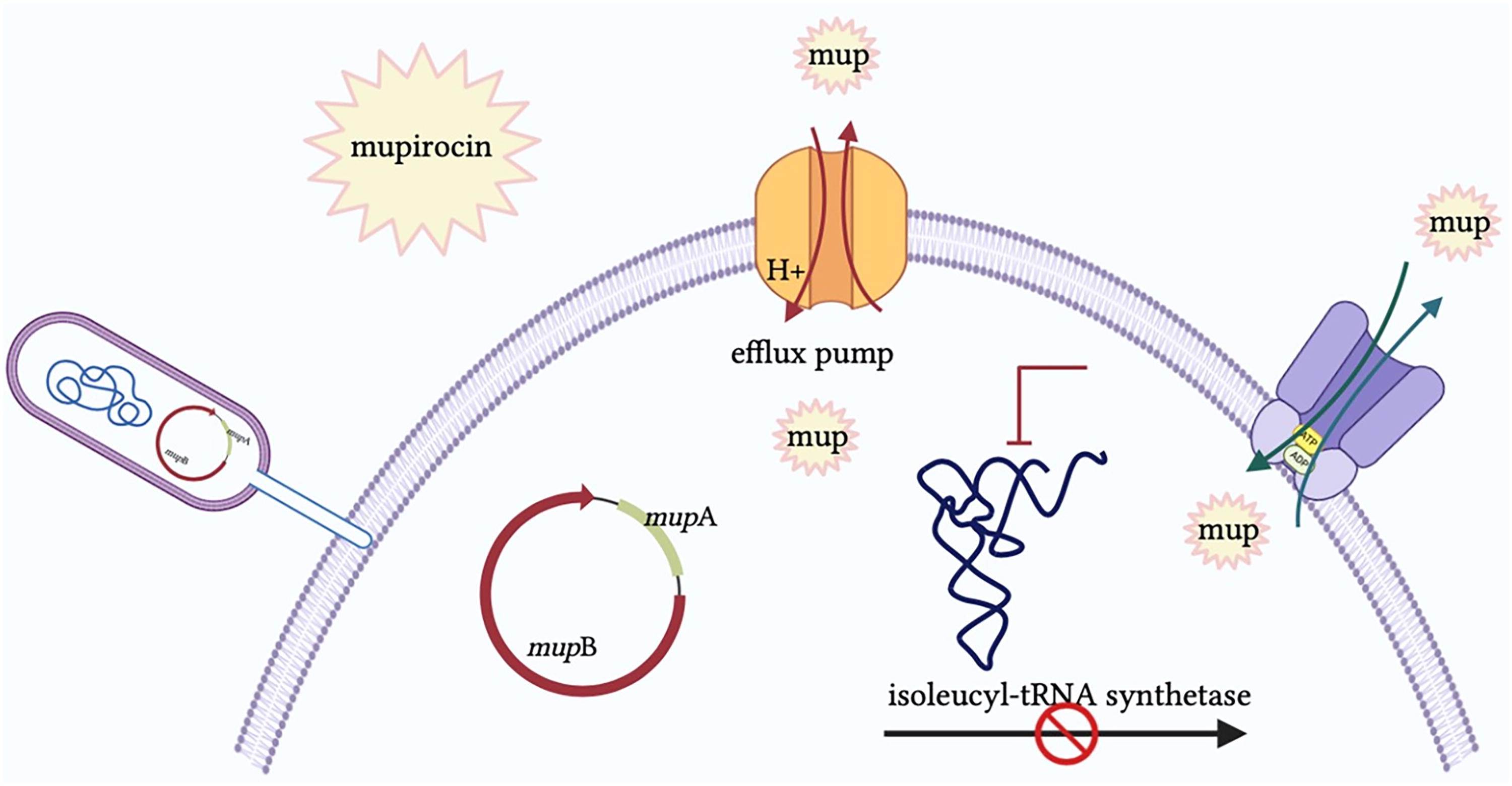
Mechanisms of MUP resistance. 46
Epidemiology of Resistance
To provide an up-to-date overview of MUP resistance patterns since the most recent comprehensive review by Khoshnood et al. (2019), we performed a structured literature search covering the period from 2017 to 2024. The search was conducted using the PubMed, Scopus, and Google Scholar databases, applying combinations of relevant keywords such as “mupirocin resistance,” “methicillin-resistant Staphylococcus aureus (MRSA),” “Staphylococcus aureus,” and “epidemiology.” Original research articles published in English that reported MUP resistance rates, molecular mechanisms, or susceptibility testing results were included. Studies were excluded if they focused solely on clinical treatment outcomes without microbiological data or if they lacked a defined sample size. Particular emphasis was placed on incorporating geographically diverse studies to assess global trends in resistance prevalence and the distribution of resistance determinants, such as mupA and mupB.
Global MUP resistance patterns have shifted notably since 2017. Overall, the prevalence of MUP-resistant staphylococci has increased in many regions, although significant geographic disparities exist. Recent surveillance confirms that Asia and Africa now report some of the highest MUP resistance rates, while the Americas and Oceania have remained comparatively low.8,47 For instance, between 2017 and 2023, the proportion of MRSA isolates resistant to MUP averaged 16.5% in Asia, 15.2% in Africa, but only 2–3% in the Americas.48–50 Europe has also seen localized surges; for example, community S. aureus in Greece reached 20% high-level resistance by 2017, and a Polish study from 2014 to 2016 detected MUP resistance in only 0.8% of isolates.51,52 In contrast, Australia's national data indicate that MUP resistance remains rare (around 1–2% of MRSA isolates with high-level resistance), reflecting successful stewardship and restricted use policies. 47 Notably, North America had historically low rates (often <5%), but recent evidence suggests emerging increases: a 2023–2024 survey of U.S. hospital MRSA found 22% of isolates carried the high-level resistance gene mupA.48,53 This sharp uptick, concentrated in a new USA300 clone variant, underscores that even regions with previously minimal resistance are at risk when MUP use is widespread.
High-level versus low-level resistance patterns have also evolved. In many areas, HLMR, typically defined by MIC ≥512 μg/mL due to the plasmid-borne mupA or mupB genes, has become more prevalent relative to low-level resistance (due to point mutations in the native ileS gene). For example, in India, the reported frequency of HLMR in MRSA rose from ∼12% in 2018 to over 21% by 2023 in some centers.43,44,54–59 Several countries reported their first detections of high-level resistant strains after 2017: Argentina documented a 2.3% HLMR rate (its first MUP-resistant MRSA cases) by 2015, and Nepal's earliest survey (2014–2015) found >50% of hospital MRSA were MUP-resistant, mostly high-level.48,60 In Europe, an alarmingly high HLMR rate of 29.6% was observed in MSSA isolates from Greece (2014–2018), suggesting heavy selective pressure even outside MRSA-focused decolonization programs. 61 Conversely, some settings have achieved declines in resistance with stewardship: one Brazilian hospital saw MUP resistance drop significantly after restricting MUP use. 62
Genetic mechanisms underlying MUP resistance have diversified slightly since 2017, although mupA remains the dominant mechanism. The mupA gene (also known as ileS2), carried on transferable plasmids, is still the primary driver of high-level resistance worldwide. 63 The mupB gene, a homolog that confers high-level resistance, has been reported infrequently but continues to emerge sporadically. For example, a novel mupB-positive MRSA isolate was identified in a 2017 study in Nepal. 60 In 2023, an Egyptian hospital identified mupB in 18% of coagulase-negative staphylococci isolates, alongside mupA in 13%, suggesting that non-aureus staphylococci may serve as reservoirs for MUP resistance genes. 50 Low-level resistance (MIC 8–256 μg/mL) due to chromosomal mutations in ileS continues to be observed, often accompanying high-level strains or in isolates lacking plasmid genes. Importantly, recent studies have shown that MUP resistance is increasingly associated with multidrug-resistant strains. 63 HLMR tends to co-occur in MRSA lineages that are already resistant to multiple antibiotics, compounding treatment challenges. Furthermore, the spread of MUP resistance genes into community-associated clones (e.g., MRSA ST8/USA300 in the United States) and into MSSA populations (as seen in Greece) is a new epidemiological concern in the post-2017 era.53,61
Table 3 provides a geographically diverse summary of MUP resistance epidemiology from 2017 to 2024, highlighting regional differences and the number of isolates examined in representative studies. Notably, high-level resistance rates are rising in several Asian and African countries while remaining low in regions such as North America and Oceania, except in specific high-use settings. The table also details the presence of mupA/mupB genes and MIC ranges, underscoring how high-level resistance has become widespread in specific locales. These diverse data highlight the importance of ongoing global surveillance and prudent use of MUP to mitigate the spread of resistance.
Epidemiology of Mupirocin Resistance in Staphylococcus aureus (2017–2024)
Low-level resistance generally corresponds to MUP MIC in the 8–256 μg/mL range, while high-level resistance corresponds to MIC ≥512 μg/mL (often >1,024 μg/mL). Where a dash (–) is shown, data were not reported in the study.
CoNS, coagulase-negative staphylococci; H, high-level resistant; L, low-level resistant; MIC, minimum inhibitory concentration; MUP, mupirocin; MRSA, methicillin-resistant S. aureus; MSSA, methicillin-susceptible S. aureus; MUP, mupirocin; NR, not reported.
Recent Advances in MUP Research
Synergistic combinations and antibiofilm strategies
Facing rising resistance, researchers have investigated combination therapies to potentiate MUP's activity. One promising approach is pairing MUP with antimicrobial peptides or natural products that have complementary effects. Melittin, a honeybee venom peptide, has shown synergistic activity with MUP. In vitro studies have demonstrated that melittin reduces the effective concentration of MUP required to inhibit MRSA, with a fractional inhibitory concentration index of ∼0.75, indicating partial synergy. The MUP–melittin combination not only enhances bacterial growth inhibition but also suppresses staphylococcal biofilm formation, in part by downregulating biofilm-associated genes such as hld. 82
Similarly, plant-derived essential oils and their constituents have exhibited synergistic effects with MUP. For example, the monoterpene α-pinene (from rosemary oil) in combination with MUP produced a marked increase in anti-MRSA efficacy. In a recent study, the combination of MUP and α-pinene demonstrated significantly improved antibacterial and antibiofilm performance, reducing the MUP minimum bactericidal concentration by 33-fold. It cleared MRSA infections in a mouse wound model more effectively than MUP alone. 83 Additionally, a formulated cream combining MUP with specific essential oils was able to eradicate S. aureus biofilms in vitro, highlighting that phytochemicals can synergize with MUP to overcome biofilm-mediated resistance. 84 These findings suggest that strategic combinations, whether with peptides such as melittin or bioactive plant oils, can amplify MUP's bactericidal and antibiofilm effects, offering potential new avenues for MRSA decolonization therapy.
Innovative formulations (nanocarriers and sprays)
Parallel to combination strategies, significant progress has been made in developing novel MUP delivery systems. The goal of these formulations is to enhance drug stability, penetration, and activity at infection sites while mitigating the development of resistance. Nanocarriers, in particular, have shown promise. MUP-loaded nanoparticles (e.g., silver nanoparticles functionalized with antibiofilm agents) have demonstrated improved antimicrobial efficacy against S. aureus compared with conventional ointment forms. In one study, MUP adsorbed on silver nanoparticles (synthesized with tannic acid or other reducing agents) achieved high drug entrapment and sustained release, and the optimized nanoformulation exhibited superior synergistic activity against planktonic MRSA when compared with MUP alone. 85 Such nano delivery systems not only directly inhibit bacteria but also disrupt biofilms through the incorporated antibiofilm compounds, addressing a key challenge in chronic wound infections. 41
Another cutting-edge approach is the development of MUP topical sprays. A film-forming MUP spray has been formulated using polymeric agents that create a thin adhesive layer over the wound or nasal mucosa. This spray demonstrated remarkable activity in preclinical tests, maintaining drug stability for up to 1 year and, upon application, forming a uniform film that ensured close contact with the infected surface. Consequently, the MUP spray achieved significantly enhanced antimicrobial effects (up to 18-fold greater killing of certain bacteria in one model) compared with standard MUP ointment. Notably, the spray formulation was shown to prevent in vitro biofilm formation and even to disrupt established biofilms on wound surfaces, thereby markedly improving MUP's antibiofilm efficacy. Other nanoformulations, such as nanostructured lipid carriers and liposomal gels, are also under exploration for MUP delivery; these have demonstrated higher drug loading capacity, controlled release profiles, and good biocompatibility in preliminary studies. 41 Together, these recent advances in formulation technology, from nanoparticle systems to novel topical delivery methods, are expanding the therapeutic toolkit for MUP. By enhancing drug localization and activity at infection sites and by integrating antibiofilm properties, such innovations aim to maintain MUP's clinical utility against MRSA in the face of emerging resistance.
Conclusions
MUP remains an important topical antimicrobial agent, particularly in the treatment of localized skin infections and nasal decolonization of MRSA. Its unique mechanism and favorable safety profile make it a valuable component of infection control protocols. However, the emergence and global spread of HLMR highlight the need for continuous surveillance, stewardship, and the development of alternative decolonization strategies. This review underscores the importance of updated epidemiological data and reinforces the role of MUP in the ongoing fight against antibiotic-resistant pathogens, provided it is used appropriately and supported by resistance monitoring programs.
Footnotes
Authors’ Contributions
Y.S.: Conceptualization and writing—original draft. K.J.: Writing—original draft. M.H. and A.M.: Conceptualization, writing—original draft, and writing—review and editing.
Confirmation Statement
Each author confirms that their research is supported by an institution that is primarily involved in education or research.
Disclosure Statement
No competing financial interests exist.
Funding Information
The authors would like to express their gratitude to the Research and Technology Vice-Chancellor of Babol University of Medical Sciences. This study was financially supported by Babol University of Medical Sciences, Babol, Iran (Grant No. 724136095). Also, this study was approved by the ethics committee of Babol University of Medical Sciences (Approval No. IR.MUBABOL.HRI.REC.1403.235).