
Abstract
Purpose:
To investigate the protective effect of Qiming granules on diabetic retinopathy (DR).
Methods:
The diabetic model was established by intraperitoneal injection of streptozotocin. Histopathological alterations in the rat retina were examined using hematoxylin and eosin techniques. The TUNEL assay was utilized to identify apoptosis in retinal cells. Western blotting was used to investigate the expression of tight junction proteins and the phosphatidylinositol 3-kinase (PI3K/Protein Kinase B (Akt) signaling pathway within retinal tissues. TUNEL assay, immunofluorescence, and Western blot analysis were employed to examine cellular apoptosis, tight junction protein expression, and proteins related to the PI3K/Akt signaling pathway.
Results:
After Qiming intervention, it was alleviated compared with the model group. TUNEL staining showed an increase in the apoptosis rate of cells in retinal tissue. Western blotting analysis revealed that the Qiming group exhibited a marked upregulation in the expression levels of occludin, zonula occludens-1, p-PI3K/PI3K, and p-Akt/Akt proteins (P < 0.05). The results from the in vitro experiments indicated that the rate of apoptosis declined following Qiming intervention (P < 0.05). There was a noted reduction in the expression levels of B-cell lymphoma-2-associated X protein (Bax)/B-cell lymphoma-2 (Bcl-2) and cleaved Caspase-3 proteins, with the decrease being statistically significant (P < 0.05).
Conclusion:
Qiming granules offered protection against damage to the blood–retinal barrier by preventing apoptosis, which in turn slowed the progression of DR. Its mode of action might be associated with modulating the PI3K/Akt signaling pathway.
Introduction
Diabetic retinopathy (DR) stands as the leading eye issue for individuals with diabetes. 1 Eye diseases related to diabetes rank as the fifth leading cause of moderate to severe vision impairment and blindness worldwide. As diabetes cases continue to rise, the occurrence of DR is also growing annually. 2 Currently, the main clinical interventions for DR include laser photocoagulation and anti-vascular endothelial growth factor (VEGF) therapies, but these approaches have inherent limitations closely linked to their inability to target the PI3K/Protein Kinase B (Akt) signaling pathway, a core regulator of retinal cell survival and blood–retinal barrier (BRB) integrity.
Laser photocoagulation primarily ablates ischemic retinal tissue to reduce VEGF overexpression, yet it fails to address the underlying molecular dysfunction of the PI3K/Akt pathway. 3 Under high glucose conditions, this pathway remains suppressed, continuing to drive retinal cell apoptosis (via upregulated B-cell lymphoma-2-associated X protein [Bax]/B-cell lymphoma-2 [Bcl-2] and cleaved Caspase-3) and BRB breakdown (via downregulated occludin and zonula occludens-1 [ZO-1]) even after laser treatment. This explains why laser therapy cannot prevent progressive retinal neurodegeneration or recurrent vascular leakage, and it also carries risks of collateral damage to normal retinal tissue, further compromising visual function.
Anti-VEGF agents (e.g., ranibizumab) target pathological angiogenesis by neutralizing VEGF, but they do not modulate the PI3K/Akt pathway either. 4 Clinical studies have shown that approximately 30%−40% of DR patients exhibit poor responsiveness to anti-VEGF therapy, partly because the suppressed PI3K/Akt pathway persists to impair endothelial cell survival and tight junction stability—key factors for maintaining BRB integrity.5,6 Additionally, anti-VEGF therapy requires frequent intravitreal injections (every 4–8 weeks), leading to high treatment costs, poor patient compliance, and potential complications such as endophthalmitis or retinal detachment. Neither laser nor anti-VEGF therapy can reverse the PI3K/Akt-mediated retinal cell apoptosis and barrier damage, which are early and persistent pathological features of DR.
In contrast, the PI3K/Akt signaling pathway offers unique advantages as a therapeutic target for DR. As a central node regulating cell survival, it directly controls two core pathological processes of DR: (1) inhibiting retinal cell apoptosis by downregulating pro-apoptotic proteins (Bax, cleaved Caspase-3) and upregulating anti-apoptotic protein (Bcl-2); 7 (2) maintaining BRB integrity by promoting the expression of tight junction proteins (occludin, ZO-1). Preclinical studies have confirmed that activating the PI3K/Akt pathway can reduce retinal vascular permeability, inhibit pericyte loss, and alleviate retinal neurodegeneration in streptozotocin (STZ)-induced diabetic ratseffects that cannot be achieved by existing treatments. 8
Qiming granules, a traditional Chinese medicine with anti-inflammatory and antioxidative properties, has been shown to promote Akt phosphorylation in microvascular cells of diabetic ulcers.9,10 Given that DR shares similar microvascular dysfunction mechanisms, we hypothesize that Qiming granules may ameliorate DR by targeting the PI3K/Akt pathway—addressing the unmet needs of existing therapies by reversing the core molecular pathology of DR. This study thus aims to verify this hypothesis through in vivo and in vitro experiments, providing a new mechanistic basis for developing more effective DR treatments.
Materials
Animals
A group of 48 male Sprague–Dawley rats, aged 8 weeks, were procured from Beijing SPI-BE Co., Ltd. The animal permit is recorded as SYXK (Jing) 2020-0001, with the quality certification being 110324221. The experimental animal center at Zhengzhou University, which is maintained at an SPF level, was responsible for raising the rats. The environmental conditions in the animal facility were regulated to sustain a temperature range of 22–25°C, with humidity levels kept between 50% and 60%. Additionally, the lighting was set to alternate between light and dark periods, each lasting 12 h.
Cells
Adult retinal pigment epithelial cells (cell line Retinal Pigment Epithelial Cells (ARPE)-19 were obtained from Beijing Beina Biotechnology Co., Ltd.
Drugs and reagents
Qiming Granules, batch number A2110071, were acquired from Guangdong Yifang Pharmaceutical Co., Ltd.; Calcium dobesilate (H20030088) was obtained from Shanghai Zhaohui Pharmaceutical Co., Ltd. STZ (lot number 572201) was sourced from Sigma Corporation. Sodium citrate buffer (C1013), RIPA tissue/cell lysis buffer (R0010), hematoxylin stain (G1121), and eosin stain (G1121) were sourced from Beijing Solarbio Science and Technology Co., Ltd. A nitrocellulose membrane (HATF00010) was sourced from Millipore Corporation in the United States. A kit for ECL chemiluminescence (catalog number 34095) was obtained from Invitrogen, a US-based company. The TUNEL detection kit, model C1090, was obtained from Beyotime Biotechnology, located in Shanghai., Ltd. The three-color prestained protein marker (WJ102) and the PAGE gel quick preparation kit (PG213) were sourced from Shanghai Yaji Biotechnology Co., Ltd. A rabbit polyclonal antibody targeting cleaved caspase-3 (catalog number 9661S), as well as a PI3K rabbit polyclonal antibody (catalog number 4292S), a phosphorylated PI3K (p-PI3K) rabbit polyclonal antibody (catalog number 17366S), an Akt rabbit polyclonal antibody (catalog number 9272S), and a phosphorylated Akt (p-Akt) rabbit polyclonal antibody (catalog number 4060S) were sourced from CST Company, located in the United States. Rabbit polyclonal antibodies targeting B-cell lymphoma-2 (Bcl-2) (26593-1-AP), occludin (27260-1-AP), and ZO-1 (21773–1-AP) were obtained from Wuhan Sanying Biotechnology Co., Ltd. The rabbit polyclonal antibody for Bax with catalog number A0207 was obtained from Wuhan ABclonal Biotechnology Co., Ltd. The PI3K inhibitor, LY294002 (catalog number HY-10108), was acquired from MCE Company, based in the United States.
Instruments
The Tissuelyser homogenizer manufactured by Shanghai Jingxin Industrial Development Co., Ltd. The Eppendorf 5425 R tabletop centrifuge made in Germany. 3K30 refrigerated centrifuge manufactured by Heraeus, Germany. PowerPac all-purpose electrophoresis device and Mini-PROTEAN Tetra gel electrophoresis apparatus, both from Bio-Rad Corporation, USA. The eBlot L1 transfer system, produced by Nanjing GenScript Biotechnology Co., Ltd. G BOX gel documentation system, developed by SYNGENE located in the United Kingdom. The BX53 inverted optical microscope, equipped with phase contrast features, is produced by the Japanese firm Olympus. The DM2500 by Leica is a fluorescence microscope produced in Germany.
Methods
In vivo experiment
Preparation, grouping, and administration of animal models
Following a 1-week acclimatization period, the body weights of 48 male Sprague–Dawley rats, each 8 weeks old, were measured. A selection of 12 rats was randomly made to form the control group, and a separate set of 36 rats was chosen to constitute the model group. Following a 12-hour fasting period, the diabetic model was established by a single intraperitoneal injection of 60 mg/kg STZ solution. Random blood glucose levels were measured three times; the model was considered successful if blood glucose was ≥16.7 mmol/L in all three measurements within 72 h after STZ injection, with a modeling success rate of 86.1%. 11 In this research, 5 of the 36 rats were not included in the modeling process: four rats did not meet the required blood glucose levels, and one unfortunately passed away early as a result of severe hypoglycemia and an associated infection. The remaining 31 rats were sorted into different groups according to their weight. The exclusion protocol complied with ethical guidelines for animal experimentation and maintained statistical integrity.
Randomization procedure: Upon effective modeling (blood glucose levels ≥16). With three consecutive measurements of 7 mmol/L, a total of 31 diabetic rats were allocated into groups through a computer-generated random number table, ensuring that stratification by body weight was maintained to achieve balanced group distribution. Preparing Qiming granules: the dosage of 1.25 g/kg per dose is derived from converting the equivalent dose from rats to humans, using a conversion ratio of 1:6.25). Stock solution: 125 mg/mL prepared by dissolving Qiming Granules in 0.5% sodium hydroxymethyl cellulose (Sigma, C5678), filtered through 0.22 μm syringe filter, and stored at 4°C for ≤24 h. Calcium Dobesilate Preparation Solvent: 0.9% NaCl solution (sterile), prepared fresh daily. Dosage confirmation: 135 mg/kg aligns with previous DR studies.
After assessing the 31 effectively modeled rats, they were randomly assigned into three groups: the model group (n = 11), the Qiming group (n = 10), and the calcium dobesilate group (n = 10). The Qiming group’s dosage was determined by referencing findings from the group’s earlier research. The rats received an oral dose of 1.25 g/kg Qiming per day, while calcium dobesilate was given through intragastric administration at a dosage of 135 mg/kg/day for a continuous period of 12 weeks. The medications were mixed into a solution of sodium hydroxymethyl cellulose.
This study adheres to the Association for Research in Vision and Ophthalmology Statement for the Use of Animals in Ophthalmical and Vision Research as required by JOPT.
Observation of retinal histopathology
Hematoxylin and eosin (H&E) staining: Following the final treatment, the rats were euthanized, their eyeballs excised and preserved in 4% paraformaldehyde solution. Following paraffin embedding, the sections were stained using H&E and examined under a microscope for any pathological alterations.
Detection of retinal cell apoptosis by TUNEL staining
Following the removal of wax and dehydration of the tissue sections, apply proteinase K and incubate at 37°C for 20 min. Rinsed with PBS thrice, leaving each rinse for a duration of 10 min. Applied 50 μL of the TUNEL detection mixture and place it in a dark setting at 37°C for an hour. Rinsed three times with PBS, allowing 10 min for each rinse. After sealing with an antifluorescence quenching mounting solution containing DAPI, observed and took pictures under a microscope.
Detection of bax, bcl-2, cleaved caspase-3, occludin, ZO-1, p-PI3K, PI3K, p-akt, and akt protein expression in retinal tissue by Western blotting
Collected the retinal tissues from each group and applied a lysis solution to extract proteins. Protein samples were subjected to SDS-PAGE prior to being moved onto a PVDF membrane for further analysis. Incubated with 5% milk solution and agitated using a shaker at ambient temperature for a duration of 1 hour. Once the membrane had been washed three times with TBST, introduce the primary antibody dilution solution. Allowed it to incubate overnight on a shaker at a temperature of 4°C. Acquired the main antibody. Followed the TBST wash, introduced the secondary antibody and let it incubate for an hour. Eliminated the second antibody. Once the membrane had been washed using TBST, proceed to develop with ECL reagent.
In vitro experiment
Cell culture
ARPE-19 cells were grown in a DMEM/F12 medium that included 10% fetal bovine serum, with the addition of 100 μg/mL streptomycin and 100 μg/mL penicillin. The incubation environment was maintained at 37°C with 5% CO2.
Detection of ARPE-19 cell apoptosis by TUNEL staining
During the logarithmic growth phase, introduced ARPE-19 cells into a 96-well plate at a density of 1 × 10⁵ cells per well, allowing them to incubate for a 24-hour period. Set up control group, hyperosmotic group, high glucose group, Qiming group, and calcium dobesilate group. The control group was grown in a setting with a concentration level of 5.5 mmol/L glucose. The hyperosmotic group was grown in a setting with a concentration level of 5.5 mmol/L glucose and 25 mmol/L mannitol. The high glucose group was grown in a setting with a concentration level of 30 mmol/L glucose. The Qiming group was cultured in medium containing 30 mmol/L glucose and Qiming (10, 50, 100, 150, 200 μg/mL) was added for continuous culture. The calcium dobesilate group was cultured in medium containing 30 mmol/L glucose, and calcium dobesilate (5, 10, 20, 40, 80 μmol/L) was added for 24 h continuous culture.
Cell Counting Kit-8 (CCK-8) assay: Cell viability was measured at 450 nm using a microplate reader (Bio-Rad), with viability calculated as (OD-treatment/OD-control) × 100%. 150 μg/mL Qiming was chosen as it showed maximal cytoprotection (viability: 82.3% ± 4.1% vs. high glucose group: 56.7% ± 3.2%, P < 0.01) with no cytotoxicity. Twenty micromole per liter calcium dobesilate was optimal (viability: 79.5% ± 3.8% vs. high glucose group, P < 0.01).
Based on the CCK-8 experiment results, the administration concentration for Qiming was found to be 150 μg/mL, while for calcium dobesilate, it was determined to be 20 μmol/L. After treating each group of cells for 24 h, the cells were rinsed once with PBS, followed by the addition of 4% paraformaldehyde to fix the cells for 30 min. Following a PBS rinse, added PBS that included 0.3% Triton X-100 solution and allowed to sit at ambient temperature for 5 min. Dispensed 50 μL of the TUNEL detection solution and kept it at 37°C in a dark environment for an hour. After rinsing with PBS thrice, applied an antifade mounting medium that included DAPI, then viewed and captured images using a fluorescence microscope.
Detection by Western blotting
Evaluation of the protein levels of Bax, Bcl-2, active caspase-3, occludin, and ZO-1 in ARPE-19 cells. Handled cells by following the procedure outlined in section “2.2.2.” Gathered cells and isolate proteins. Identified the presence of Bax, Bcl-2, and cleaved caspase-3 proteins using the procedure outlined in section “2.1.4.”
Immunofluorescence detection
Occludin and ZO-1 levels in the ARPE-19 cell line follow the procedure outlined in step “2.2.2” to process the cells. Wash using a saline solution with phosphate buffer. Apply 1 mL of 4% fixative solution for tissue cells to each well and allow it to fix at room temperature for half an hour. After rinsing with PBS, apply a 1% bovine serum albumin solution for blocking, and let it incubate at 37°C for half an hour. Remove the blocking solution. Following the PBS shaking and washing steps, introduce 200 μL of the primary antibody at a 1:500 dilution into each well. Let the plate sit on a shaker at 4°C overnight for incubation. Recover the primary antibody. Once the cells have been washed, introduce 200 μL of a fluorescent secondary antibody at a dilution of 1:200 into every well. After letting the sample sit in the dark for one hour, remove the secondary antibody. Add 200 μL of DAPI solution to each well and incubate in the dark for 15 min to stain the nucleus. Capture and photograph samples via a microscope for archival purposes.
Detection by Western blotting
Impact of the PI3K blocker LY294002 on Bax, Bcl-2, cleaved Caspase-3, and proteins within the PI3K/Akt signaling pathway. Establish a control group, a high glucose group, a Qiming group at a concentration of 150 μg/mL, and a Qiming group at 150 μg/mL combined with LY294002 at 10 μmol/L. Collect cells and extract proteins following a 24-hour period of drug intervention. Identify how related proteins are expressed using the approach outlined in section “2.1.4.”
Statistical analysis
GraphPad Prism version 9.0 was utilized for conducting statistical analysis and generating charts. The results of the experiments were presented as x ± s. A two-way ANOVA was supplemented to analyze the independent and interactive effects of two factors on ARPE-19 cell indicators. An independent samples t test was employed to assess the differences between two separate groups. Meanwhile, a one-way ANOVA was utilized to analyze the variations among several groups in vivo. P < 0.05 was considered to be statistically significant.
Results
Baseline body weight and blood glucose levels of rats in each group
Baseline data (body weight and random blood glucose) of the 31 modeled rats (model group n = 11, Qiming group n = 10, calcium dobesilate group n = 10) were analyzed. No significant differences were observed among groups (P > 0.05, Table 1), confirming the rationality of randomization.
Baseline Body Weight and Blood Glucose Levels of Rats in Each Group (x ± s)
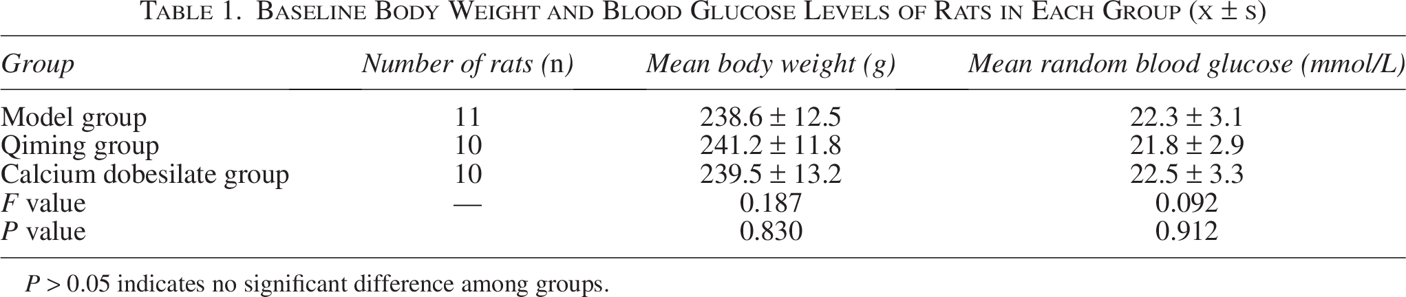
P > 0.05 indicates no significant difference among groups.
Effect of Qiming granules on pathological changes of retina in diabetic rats
The results of H&E staining are shown in Figure 1. In the control group, the retinal architecture of the rats was well-defined: the inner nuclear layer thickness was 32.5 ± 3.6 μm, the outer nuclear layer thickness was 38.2 ± 3.3 μm, and the ganglion cell density was 32.6 ± 3.8 cells/field. Within the model group, the retinal tissue in rats exhibited obvious pathological damage: retinal capillaries were dilated (diameter: 18.7 ± 2.4 μm vs. control group: 10.2 ± 1.3 μm, P < 0.05), the inner nuclear layer (19.3 ± 2.5 μm) and outer nuclear layer (24.7 ± 2.1 μm) were significantly thinned (both P < 0.05 vs. control group), and the ganglion cell density (16.5 ± 2.8 cells/field) was significantly reduced (P < 0.05 vs. control group), with disorganized cell arrangement. Compared with the model group, the pathological alterations in the Qiming group and the calcium dobesilate group were significantly improved: the inner nuclear layer thickness (Qiming group: 28.6 ± 3.2 μm; calcium dobesilate group: 27.3 ± 2.9 μm), outer nuclear layer thickness (Qiming group: 35.1 ± 2.9 μm; calcium dobesilate group: 33.8 ± 2.7 μm), and ganglion cell density (Qiming group: 28.3 ± 3.5 cells/field; calcium dobesilate group: 26.9 ± 3.2 cells/field) were all significantly increased (all P < 0.05), and the capillary diameter (Qiming group: 12.5 ± 1.8 μm; calcium dobesilate group: 13.1 ± 1.9 μm) was significantly reduced (both P < 0.05).
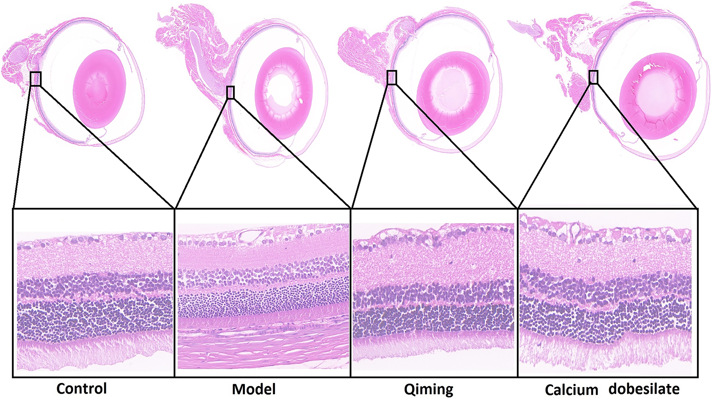
Histopathologic morphological changes in retina of rats in each group (H&E, × 400; n =12 in control group; n = 11 in model group; n = 10 in Qiming group; n = 10 in calcium dobesilate group). H&E, hematoxylin and eosin.
Effect of Qiming granules on apoptosis of retinal cells in diabetic rats
As illustrated in Figure 2, the TUNEL-positive cell rate (apoptosis rate) in the model group (27.6 ± 4.2%) was significantly higher than that in the control group (3.1 ± 1.5%) (P < 0.05), with a significant increase in red fluorescence intensity. In comparison with the model group, both the Qiming group and the calcium dobesilate group exhibited a notable reduction in apoptosis rate (Qiming group: 9.8 ± 2.3%; calcium dobesilate group: 11.5 ± 2.6%) (both P < 0.05) and red fluorescence intensity. This indicated that the administration of these drugs could significantly lower retinal cell apoptosis levels in diabetic rats.
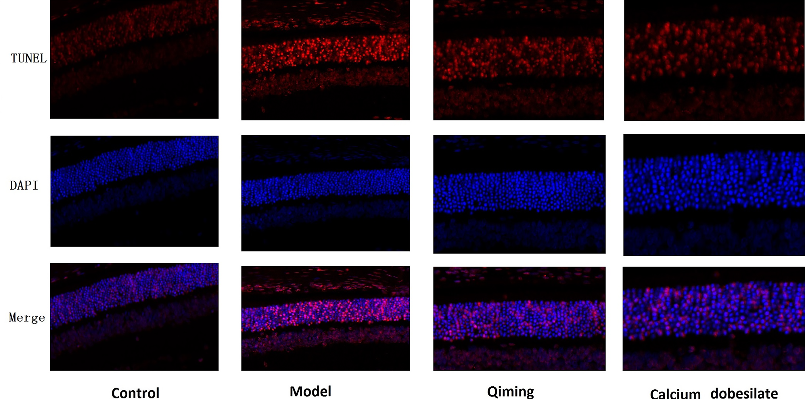
Apoptosis of retinal cells in each group of rats (×400; n =12 in control group; n = 11 in model group; n = 10 in Qiming group; n = 10 in calcium dobesilate group).
Effect of Qiming granules on the expression of apoptosis-related proteins in retinal tissue of diabetic rats
As illustrated in Figure 3A, the model group rats exhibited notably elevated expression of Bax/Bcl-2 (2.84 ± 0.43) and cleaved caspase-3 (0.91 ± 0.12) proteins in their retinal tissues when compared with the control group (Bax/Bcl-2: 0.26 ± 0.09; cleaved caspase-3: 0.39 ± 0.08) (both P < 0.05). In comparison with the model group, the Qiming group and the calcium dobesilate group exhibited a significant reduction in the expression levels of Bax/Bcl-2 (Qiming group: 0.97 ± 0.15; calcium dobesilate group: 0.68 ± 0.07) and cleaved caspase-3 (Qiming group: 0.81 ± 0.07; calcium dobesilate group: 0.57 ± 0.09) proteins (all P < 0.05), indicating that Qiming granules might effectively inhibit retinal cell apoptosis in diabetic rats.

Proteins in retinal tissues of rats in each group.
Effect of Qiming granules on the expression of occludin and ZO-1 proteins in retinal tissue of diabetic rats
As shown in Figure 3B, in comparison with the control group (occludin: 0.91 ± 0.12; ZO-1: 0.90 ± 0.17), the retinal tissue of rats in the model group exhibited a marked reduction in the expression levels of occludin (0.33 ± 0.08) and ZO-1 (0.28 ± 0.08) proteins, with statistical significance (both P < 0.05). In the Qiming and calcium dobesilate groups, the levels of occludin (Qiming group: 0.60 ± 0.09; calcium dobesilate group: 0.91 ± 0.06) and ZO-1 (Qiming group: 0.65 ± 0.11; calcium dobesilate group: 0.88 ± 0.09) proteins were notably higher compared with the model group (all P < 0.05). This implied that Qiming granules may effectively protect the retinal barrier integrity in diabetic rats.
Effect of Qiming granules on the expression of PI3K/Akt pathway-related proteins in retinal tissue of diabetic rats
As shown in Figure 3C, the levels of p-PI3K/PI3K (0.15 ± 0.02) and p-Akt/Akt (0.06 ± 0.02) in the retinal tissues of rats from the model group were substantially lower than those observed in the control group (p-PI3K/PI3K: 1.25 ± 0.08; p-Akt/Akt: 0.90 ± 0.08) (both P < 0.05). Compared with the model group, the levels of p-PI3K/PI3K (Qiming group: 0.41 ± 0.06; calcium dobesilate group: 0.82 ± 0.07) and p-Akt/Akt (Qiming group: 0.40 ± 0.08; calcium dobesilate group: 0.75 ± 0.09) in the retinal tissue of rats in the Qiming group and the calcium dobesilate group were notably elevated (all P < 0.05). This implied that Qiming granules might mitigate DR damage through the activation of the PI3K/Akt signaling pathway.
Effect of Qiming granules on apoptosis of ARPE-19 cells induced by high glucose
Two-way ANOVA analysis showed that both glucose concentration (F = 189.62, P < 0.01) and drug intervention (F = 98.35, P < 0.01) had significant independent effects on ARPE-19 cell apoptosis rate, and their interaction was also significant (F = 27.58, P < 0.01). As shown in Figure 4, exposure to high glucose levels notably elevated the apoptosis rate of ARPE-19 cells (11.4 ± 0.54%) in comparison with the control group (0.54 ± 0.02%) (P < 0.05), while a notable enhancement in red fluorescence was observed. In contrast to the high glucose group, both the Qiming group and the calcium dobesilate group showed a notable decrease in cell apoptosis rates (Qiming group: 4.35 ± 0.08%; calcium dobesilate group: 0.68 ± 0.04%) (both P < 0.05). The results indicated that Qiming granules had the ability to significantly inhibit the apoptosis of ARPE-19 cells induced by high glucose.
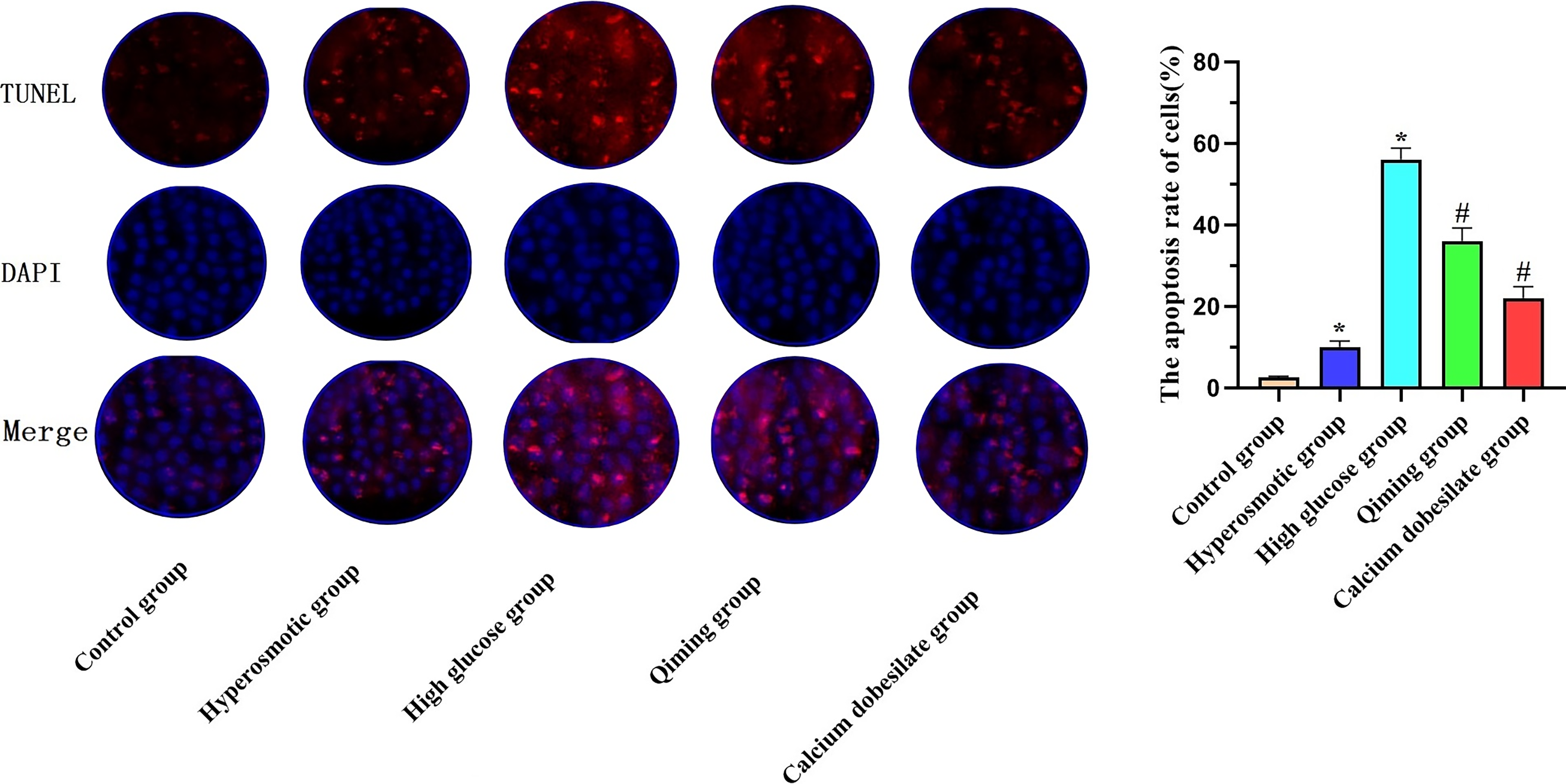
Effect of Qiming granules on apoptosis of high glucose-induced ARPE-19 cells (×200; n = 3 in each group).
Effect of Qiming granules on the expression of apoptosis-related proteins in ARPE-19 cells induced by high glucose
As shown in Figure 5, the high glucose group cells exhibited a notable increase in the levels of Bax/Bcl-2 (11.40 ± 0.54) and cleaved caspase-3 (0.79 ± 0.05) proteins when compared with the control group (Bax/Bcl-2: 0.54 ± 0.02; cleaved caspase-3: 0.03 ± 0.01) (both P < 0.05). In contrast to the high glucose group, the Qiming and calcium dobesilate groups exhibited a significant reduction in the expression levels of Bax/Bcl-2 (Qiming group: 4.35 ± 0.08; calcium dobesilate group: 0.68 ± 0.04) and cleaved caspase-3 (Qiming group: 0.31 ± 0.01; calcium dobesilate group: 0.50 ± 0.07) proteins (all P < 0.05), suggesting that Qiming granules had the potential to effectively reduce apoptosis in ARPE-19 cells, thereby mitigating retinal damage under high glucose conditions.
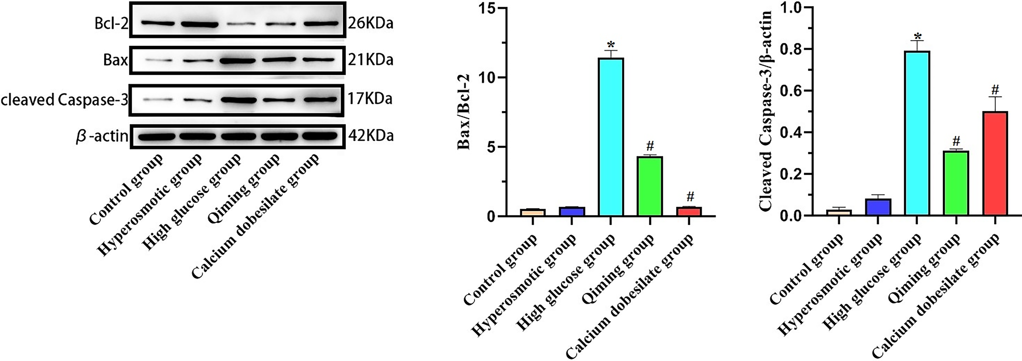
Effect of Qiming granules on apoptosis-related protein expressions in high glucose-induced ARPE-19 cells (*P < 0.05 vs. control group; #P < 0.05 vs. high glucose group; n = 3 in each group).
Effect of Qiming granules on the expression of occludin and ZO-1 in ARPE-19 cells induced by high glucose
To confirm Qiming’s protective impact on the retinal barrier, researchers employed immunofluorescence techniques to analyze how Qiming affects the expression of occludin and ZO-1 in ARPE-19 cells exposed to high glucose levels. Two-way ANOVA results for fluorescence intensity showed significant effects of glucose concentration (F = 156.47, P < 0.01), drug intervention (F = 82.63, P < 0.001), and their interaction (F = 21.39, P < 0.01). Figure 6 illustrates that, relative to the control group (occludin fluorescence intensity: 72 ± 3; ZO-1 fluorescence intensity: 49 ± 3), the cells in the high glucose group exhibit disrupted intercellular junctions, with a notable decrease in the fluorescence intensity of occludin (42 ± 3) and ZO-1 (26 ± 2) (both P < 0.05). In comparison with the high glucose group, the Qiming group and the calcium dobesilate groups exhibited a partial restoration of tight junctions within the cells and a notable enhancement in the fluorescence levels of occludin (Qiming group: 62 ± 1; calcium dobesilate group: 64 ± 3) and ZO-1 (Qiming group: 37 ± 3; calcium dobesilate group: 41 ± 2) (all P < 0.05).
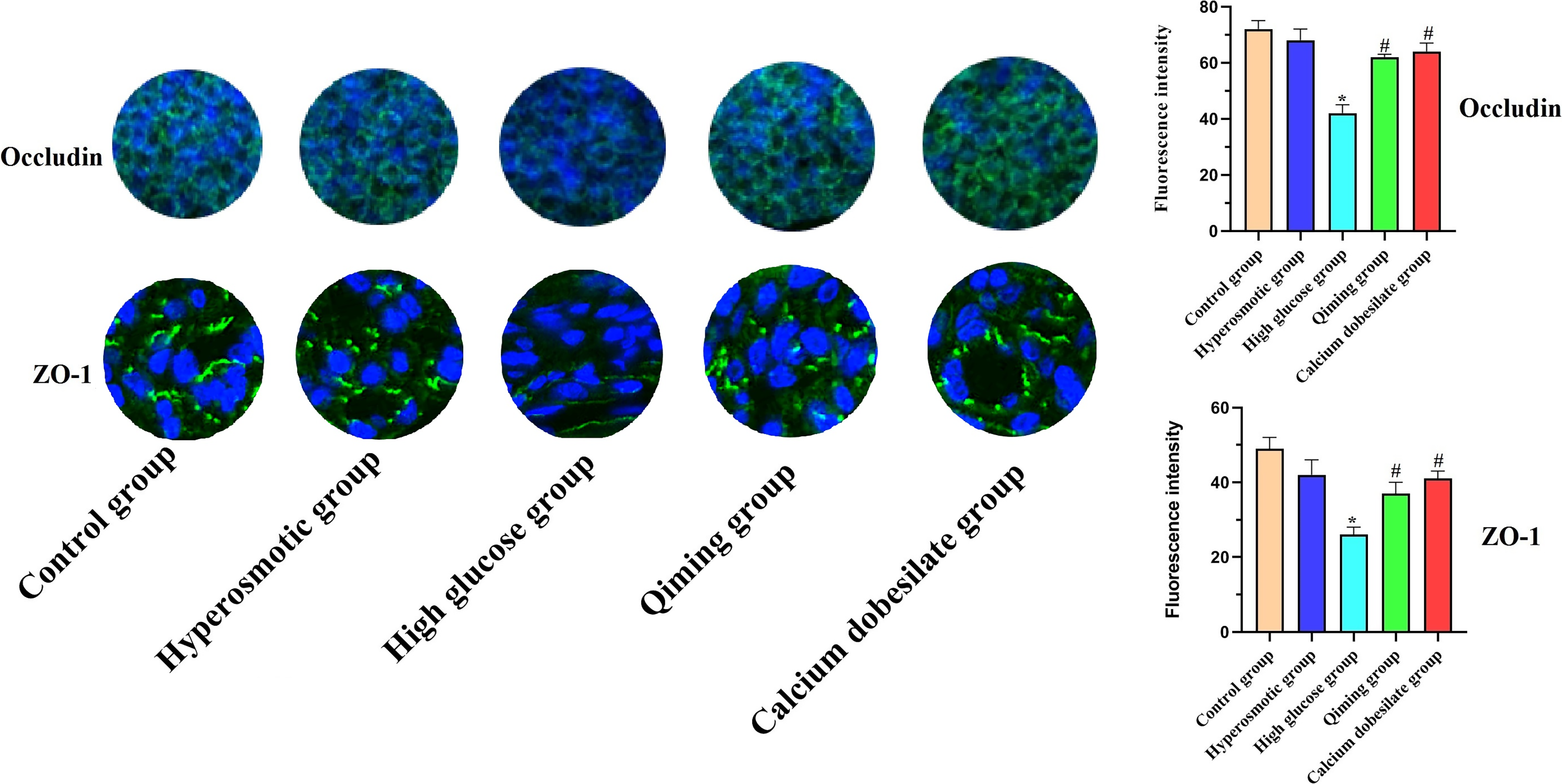
Effect of Qiming granules on expressions of occludin and ZO-1 in high glucose-induced ARPE-19 cells (*P < 0.05 vs. control group; #P < 0.05 vs. high glucose group; n = 3 in each group).
Effect of Qiming granules on the expression of tight junction-related proteins in ARPE-19 cells induced by high glucose
As illustrated in Figure 7A, Occludin (0.13 ± 0.03) and ZO-1 (0.31 ± 0.04) protein levels in the high glucose group cells were considerably lower compared with those in the control group (occludin: 0.85 ± 0.03; ZO-1: 0.98 ± 0.07) (both P < 0.05). In comparison with the high glucose group, the Qiming group and the calcium dobesilate group showed a marked increase in the levels of occludin (Qiming group: 0.19 ± 0.02; calcium dobesilate group: 0.25 ± 0.03) and ZO-1 (Qiming group: 0.43 ± 0.01; calcium dobesilate group: 0.33 ± 0.06) proteins (all P < 0.05), aligning with the findings from the immunofluorescence analysis. It indicated Qiming granules could effectively safeguard ARPE-19 cells from high glucose-induced damage by strengthening intercellular tight junctions.
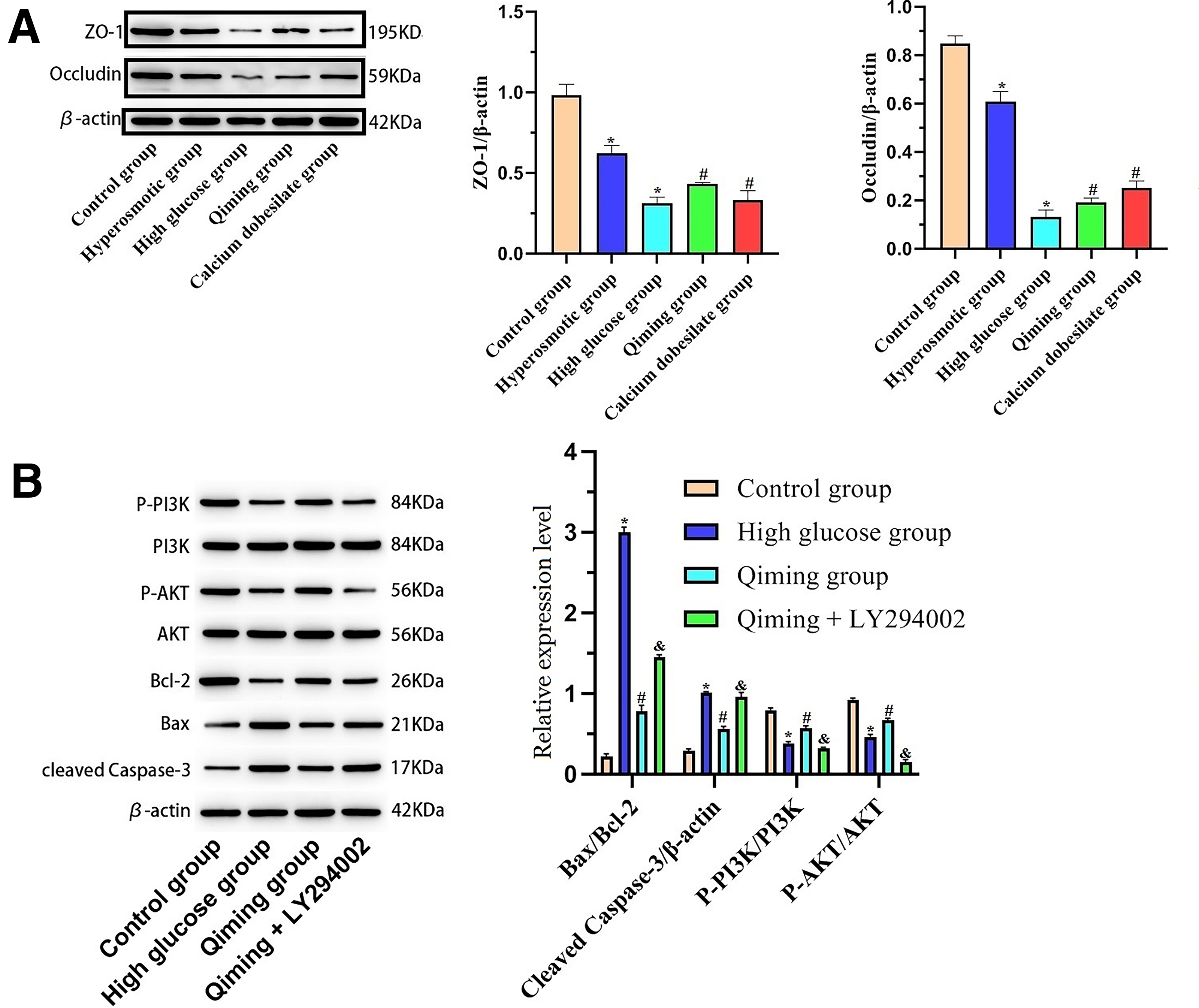
Proteins in high glucose-induced ARPE-19 cells.
Qiming granules reduce apoptosis and protect retinal barrier damage by activating the PI3K/Akt pathway
Two-way ANOVA for the p-Akt/Akt ratio showed significant effects of glucose concentration (F = 172.85, P < 0.01), drug intervention (F = 91.52, P < 0.01), and their interaction (F = 24.76, P < 0.01). As shown in Figure 7B, there was a notable reduction in the p-PI3K/PI3K (0.38 ± 0.02) and p-Akt/Akt (0.46 ± 0.03) ratios in the high glucose group when compared with the control group (p-PI3K/PI3K: 0.79 ± 0.03; p-Akt/Akt: 0.92 ± 0.02) (both P < 0.05), leading to the suppression of the PI3K/Akt signaling route. In the Qiming group, the levels of p-PI3K/PI3K (0.57 ± 0.03) and p-Akt/Akt (0.67 ± 0.02) were markedly elevated when compared to the high glucose group (both P < 0.05). Following treatment with the PI3K inhibitor LY294002, the phosphorylation of Akt was suppressed (p-Akt/Akt: 0.15 ± 0.03, P < 0.05 vs. Qiming group). The levels of Bax/Bcl-2 (1.45 ± 0.03) and the activated form of caspase-3 (0.96 ± 0.05) proteins exhibited a notable rise (both P < 0.05 vs. Qiming group). This indicated that Qiming granules might prevent apoptosis by activating the PI3K/Akt pathway, consequently minimizing damage to the retinal barrier.
Discussion
DR represents the primary microvascular complication found in individuals with diabetes, with its pathological development being intimately connected to the BRB. 12 Prolonged high blood sugar levels could harm the retinal barrier by causing the loss of pericytes in capillaries and thickening the basement membrane. This process increased vascular permeability in the retina, which eventually resulted in BRB breakdown. 13 The BRB primarily consists of an internal barrier and an external barrier. The outer barrier was chiefly formed by the retinal pigment epithelium (RPE) and its related junctions. 14 The link between cells was fundamental to the proper operation of the barrier function. Tight junctions primarily consist of proteins such as Occludin and ZO-1, among others. 15 In individuals with diabetes, Occludin was strongly associated with damage to the BRB. Elevated glucose levels could specifically decrease occludin expression, leading to enhanced BRB permeability. Studies had demonstrated that in the STZ-induced diabetic rat model, there was a 10% increase in Evans blue leakage after 8 weeks when compared with the control group. Results from the immunofluorescence study demonstrated a marked reduction in occludin levels in rats with diabetes. 16 Within the ZO group, ZO-1 was found in the zonula occludens region of both epithelial and endothelial cells. 17 In the initial phase of DR, oxidative stress along with inflammation could trigger an increase in inflammatory factors and chemokines. This process caused a reduction in ZO-1 expression and damages the retinal barrier, which subsequently led to symptoms such as bleeding. 18 This study’s findings indicated that, in comparison with the control group, there was a notable decrease in the expressions of Occludin and ZO-1 in the rats of the model group. This suggested that the tight junctions in the retinal tissue of diabetic rats were compromised, leading to damage to the BRB. In comparison with the model group, the administration of Qiming resulted in a notable increase in the expression levels of Occludin and ZO-1. This suggested that Qiming exerts a protective effect on BRB function to some degree. In in vitro studies, researchers used immunofluorescence and western blotting to examine the protein expression and tight junctions in ARPE-19 cells.
The apoptosis of retinal cells triggered by elevated glucose levels was a significant contributing factor to the pathogenesis of DR. 19 Elevated blood sugar levels could result in a buildup of reactive oxygen species (ROS) within the mitochondria, enhancing the permeability of the mitochondrial membrane. This process facilitated the release of cytochrome C from the mitochondria in the retina, subsequently activating Caspase-9. This activation then triggered a cascade of biological events that activated Caspase-3, ultimately causing cell apoptosis. 20 The PI3K/Akt signaling cascade was essential in controlling retinal cell programmed cell death. 21 Activated Akt initiated phosphorylation cascades downstream, leading to enhanced cell survival. 22 In an environment with elevated glucose levels, the PI3K/Akt signaling pathway experienced blockage, leading to an increase in the expression of the pro-apoptotic factor Bax, while the expression of the antiapoptotic factor Bcl-2 was suppressed. When caspase 3 was further activated, there was an increase in cell apoptosis. 23 The research concluded that Qiming granules could lower the protein levels of Bax/Bcl-2 and cleaved caspase-3 in the retinal tissue of diabetic rats and in ARPE-19 cells. This was also verified using TUNEL staining. This research also investigated the levels of the PI3K/Akt signaling pathway and associated apoptosis proteins in ARPE-19 cells. The findings indicated that following Qiming intervention, the protein expression levels of p-PI3K/PI3K and p-Akt/Akt were notably higher in comparison with the high glucose group. After treating with the PI3K inhibitor LY294002, Akt phosphorylation was suppressed. Meanwhile, there was an increase in the expression of downstream Bax/Bcl-2 proteins as well as in cleaved caspase-3 proteins. It was proposed that Qiming granules might prevent cell apoptosis by influencing the PI3K/Akt signaling pathway. We compared the similarities and differences between Qiming granules and other traditional Chinese medicines in the treatment of DR, highlighting their distinct targets and therapeutic pathways, shown in Table 2.
The Mechanisms of Qiming Granules and Other Representative Chinese Herbs in Treating Diabetic Retinopathy
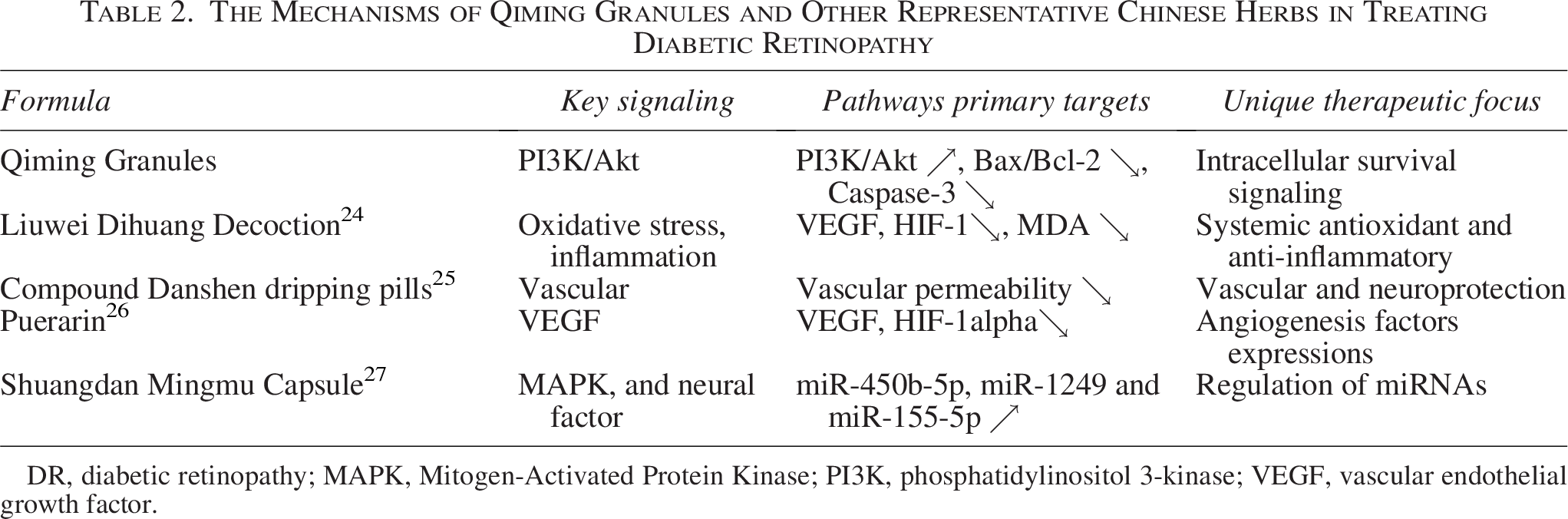
DR, diabetic retinopathy; MAPK, Mitogen-Activated Protein Kinase; PI3K, phosphatidylinositol 3-kinase; VEGF, vascular endothelial growth factor.
The findings indicated that following the Qiming treatment, the levels of occludin and ZO-1 between cells were elevated, contributing to the protection of ARPE-19 cells. As an essential element of tight junctions, ZO-1 was located at the zonula occludens in RPE cells. It played a crucial role in facilitating interactions between transmembrane proteins, such as occludin, and the actin cytoskeleton to preserve the integrity of the BRB. In DR, the oxidative stress caused by elevated glucose levels led to a decrease in ZO-1 expression. This resulted in the disruption of RPE tight junctions and an increase in BRB permeability. This research revealed that Qiming granules enhanced ZO-1 expression in both ARPE-19 cells and retinal tissues. This upregulation directly supported their contribution to maintaining the integrity of the outer BRB through modulation of the PI3K/Akt pathway, highlighting a crucial mechanistic connection in the development of DR.
The 30 mmol/L glucose concentration for the high-glucose group was determined based on clinical pathology and literature. Clinically, retinal interstitial glucose in poorly controlled diabetic patients (e.g., Glycated Hemoglobin A1C (HbA1c) ≥7.0%) ranges from 25 to 35 mmol/L, so 30 mmol/L simulates the actual retinal high-glucose microenvironment in DR. Literarily, it aligns with mainstream studies (Su et al., 2023; Liu et al., 2025)5,6 which used 30 mmol/L glucose to induce ARPE-19 cell damage (barrier disruption, apoptosis) and PI3K/Akt pathway inhibition—consistent with this study’s focus. A hyperosmotic control group (5.5 mmol/L glucose + 25 mmol/L mannitol) confirmed no osmotic interference (P > 0.05), ensuring model reliability.
This study found that Qiming granules ameliorated DR by regulating the PI3K/Akt pathway to inhibit retinal cell apoptosis and protect BRB integrity has potential applicability across DR stages, with clear correspondence to clinical progression. For nonproliferative DR (NPDR)-characterized by BRB breakdown and retinal microvascular damage—our STZ-induced diabetic rat model (showing capillary dilation, reduced occludin/ZO-1, and increased apoptosis) and high glucose-induced ARPE-19 cell model recapitulate NPDR’s core pathology; Qiming granules’ restoration of PI3K/Akt activation, tight junction proteins, and apoptosis-related proteins directly targets NPDR’s key events, potentially delaying progression. For proliferative DR (PDR)-driven by retinal ischemia/hypoxia and neovascularization—though not directly assessing neovascularization, our results (PI3K/Akt reactivation) plus prior evidence of Qiming granules reducing VEGF suggest indirect mitigation of PDR by alleviating ischemia/hypoxia (the root of PDR). Our validated models (STZ rats for NPDR, ARPE-19 cells for early DR dysfunction) align with clinical stages, strengthening translational value: Qiming granules could be tested as a disease-modifying agent for NPDR and a progression preventer for pre-PDR, supporting its promise for both early and advanced DR.
In summary, the research indicated that Qiming Granules might decrease the apoptosis of retinal cells and safeguard the integrity of the BRB by modulating the PI3K/Akt signaling pathway. This action could potentially delay the progression of DR, offering insights into the mechanism by which Qiming could be beneficial in DR treatment.
Limitations
A notable limitation of this study lies in its relatively limited mechanistic depth, particularly the lack of exploration into downstream signaling pathways of the PI3K/Akt pathway. While the findings confirm that Qiming granules activate the PI3K/Akt pathway to inhibit retinal cell apoptosis and protect BRB integrity, key downstream effectors known to mediate the pathway’s regulatory roles in DR—such as mTOR (which modulates cell survival and metabolism), Forkhead Box O3a (FoxO3a) (a transcription factor governing apoptotic gene expression), and NF-κB (a central mediator of inflammation linked to BRB disruption)-were not investigated. Elucidating whether Qiming granules further regulate these downstream molecules (e.g., via PI3K/Akt-dependent phosphorylation of mTOR or FoxO3a, or suppression of NF-κB activation) would have provided a more comprehensive understanding of the hierarchical signaling cascade underlying its therapeutic effects, and could have uncovered additional mechanistic layers that enhance the translatability of the findings to clinical DR intervention.
From a translational perspective, this study’s dosage setting, bioavailability-related findings, and long-term application potential of Qiming granules lay a foundation for its clinical translation in DR, though key details need clarification. For dosage, the 1.25 g/kg rat dose (based on 1:6.25 human–rat conversion) aligns with preclinical standards and the positive control calcium dobesilate (135 mg/kg, consistent with prior DR studies). Future clinical translation could optimize adult doses via escalation trials, considering human pharmacokinetics (e.g., gastrointestinal absorption, liver metabolism) to develop individualized regimens for patients with varying weights or blood glucose levels.
Regarding bioavailability, indirect evidence (in vitro regulation of occludin/ZO-1 and PI3K/Akt in high glucose-induced ARPE-19 cells; in vivo changes in retinal apoptosis/PI3K/Akt proteins) suggests Qiming granules’ active components penetrate the BRB. Subsequent studies could use HPLC-MS to quantify key active components’ retinal concentration and retention time in rats/nonhuman primates, clarifying BRB penetration efficiency for clinical dosing frequency.
For long-term safety, the 12-week rat study showed no retinal toxicity and reduced apoptosis, supporting short-term safety. Long-term clinical use requires more data: extend animal trials (6 months/1 year) to monitor liver/kidney function, blood routine, and retinal pathology; conduct multicenter, long-term clinical follow-ups to track adverse reactions (e.g., gastrointestinal issues, liver enzyme changes) and interactions with hypoglycemic/anti-VEGF drugs, ensuring its place in long-term DR management.
Authors’ Contributions
T.Q. designed research. L.Z. wrote the paper. Y.L. analyzed data. Z.W. performed research.
Footnotes
Author Disclosure Statement
The authors declare that they have no competing interests.
Funding Information
This study was funded by the Key Basic Research Program of Henan Colleges and Universities, with grant numbers 26A320064 and 26A360023.
Ethical Statement
This study involved the use of established human cell lines. The cell lines used in this research were obtained from Beijing Beina Biotechnology Co., Ltd. and were used in accordance with institutional and national ethical standards. The cell lines have been previously published, and no new human tissues were used in this study. This experiment and related experimental operations have been approved by the Ethics Committee for Scientific Research and Clinical Trials of the First Affiliated Hospital of Zhengzhou University (approval number 2023-KY-0050).