



Abstract
Epilepsy is a neurological condition that causes unprovoked and recurring seizures. Electroencephalography (EEG) is essential for diagnosing epilepsy, as EEG records the brain's electrical activity. However, the prevailing works are not focused on detecting the multiple seizure occurrences in one person and the Seizure Clusters (SC). So, a significant epilepsy detection framework using Meta-step Rootsig Long Short-Term Memory (MsRs-LSTM) and Fractional integro-differential Duffing Oscillator (Fid-DO) is proposed in this framework. The EEG signals are initially preprocessed and decomposed using Extrema mirror Empirical mode Decomposition (EED). Then, the signals’ peaks are detected, sub-bands are identified, and features are extracted from them. Similarly, from the preprocessed signal, Fid-DO detects weak periodicity, and the features are extracted from the signals being detected. Afterwards, the extracted features’ dimensionality is reduced and given to MsRs-LSTM for epilepsy classification. The evaluation outcomes stated the robustness of the proposed framework in epilepsy detection with 97.56% classification accuracy.
Keywords
Introduction
Epilepsy is a neurological disorder that is exemplified by recurrent seizures. 1 These seizures are due to the brain's abnormal electrical activity. 2 Epilepsy can affect people of all ages, and its seriousness can vary extensively from person to person. 3 Epilepsy can have numerous causes, including brain tumours, genetic factors, developmental disorders, brain injury, infections, and prenatal injuries. 4 In many cases, the cause may not be identified, and it is named idiopathic epilepsy. 5 There are 2 sorts of seizures namely, partial seizure, which affects the brain's particular area, and generalized seizure, which affects the entire brain. 6 The symptoms of epilepsy differ based on the seizure types, and some common symptoms are temporary confusion, loss of consciousness, sensory changes, and automatic behaviours. 7 Epilepsy is diagnosed by neurological examination through EEG.
Nevertheless, epilepsy cannot be cured in most cases. 8 Several people effectively manage their condition with treatment plans such as getting enough sleep, managing stress, avoiding triggers, and taking medications as prescribed to minimize the risk of seizures.9,10 A neurological condition that affects more than 50 million individuals worldwide, epilepsy is characterized by recurring seizures brought on by aberrant brain activity. Treatment options include anti-epileptic medications, surgery, ketogenic diets, and therapies; diagnosis is made using EEG, imaging, and medical history. Wearable technology, genetic research, and artificial intelligence advancements enhance diagnosis and treatment.
Early diagnosis and appropriate treatment are crucial for optimizing and minimizing the impact of seizures. 11 Several Machine Learning (ML) and Deep Learning (DL) methodologies like Convolutional Neural Network (CNN), Support Vector Machine (SVM), Artificial Neural Network (ANN), Recurrent Neural Network (RNN), and Random Forests (RF) are developed and employed for an early diagnosis of epilepsy.12,13 However, the existing works didn’t effectively detect the epileptic signals from the weak periodicity, 14 and all sub-bands were also not analyzed, 15 which is insufficient for epileptic detection. So, an effective epilepsy detection model using MsRs-LSTM and Fid-DO is proposed in this framework.
Problem statement
Some major challenges of the existing works are as follows, None of the existing works focused on detecting multiple seizures occurring in one person and seizure clusters. The existing work utilized the raw EEG signal for detecting epilepsy, which resulted in inaccurate results.
16
The existing work concentrated only on the alpha and delta sub-bands for epilepsy detection but failed to focus on the beta and gamma sub-bands. The existing works didn’t effectively detect the epileptic signals from the weak periodicity, which is insufficient for epileptic detection.
17
Objectives
The contributions of the proposed framework in epilepsy detection are described below, The proposed framework effectively detected multiple seizures and seizure clusters by using MsRs-LSTM. The EEG signals are preprocessed under normalization, noise removal by DE-NF, and contrast enhancement processes. This framework detects the alpha, beta, gamma, and delta sub-bands for epilepsy detection. Here, the weak periodicity is detected by using Fid-DO.
The paper's remaining organization is as follows: section 2 presents the literature survey, section 3 defines the proposed model, section 4 demonstrates the results, and section 5 winds up the paper.
Literature survey
Mitsis et al. 18 explained the epilepsy patients’ Functional Brain Networks (FBNs), exposing pronounced multiscale periodicities. The hypothesis was assessed by utilizing long-duration scalp EEG data (21–94 h.) in 9 patients with epilepsy regarding which FBNs were developed. The outcomes showcased that these networks regularly differ over time, thus exhibiting several peaks at intervals ranging from 1 to 24 h. The seizure onset effects on the FBN features were established to be significantly smaller in magnitude in contrast to the alteration that happened owing to these inherent periodic cycles. In many of these subjects, a moderately lower number of seizures was documented.
Durga Praveen Devi (2024) uses machine learning approaches to increase the precision of MRI data for brain tumour diagnosis. The method uses a multilayer perceptron for classification, local phase quantization for texture extraction, graphing for structural analysis, and K-nearest neighbours preprocessing. On a test set of 1104 photos, the model's accuracy was 87%, with precision and recall scores of 0.86 and 0.93, respectively. This demonstrates how integrated machine learning may be used to diagnose illnesses. 19
Akbarian and Erfanian 20 described a technique for seizure identification utilizing graph theory, effectual connectivity, and Multi-Level Modular Networks (MLMN). Regarding the amalgamation of numerous EBC classification outcomes at varied frequencies, the MLMN was implemented. The classification outcomes of 3 varied EBCs were combined at a particular frequency by utilizing another methodology termed Modular Effective Neural Network (MENN). Results revealed that the MLMN had the highest mean accuracy. In contrast to the other studies, which utilized the MIT-CHB database, the applied methodology offered higher accuracy. Even though the applied method had numerous benefits, the MLMN method indicated more time during the training.
Supriya et al. 21 individualized the description of SC regarding temporal clustering evaluation. For the detection of automated individualized seizure clusters, an algorithm has been developed that amalgamated cumulative sum change-point evaluation with bootstrapping as well as aberration identification, thus providing a model for personalized seizure cluster detection at the clinical significance's user-specified levels. More seizures were identified as clusters by using the applied algorithm on the basis of routine definitions, which had the better probability. The limitation was that there were errors in identifying SC that might not satisfy a pre-specified threshold (poor sensitivity).
Patient care has improved due to the adoption of IoT technology in healthcare systems. Naresh Kumar Reddy Panga (2022) investigates how an Internet of Things-based health monitoring system processes ECG signals using the Discrete Wavelet Transform (DWT). The DWT's time-frequency localization capabilities are essential for effective non-stationary signal analysis. Preprocessing, feature extraction, signal capture, and real-time IoT-based transmission to cloud servers are all included in the system design. According to performance measurements, data reduction and signal clarity have improved. 22
Zhan and Hu 23 explained the feature extraction process for the epileptic seizures’ automated detection utilizing a complex network structure. The study's objective was to implement a graph theory-centric innovative model together with a complex network system that was effectual for the EEG signals’ automated classification to identify epilepsy. The evaluation showed that for the Bern-Barcelona database as well as the Bonn University database, a better accuracy level has been obtained. The experiential outcomes revealed that the applied model was more effective in distinguishing epileptic seizure signals as of varied EEG signals. Since the graph equation approach was included, there will be a possible information loss for the reason that the criteria forming the valuable nodes could be ignored.
Using the Hadoop Distributed File System (HDFS) Hadoop architecture and Improved You Only Look Once version four (YOLO v4), a novel technique for identifying small blood cells in acute lymphoblastic leukaemia (ALL) has been created. The enhanced YOLOv4 method resolves the ALLIDB1 dataset's class imbalance problems, increasing the accuracy of healthy and blast cell identification and recognition. 24
Sukriti et al. 25 described the epilepsy identification methodology utilizing a Multiview clustering algorithm together with deep features. This study applied a classification methodology regarding unsupervised Multiview clustering outcomes to improve the effect of automatic detection. The experiential outcomes demonstrated that, in contrast to the existing EEG identification, the applied system was highly intelligent, automated, and effective in terms of ML techniques. Analyzing the brain signals posed challenges because of their high volume of data.
Najafi et al. 26 explained the localization methodology for epileptic seizure onset zones regarding time frequency and clustering exploration. A threshold methodology was utilized to identify Events of Interest (EoIs). Next, for the purpose of acquiring Channels of Interest (CoIs) by computing the average power of EoIs on every single channel, a time-frequency evaluation model was employed. The localization model's better performance was displayed by analogizing its sensitivity and specificity with the other prevailing methodologies. Relying solely on seizure semiology (observed clinical features) may not accurately determine the site of seizure onset.
Aslam et al. 27 described the automated identification of epileptic seizures utilizing multiscale and refined composite Multiscale Dispersion Entropy (MDE). MDE and Refined Composite MDE (RCMDE), the 2 entropy features being established recently, were utilized to detect seizures. The experimental outcomes illustrated that both the MDE and RCMDE were regarded as promising feature extraction methodologies since they successfully quantify the EEG signals’ complications and obtain better classification accuracies whilst c = 5 and m = 3 for both MDE and RCMDE models. Accurate seizure detection required prolonged monitoring, introducing complexities due to multiple channels and storage.
Proposed methodology for detecting epilepsy using msrs-lstm and fid-do
In this framework, the epilepsy disease is detected using MsRs-LSTM and Fid-DO. EEG signals and other sequential data are processed by the recurrent Long Short-Term Memory (LSTM) neural network. It uses sophisticated processes to increase performance, decrease overfitting, and boost convergence and learning efficiency, including meta-step gradient pruning and rootsig activation. The model categorizes EEG data using sequential patterns into normal and epileptic states. Fid-DO is a mathematical model for detecting weak periodicities in EEG signals that is based on the Duffing Oscillator and improved with fractional integro-differential equations. It assists in detecting faint periodic patterns in EEG data frequently linked to seizures that linear approaches could overlook. Fractional derivatives improve the model's sensitivity to long-term dependencies and minute changes. Here, multiple seizures and seizure clusters are also detected. The structure of the proposed framework is depicted in Figure 1. The proposed approach employs cutting-edge methods to identify seizure and epilepsy clusters in EEG recordings. Signals are preprocessed using normalization, noise reduction, and contrast enhancement. They are then broken down into smaller parts and peaks and frequency sub-bands are identified. PCA is used to extract and minimize important characteristics, while a Fractional Integro-Differential Duffing Oscillator (Fid-DO) is used to identify weak periodicities. Features of the Meta-step Rootsig Long Short-Term Memory (MsRs-LSTM) model are categorized as either normal or epileptic.
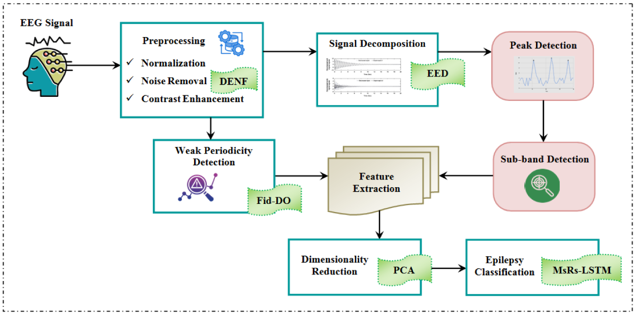
Architecture of the proposed framework.
The epilepsy detection process by the proposed framework is summarized as follows. Initially, the EEG signals are preprocessed and decomposed using EED. Then, the peaks and sub-bands are detected, followed by extracting features from them. Similarly, the Fid-DO detects weak periodicity from the preprocessed signal, and features are extracted from it. Next, the dimensions of the extracted features are reduced by PCA and given to MsRs-LSTM for epilepsy detection. Feature dimension suppression improves computational efficiency, model performance, and robustness by reducing the number of features in a dataset while maintaining important information. It reduces noise and redundancy, streamlines models, lessens the impact of the curse of dimensionality and facilitates visualization. By concentrating on key seizure-related characteristics, PCA is utilized in the suggested framework to optimize the EEG data for effective and precise epilepsy identification.
Initially, the EEG signals are collected from the dataset and the data are preprocessed to augment the epilepsy detection's efficacy. EEG signals are scaled to a standard range using normalization, guaranteeing signal consistency and comparability. For example, it modifies the EEG signal's amplitudes to fall within a specified range (0 to 1 or −1 to 1). By doing this, biases brought on by differences in signal amplitudes are removed, and algorithms operate more consistently across datasets. The utilized e number of EEG signals

The evaluation of the noise reduction stage guarantees that undesirable artefacts are eliminated while maintaining crucial characteristics, improving the quality of the EEG data. As a result, there are fewer false positives or negatives, enhanced detection accuracy, and dependable feature extraction. A key component of the suggested epilepsy detection architecture, noise reduction, is validated by SSIM, MAE, and PSNR metrics. The EEG signals are then preprocessed under normalization, noise removal, and contrast enhancement processes described below.
28

Here,

Here,

Where,

Then, a bilinear transform

Afterwards, the notch filter transfer function

Where, z denotes the complex variable in z-domain,
After that, the contrast-enhanced signal

Here,

Where,


Here, p denotes the total P number of frequency components. Then, from the envelopes, get the middle value

The above process results in the first IMF

Finally, the decomposed signal

Then, the decomposed EEG signal is further processed for detecting the peaks to determine the sub-bands of the EEG signal.
Here, the peaks of the decomposed signals
Sub-band detection
From the peak detected signal

Similarly,

Here,

Here, x denotes the displacement of the oscillator,

Next, the numerical solution for Fid-DO is obtained by discretizing this system using a numerical integration method to get the displaced x. Then, the frequency spectrum

Afterwards, weak periodicity is detected by analyzing the frequency peaks of the signals. The peak detection

The amplitudes of the detected peaks are analyzed to assess the strength of periodicity. The low amplitude indicates the weak periodicity

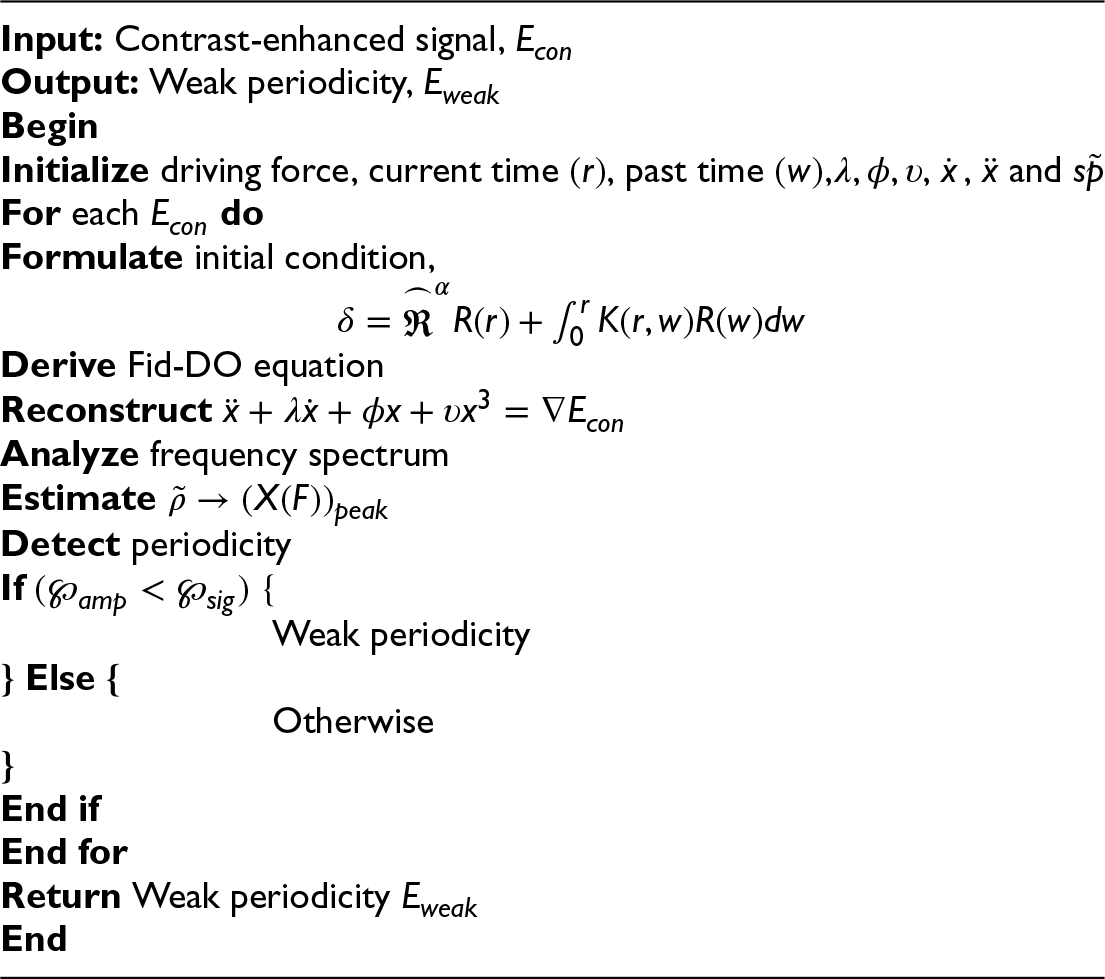
Hereafter, the features of the weak periodicity signals and sub-bands are extracted to train the epilepsy classification model.
Feature extraction is extracting important features from EEG signals, such energy, mean and standard deviation, fracture dimensions, and Lyapunov exponents, to precisely determine the underlying patterns and differentiate between epileptic and normal states. From
Dimensionality reduction
Afterwards, the extracted features’ dimensions 
Next, the mean 

After that, the covariance matrix 
Then, the Eigenvalues 
Here, 
Where, 
Here, d denotes the total number of features. Then these features are given for epilepsy detection.
Based on

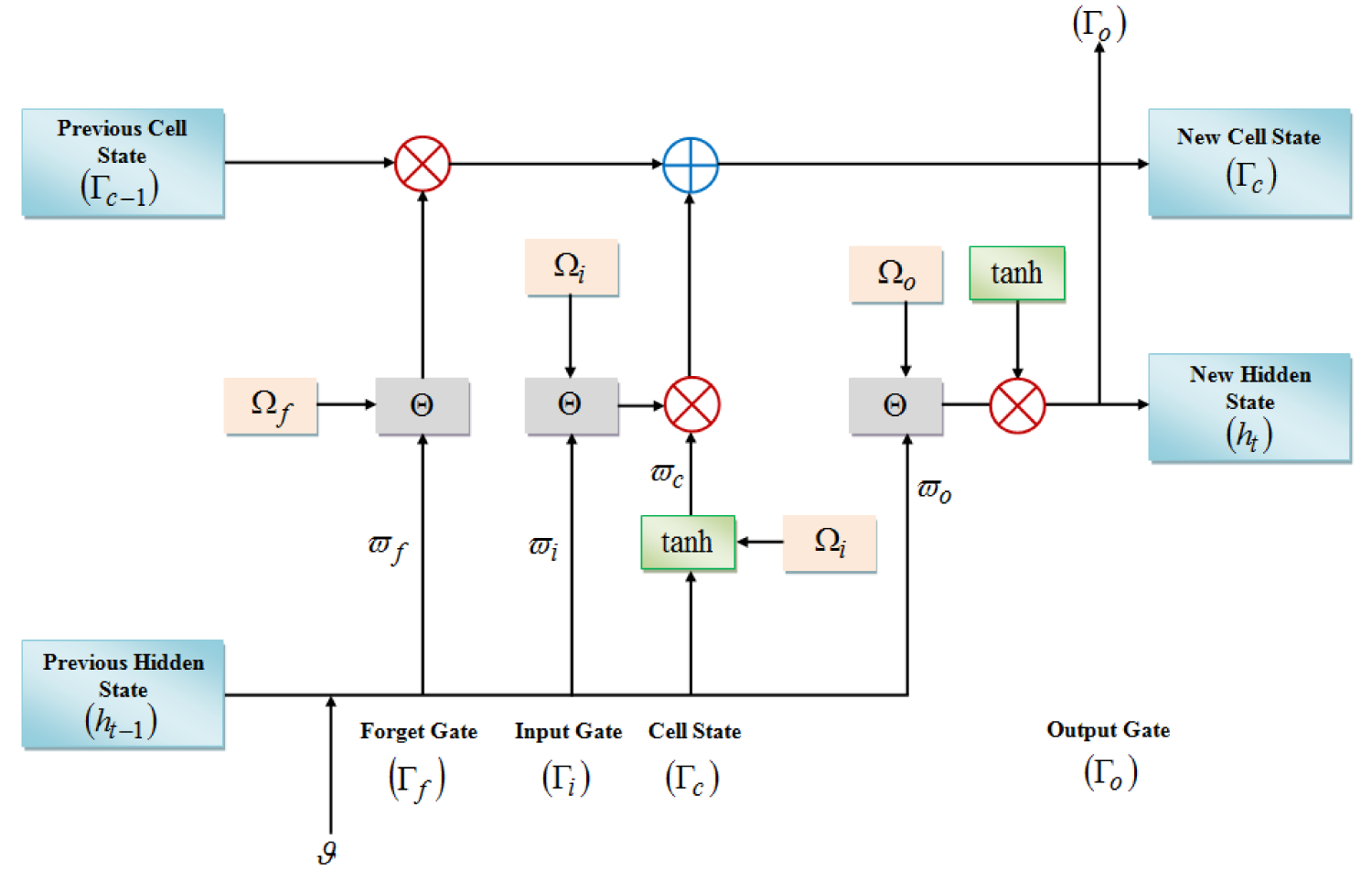
Structure of proposed MsRs-LSTM.
Here, 
Then, the meta-step gradient pruning regularization is expressed as,

Here,

Here,


Where,


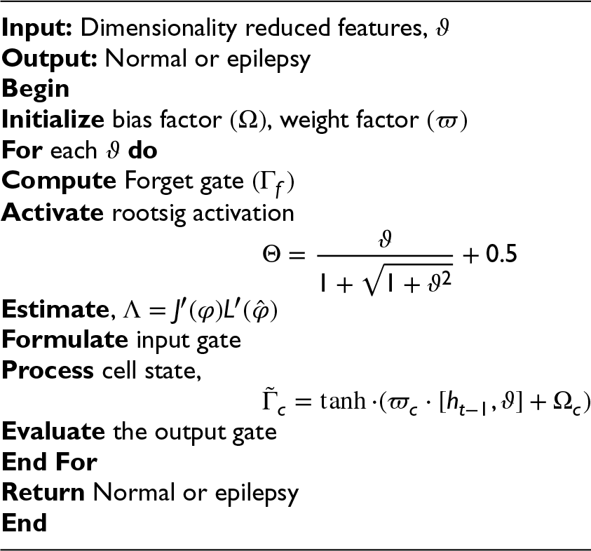
Thus, the framework effectively detected epilepsy, and the results of the proposed framework are discussed further.
This section analyzes the proposed model's performance by comparing it with the prevailing methodologies for different metrics. The proposed model is executed in the MATLAB platform with wider applicability.
Dataset description
The Children's Hospital Boston-Massachusetts Institute of Technology (CHB-MIT) Scalp EEG database analyses the proposed system's performance. This dataset comprises the collection of EEG recordings of the 22 pediatric subjects having untraceable seizures. EEG data analyses brain electrical activity to identify seizure clusters and epilepsy. The CHB-MIT Scalp EEG database confirms the suggested framework, acts as a diagnostic tool, and offers information for recognizing seizure patterns. The framework's usefulness is demonstrated by the fact that EEG makes feature extraction for machine learning possible and makes it easier to compare performance with current techniques. Overall, 182 number of seizures are annotated. From the dataset, 80% of the data is utilized for training, whereas the remaining 20% is utilized for testing the proposed model. The dataset link used for the analysis is mentioned below in the reference list. The image results attained by the proposed technique are shown below.
Table 1 shows the image outcome of the normal EEG signal and the epileptic EEG signals.
Image results.
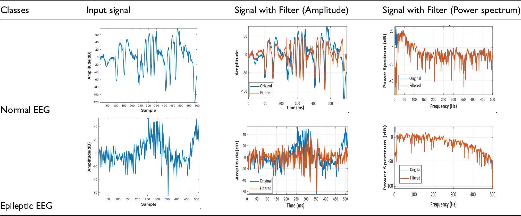
Image results.
Here, the proposed MsRs-LSTM's performance is analyzed by comparing it with the existing techniques, namely LSTM, Bi-directional LSTM (Bi-LSTM), Gated Recurrent Unit (GRU), and CNN (Figure 3).

Effectiveness evaluation for the proposed MsRs-LSTM.
This figure compares the proposed MsRs-LSTM's effectiveness based on sensitivity, specificity, accuracy, precision, and f-measure with several existing algorithms. Here, the proposed MsRs-LSTM attained 98.78%, 98.75%, 97.56%, 98.67%, and 98.68% of sensitivity, specificity, accuracy, precision, and f-measure, respectively. Thus, due to meta-step gradient pruning regularization integration, the proposed framework enhances the system's performance more than the other prevailing techniques by accurately identifying epileptic seizures while minimizing false positives and negatives.
Table 2 represents the proposed MsRs-LSTM's performance for epilepsy prediction regarding Negative Predictive Value (NPV), False Positive Rate (FPR), False Negative Rate (FNR), True Positive Rate (TPR), and True Negative Rate (TNR). The proposed model attained an NPV of 98.69%, FPR of 0.0068, FNR of 0.0056, TPR of 98.78% and TNR of 98.75%. As the introduced Rootsig activation function augments the learning efficiency of the network, the proposed network detected epilepsy with higher performance. Meanwhile, the existing LSTM attained 95.89% of NPV; Bi-LSTM attained FPR of 0.0304; GRU attained TPR of 88.44%; and CNN attained TNR of 82.44%, which are inferior to the proposed model. Thus, the proposed network's performance is better than the existing techniques.
Performance comparison for MsRs-LSTM.

This section analyses the proposed framework's performance in evaluating the proposed De-NF regarding Structural Similarity Index Measure (SSIM), Mean Square Error (MSE), Mean Absolute Percent Error (MAPE), Root Mean Square Error (RMSE), and Peak Signal-to-Noise Ratio (PSNR) (Figure 4).

Performance analysis for the proposed De-NF.
Due to the damping error integration, the proposed framework's performance increases regarding SSIM, MAE, MAPE, and RMSE. The performance is evaluated by comparing with traditional methods, namely Notch Filter (NF), Wiener Filter (WF), Gaussian Filter (GF), and Bilateral Filter (BF). This figure indicates that the proposed De-NF achieved 0.9264, 0.0034, 00046, and 0.0045 for SSIM, MAE, MAPE, and RMSE, respectively. Thus, compared to the other conventional techniques, the proposed framework performs better by providing accurate and reliable assessments of image quality and predictive accuracy.
Figure 5 showcases the proposed De-NF's performance based on validating the PSNR. This figure also indicates that the proposed De-NF has high PSNR, which enhances the system performance by minimizing distortion, ensuring superior fidelity, and maintaining high-quality visual information. Moreover, the proposed De-NF attained 65.77 PSNR, whereas the conventional NF attained 61.45 PSNR. Thus, the proposed De-NF outperforms the other prevailing techniques due to the integration of damping error. Using MsRs-LSTM and Fid-DO techniques, the proposed framework for epilepsy detection increases accuracy and reliability over current approaches. It achieves great accuracy and sensitivity by addressing noise problems in EEG signals. The sensitivity of the Damping Error Noise Filter (De-NF) is 98.8%, indicating its effectiveness. Future research should concentrate on the generalisability of datasets and sophisticated methodologies.

PSNR validation of the proposed De-NF.
Table 3 indicates the comparative evaluation of the proposed work with the prevailing works. The existing works developed models like RNN-LSTM, CNN-RNN, ANN, SVM, and deep forest classifiers for diagnosing focal and generalized epilepsy, epileptic seizure prediction, and ESD. The existing works achieved sensitivity rates like 96.8%, 93.8%, 97.5%, 95.2%, and 98% for epilepsy detection, which are considerably lower than the proposed model. Because the proposed model overcomes the vanishing gradient issue by integrating meta-step gradient pruning regularization and rootsig activation function, the proposed model's sensitivity rate is 98.8%. Hence, the comparative analysis states that the proposed methodology is a significant model for identifying epilepsy.
Comparative analysis with related works.

Using MsRs-LSTM and Fid-DO techniques, the suggested framework for epilepsy detection has demonstrated notable gains in accuracy and dependability when compared to current approaches. In addition to achieving great sensitivity and accuracy, the model successfully handles EEG signal noise. One significant addition is the thorough preprocessing of EEG data, which includes noise reduction, contrast enhancement, and normalization. High SSIM and low MAE and RMSE readings demonstrate the effectiveness of the Damping Error Noise Filter (De-NF). With a sensitivity of 98.8%, the model outperforms other models, such as RNN-LSTM (96.8%) and CNN-LSTM (93.8%). The model integrates the rootsig activation function with meta-step gradient pruning regularization. This enhancement lessens the vanishing gradient issue frequently arising in deep learning models. One important component for clinical applications in epilepsy management is the ability of the MsRs-LSTM architecture to improve the identification of multiple seizures and seizure clusters.
This work developed an effective epilepsy detection framework by detecting the presence of multiple seizures using the proposed Fid-DO and MsRs-LSTM methods. The model preprocessed the EEG signals, by which the noise was removed using De-NF with SSIM of 92.64%, MAE of 0.0034, and RMSE of 0.004. Further, the signal peaks were identified and the features were extracted. Also, the weak periodicity of the signal was analyzed using Fid-DO. Subsequently, the feature dimension was suppressed, and then the epilepsy was detected using the proposed MsRs-LSTM with 97.56% accuracy, 98.67% precision, 98.68% f-measure, and 0.0068 FPR. The implemented results exhibited the enhanced performance of the proposed model over the existing techniques. Hence, a significant detection framework for Epilepsy detection was presented by the proposed methodology.
Future work
Although multiple seizures were detected in this work, the localization of epilepsy was not analyzed. Thus, in future work, the epilepsy localization will be identified with advanced techniques to enhance the epilepsy diagnosis further.
Footnotes
Authors’ contributions
Dr Priyan Malarvizhi Kumar, Dr Wael Korani is responsible for designing the framework, analyzing the performance, validating the results, and writing the article. Tayyaba Shahwar, Dr Gokulnath C is responsible for collecting the information required for the framework, provision of software, critical review, and administering the process.
Funding
The authors received no financial support for the research, authorship, and/or publication of this article.
Declaration of conflicting interests
The authors declared no potential conflicts of interest with respect to the research, authorship, and/or publication of this article.
Data availability statement
No datasets were generated or analyzed during the current study.