
Abstract
Upon adequate stimulation, real-time maps of cortical hemodynamic responses can be obtained by functional near-infrared spectroscopy (fNIRS), which noninvasively measures changes in oxygenated and deoxygenated hemoglobin after positioning multiple sources and detectors over the human scalp. This review is aimed at giving a concise and simple overview of the basic principles of fNIRS including features, strengths, advantages, limitations, and utility for evaluating human behavior. The transportable/wireless commercially available fNIRS systems have a time resolution of 1 to 10 Hz, a depth sensitivity of about 1.5 cm, and a spatial resolution up to 1 cm. fNIRS has been found suitable for many applications on human beings, either adults or infants/children, in the field of social sciences, neuroimaging basic research, and medicine. Some examples of present and future prospects of fNIRS for assessing cerebral cortex function during human behavior in different situations (in natural and social situations) will be provided. Moreover, the most recent fNIRS studies for investigating interpersonal interactions by adopting the hyperscanning approach, which consists of the measurement of brain activity simultaneously on two or more people, will be reported.
Keywords
Introduction
The rapid advancement and availability of functional neuroimaging methodologies have transformed the field of cognitive neuroscience research. The processes of autoregulation and neurovascular coupling maintain cerebral blood flow (CBF) adequate for brain activity. The coupling between the neuronal activity and the CBF is fundamental for brain functions. In fact, when a specific brain region is activated, CBF increases in a temporally and spatially coordinated manner tightly linked to changes in neural activity through a complex sequence of coordinated events involving neurons, glia, arteries/arterioles, and signaling molecules. The detection of the increased cerebral oxygenation, secondary to cortical neuronal activation (neurovascular coupling) in response to specific stimuli, has been successfully used to create human brain maps. Those brain maps allow the visualization of stimulated areas in task-related brain “activation” studies. Usually, the CBF increase lasts over the period of the stimulus/task and then subsides when the stimulus/task is terminated. The mechanism that regulates the induced focal hyperemic responses has been recently reviewed (Filosa, Morrison, Iddings, Du, & Kim, 2015). So far, functional magnetic resonance imaging (fMRI) (Magistretti & Allaman, 2015), electroencephalography (EEG) (Michel & Murray, 2012), magnetoencephalography (MEG) (Hall, Robson, Morris, & Brookes, 2014), positron emission tomography (PET) (Magistretti & Allaman, 2015), and functional near-infrared (NIR) spectroscopy (fNIRS) (Scholkmann et al., 2014) represent the most widely used functional neuroimaging modalities. However, fMRI and fNIRS are the only functional neuroimaging modalities based on the neurovascular coupling. The discovery of EEG in 1929 could be considered as the founding of the functional neuroimaging. fMRI in its 24th year has grown largely because of its noninvasiveness, relative ease of implementation, and reasonably good spatial (1-10 mm) and temporal resolution (about 1 second). In contrast, the electrophysiological responses measured by EEG, MEG, and electrocorticography have effectually a better temporal resolution (i.e., <1 millisecond). However, with respect to EEG, MEG, and fNIRS, fMRI has the substantial advantage to resolve small and/or deep structures in the brain. Moreover, the robust fMRI signal is highly reproducible and reliable even though it is slow. fMRI overcomes the spatial resolution of high density EEG (HD-EEG) and MEG (<2 cm), the high production cost of radioisotopes for PET studies, as well as the ethical limitations for their use. Nevertheless, fMRI, as well as MEG, imply large-sized equipment and huge purchase/operating costs. In addition, fMRI and PET require the subject in a supine position that may yield altered hemodynamic changes rather than in seating or standing positions.
It has been proven that the functional activation of the human cerebral cortex can be successfully explored also by fNIRS, optical topography, NIR imaging, diffuse optical imaging (DOI), or diffuse optical tomography (DOT) (Ferrari & Quaresima, 2012; Scholkmann et al., 2014). Basically, fNIRS is a noninvasive vascular-based functional neuroimaging technology that measures, simultaneously from multiple measurement sites, concentration changes of oxygenated-hemoglobin (O2Hb) and deoxygenated-hemoglobin (HHb) at the level of the cortical microcirculation blood vessels. For the measurement of the two fNIRS parameters, the spectral features of O2Hb/HHb in the NIR range are exploited (Figure 1). Thus, fNIRS has become an important complement to the fMRI and HD EEG. The most significant advantages of fNIRS can be summarized as follows: (a) the use of low-cost, silent, and transportable/portable instrumentation (e.g., mobile EEG), making long-term monitoring and repeated measurements of cortical activities possible in various situations, including the natural ones; (b) the measurement of the changes in O2Hb and HHb, delivering additional information with respect to the fMRI signal, which in turn is based on the blood-oxygen-level dependent (BOLD) signal (that is related only to HHb changes); and (c) the possibility to move freely during the fNIRS measurements, allowing measurements even during an outdoor activity in a real-life situation like bicycle riding (Piper et al., 2014) or walking around in order to accomplish a real-world prospective memory task (Pinti et al., 2015) since the subject’s body is not fixed as during fMRI/PET measurements. It is noteworthy to mention that the most recent mobile EEG systems have the great advantage, with respect to fNIRS, to be miniaturized (Bleichner et al., 2015; Debener, Emkes, De Vox, & Bleichner, 2015; Debener, Minow, Emkes, Gandras, & de Vos, 2012). Although their miniaturization does not interfere with capturing active behavior in social environments, even these mobile EEG systems are not completely limitations free. On the contrary, social behaviors could be strongly affected (Pinti et al., 2015) by the visibility of the currently available mobile fNIRS systems (for an example, see Figure 5).
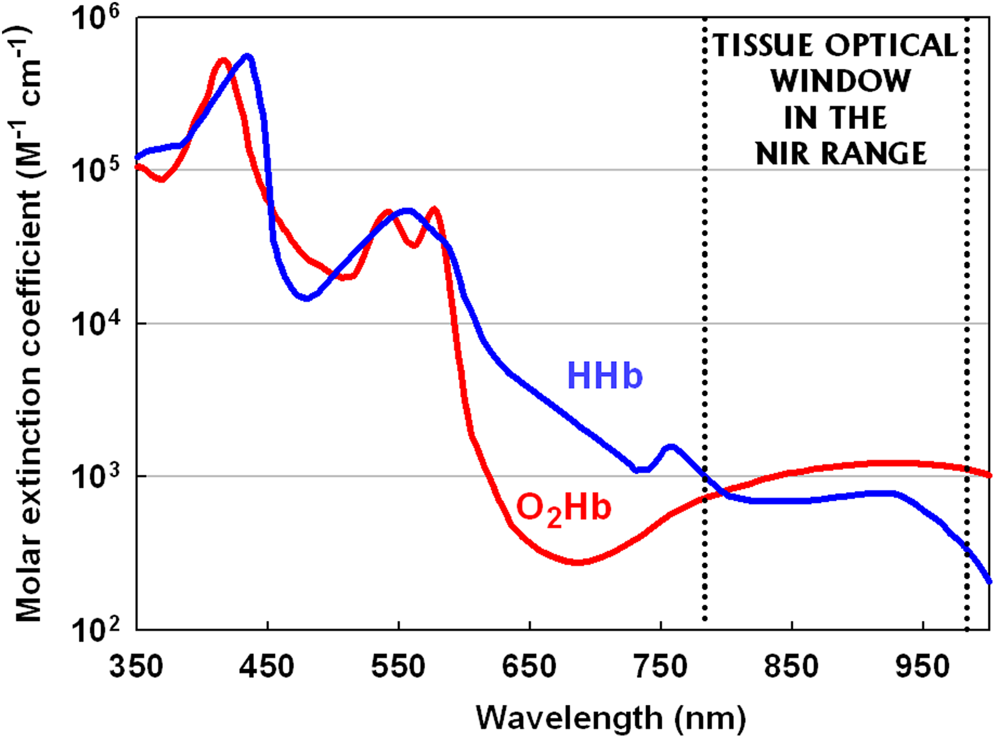
Absorption spectra of oxygenated (O2Hb) and deoxygenated (HHb) hemoglobin. The difference in their absorption spectra allows the measurement of their respective concentration. Most of the commercially available functional near-infrared spectroscopy (fNIRS) systems utilize wavelengths in the wide range (685-850 nm) in order to maximize the separation between O2Hb and HHb. Beyond 900 nm, the majority of NIR photons are absorbed by water (peak absorption centered around at 980 nm).
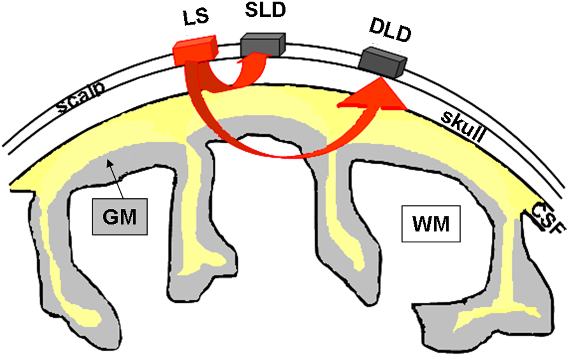
Schematic representation of the near-infrared (NIR) light traveling through the head. Spatial distribution of light flux through the different tissue layers of the head (due to complex light scattering) is simulated using the optical properties of scalp, skull, CSF, GM, WM, and so on. The penetration depth of NIR light depends on light scattering, absorption, as well as the separation distance of the light source and detector. In particular, the NIR light goes deeper and with a higher light intensity in the case of: (a) lower skull thickness compared with a higher skull thickness and (b) a greater source-detector separation. The detected emerging signal comes mainly from hemoglobin located in small vessels (<1 mm diameter) such as the capillary, arteriolar, and venular bed. The arrows represent the simplified spatial distribution of the NIR photons through the different head layers. The shorter distance between light source and detector is utilized for measuring the systemic hemodynamic fluctuations occurring in the superficial layers. These measurements can then be used as regressors in the post-experiment analysis to remove the systemic contamination and isolate the brain signal. CSF = cerebrospinal fluid; GM = gray matter of the cerebral cortex; DLD = deep light detector; LS = light source; SLD = shallow light detector; WM = white matter.
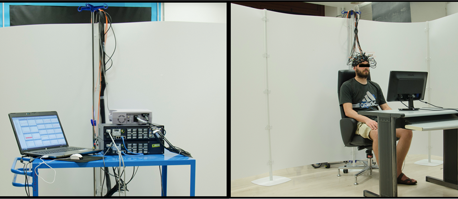
Example of a functional near-infrared spectroscopy (fNIRS) experimental setting. The subject is sitting in a comfortable high-backed chair. The illuminating optic fiber bundles and the collecting ones are assembled into a flexible probe holder fixed to the head by a Velcro brand fastener, adapting it to the size and shape of the head. The fNIRS instrumentation (Oxymon Mk III, Artinis, The Netherlands) is isolated from the subject by a white screen.
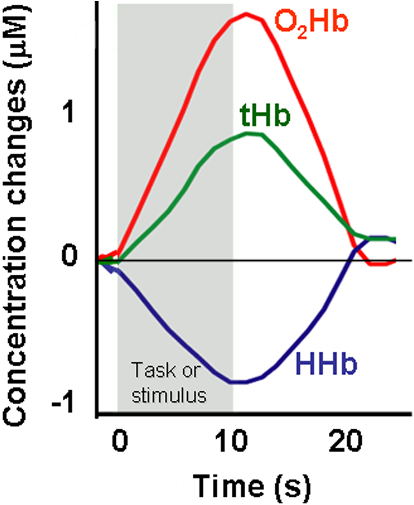
Illustration of a typical cortical hemodynamic response in an event-related paradigm, as revealed by functional near-infrared spectroscopy (fNIRS). The evoked hemodynamic response is subsequent (with about a 2-second delay) to the earlier electrical activity, which can be measured in real time by EEG/MEG. Indeed, the onset of the neural firing is almost immediate after the beginning of the paradigm, and the succeeding increase in the oxygenated hemoglobin (O2Hb, red line) and the concomitant smaller decrease in the deoxygenated hemoglobin (HHb, blue line) (as measured by fNIRS) reflect an increased local arteriolar vasodilatation in the cortex (neurovascular coupling). As a result, the local cerebral blood flow and the total hemoglobin volume [tHb = (O2Hb + HHb), green line] (strictly related to the cerebral blood volume) raise. EEG = electroencephalography; MEG = magnetoencephalography.

Mapping of the prefrontal cortex activity during face-to-face conversation by two wearable functional near-infrared spectroscopy (fNIRS) systems, each equipped with 22 channels (WOT-220, Hitachi High-Technologies Corporation, Japan). Each flexible probe unit covers the head in correspondence of the underlying dorsolateral and the rostral prefrontal cortex. The processing unit is worn on the waist. Multiple subject measurement (up to four people) is allowed. Photo courtesy of Hitachi.
Considering the well-recognized benefit of using a multimodal approach, recently many concurrent EEG-fNIRS, fNIRS-fMRI, and EEG-fMRI studies have been carried out for investigating the brain at multiple spatial and temporal scales simultaneously. Especially, concurrent EEG-fNIRS measurements can be collected without difficulty (Chen, Sandmann, Thorne, Bleichner, & Debener, 2016) compared with concurrent fNIRS-fMRI and EEG-fMRI measurements.
Interestingly, Montague et al. (2002) first performed the measurement of the brain activation simultaneously on two subjects by using two fMRI systems. This hyperscanning approach, also adopted later in the EEG and fNIRS fields, has opened new perspectives for investigating interpersonal interactions in a social context in the framework of the social cognitive neuroscience (for review, see Babiloni & Astolfi, 2014; Koike, Tanabe, & Sadato, 2015).
In summary, fNIRS is a very significant alternative/complementary functional neuroimaging methodology for investigating human cortical function under more realistic, ecologically valid parameters and in settings not compatible with fMRI, PET, and MEG.
This review is aimed at giving a concise and simple overview of the basic principles of fNIRS including features, strengths, advantages, and limitations summarized in terms that can be understood even by nonspecialists. Moreover, the main characteristics of the commercially available fNIRS systems and some examples of the present and future prospects of fNIRS for assessing cerebral cortex function during human behavior in natural and social situations, also adopting the hyperscanning approach, will be reported.
Search Strategy and Selection Criteria
Articles were identified using the stepwise approach. Papers were retrieved by the authors through different strategies. First, a search on the two databases PUBMED and SCOPUS was performed using the keywords functional near-infrared spectroscopy, functional near-infrared imaging, optical imaging, optical topography, NIR imaging, diffuse optical imaging, diffuse optical tomography, and hyperscanning. The references were screened, and the full texts of relevant publications were retrieved. Additional published reports were identified through a manual search of references in retrieved articles and in review articles. The research was restricted to literature suitable for human studies published or made available up to January 2016. Research groups known to be active in the field were contacted for gathering further information. The websites of the commercial systems were searched and visited for exploring the specifications of the instruments. After collecting all the documentation, a consensus was made by the authors to properly select material eligible for inclusion in this review. The material was sorted according to the type of fNIRS instrumentation. Tables were generated to report the origin and the properties of each instrument.
Features, Strengths, Advantages, and Limitations of fNIRS
This brief review article does not comprehensively cover all the advancements and pitfalls in all the aspects of fNIRS methods because they have been reported in detail in two recent technical review articles (Scholkmann et al., 2014; Torricelli et al., 2014). The most relevant topics and the related recent references about fNIRS (from its history to its different technical aspects) are reported in Table 1 (which includes 18 review articles). The features, strengths, advantages, and limitations of fNIRS, according to nowadays knowledge, are summarized in Box 1 and Box 2. A schematic representation of the NIR light traveling through the head is reported in Figure 2. The NIR laser or light emitting diode illuminates a selected scalp surface. The propagation of the NIR light in tissue is sufficiently complex; photons bounce off the many boundaries of cellular and subcellular substructures, changing randomly the direction of propagation. In most tissues, photons are scattered many times (even 10 times per centimeter of tissue). The light passing through the scalp/skull/brain surface is highly scattered and attenuated. Then, the emerging light can be measured by detectors positioned at different distances from the sources on the scalp surface. The penetration depth of the NIR light depends on the light scattering, absorption (mainly due to O2Hb and HHb), as well as the separation distance of the light source and detector. For a given source-detector pair, the detected photons are known to more likely have traveled through the central area of a banana shape than through its outer portions. Therefore, the fNIRS measurements are most sensitive to O2Hb and HHb molecules found in that area. Then, the intensity changes of the emerging light can be measured from 1.5 to 2.5 cm below the skull. The intensity changes revealed at the selected light wavelengths are used for calculating the concentrations of O2Hb and HHb. Because of the NIR light propagation in tissues, the exact volume of the illuminated tissue is unknown, and the spatial resolution is at around 1 cm. The poor spatial resolution depends on the geometry of the array of the sources-detectors, where the separation distance between source-detector dictates the “banana-shaped” sensing volume intrinsic to diffuse light transport.
Most Relevant References About fNIRS: From History to Different Technical Aspects.

Note. fNIRS = functional near-infrared spectroscopy; A = recent outstanding article; fNIRI = functional near-infrared imaging; Pub = publication; R = review.
Features, Strengths, and Advantages of Functional Near-Infrared Spectroscopy (fNIRS).
fNIRS is a noninvasive and safe optical technique that uses light emitting diodes or laser diodes as NIR light sources and different NIR detectors to measure human cerebral cortex oxygenation changes in response to certain stimuli/tasks (neurovascular coupling). fNIRS measurements can be performed in a natural environment without restraint and different posture. Human tissues are highly scattering media relatively transparent to light in the range 650 to 1,000 nm (Figure 1) where the dominant absorbers are oxygenated (O2Hb) and deoxygenated (HHb) hemoglobin (the oxygen transport red blood cell protein) located in small vessels (<1 mm diameter) such as the capillary, arteriolar, and venular bed. NIR light is able to penetrate human tissues because the dominant factor in its transport is represented by scattering, which is typically about 100 times more probable than absorption. As O2Hb and HHb have different absorption spectra (Figure 1), the changes in the intensity of the NIR light emerging from the head are utilized to measure O2Hb and HHb concentration changes. As consequence of the complex light scattering effect by different tissue layers, the length of NIR light path through tissue (optical path length) is longer than the physical distance between the source and the detector. The spatial distribution of NIR light through the different tissue layers is a banana-shaped region, as sketched in Figure 2. Adequate NIR light penetration depth (almost half of the source-detector physical distance) can be achieved using a source-detector distance of 2 to 3 cm and 4 to 5 cm on infants and adults’ head, respectively. The selection of the optimal source-detector separation depends on NIR light intensity, subject’s head region, and age. fNIRS allows for semiquantitative/quantitative monitoring of important physiological measures: (a) O2Hb; (b) HHb; (c) total hemoglobin (tHb), (tHb=O2Hb+HHb) (tHb is strictly related to cerebral blood volume). fNIRS parameters quantitation depends on the adopted NIRS technology. The commonly used continuous wave-based instrumentations provide only concentration changes in O2Hb and HHb (with respect to an initial value arbitrarily set equal to zero) calculated by using the modified Lambert Beer law and expressed in µmolar × cm. fNIRS measurements are repeatable and reproducible. Real-time maps of cortical hemodynamic response (optical topography) can be obtained applying an array of sources/detectors over the scalp (spatial resolution ∼1 cm). fNIRS is characterized by a relatively high temporal resolution (sampling rate up to 100 Hz; typically between 1 and 10 Hz). Locations of sources/detectors can be captured using neuronavigation systems. fNIRS signals fluctuate during the resting state reflecting physiological phenomena (i.e., fluctuations attributable to systemic arterial pulse oscillations (∼1 Hz) and respiration (∼0.2-0.3 Hz) as well as localized blood pressure fluctuations (<0.05 Hz) and functional connectivity. fNIRS instrumentation is relatively low/moderate costly, transportable or portable (Table 2). fNIRS systems can be miniaturized and even made wearable and wireless (Table 3). fNIRS is characterized by a very high experimental flexibility with respect to the other neuroimaging modalities. Compared with functional magnetic resonance imaging (fMRI), fNIRS: (a) is silent and more tolerant to subtle movement artifacts (e.g., overt speech is allowed), (b) measures O2Hb besides HHb providing a more complete evaluation of the cortical hemodynamic response, (c) allows long-time continuous measurements and repeated measurements within short intervals, and (d) has a higher temporal resolution. fNIRS technique is compatible with other electrical or magnetic monitoring systems and therapeutic devices (i.e., pacemaker, hearing aids, cochlear implants, etc.). fNIRS measurements can be integrated with fMRI, positron emission tomography (PET), electroencephalography (EEG), or event-related potentials.
Limitations of Functional Near-Infrared Spectroscopy (fNIRS).
fNIRS is unable to provide information about brain structure for anatomical reference. A stable contact between source/detector and skin is critical. The layering and dark color of hair attenuate NIR light. Cortical hemodynamic responses to cognitive stimuli that involve deep brain regions, such as basal ganglia and amygdale, cannot be investigated. Therefore, fNIRS measurements are restricted to outer cortex and have low spatial resolution (around 1 cm). Depth sensitivity of NIRS signal to cortical hemodynamic response depends on many factors (i.e., source-detector separation, source power, detector sensitivity, optical properties of the skin/skull layers, degree of white matter myelination). For most fNIRS systems, the typical depth sensitivity is about 1.5 cm. The amplitude of the changes of fNIRS signal is susceptible to the variation of source-detector position/distance. The separation of the hemodynamic changes originating either from cerebral tissue or extra-cerebral tissues/structures (scalp, temporal muscle, skull, frontal sinus, cerebrospinal fluid, and dura) is difficult. The establishment of the exact spatial origin of the cortical hemodynamic response and the precise identification of brain areas beneath the fNIRS probes are impossible without the use of three-dimensional MRI. The placement of multiple sources/detectors (in particular on the hairy scalp) is time consuming. Improvements of headgear and attaching are necessary. Sources/detectors cannot cover the entire head surface, making difficult the simultaneous exploration of all the possible cortical neural systems. Increasing the number of fNIRS measurement points, the weight, and the size of the headgear increase with subsequent greater movement artifacts and larger data exclusion from the data analysis. Continuous wave-based fNIRS systems cannot measure optical path length, then do not provide absolute quantification value of fNIRS measures. Optical path length differences across diverse head regions should be taken in consideration for interpreting fNIRS data. Technological development of time-domain and frequency-domain–based instrumentations (being able to measure the spatial and temporal variations in optical path length) will improve the fNIRS sensitivity and the quantitation of fNIRS parameters. In the case of experimental tasks inducing large systemic vascular changes and breathing pattern, the monitoring of some systemic parameters (i.e., heart rate, blood pressure, skull blood flow, partial end-tidal carbon dioxide) is desirable. No standardization is available yet for fNIRS instrumentations/signal processing/data analysis and statistics procedures. The software of fNIRS systems does not provide data stream mining, namely, real-time input that allows an interactive user interface to adapt his or her behavior. fNIRS signal artifacts (due to head motion and reduction of the grip of the sources/detectors on skull) are not automatically corrected by software. Most of the commercially available fNIRS systems are not approved by the USA Food and Drug Administration.
Commercially Available fNIRS Instrumentation
Since the middle of the 1990s, multi-channel fNIRS systems, which utilize arrays of multiple NIR sources and detectors arranged over the scalp, have been introduced (Ferrari & Quaresima, 2012). Different commercially available fNIRS instruments with the related key features, advantages and disadvantages, and parameters measurable by using different techniques have been recently reviewed in detail (Scholkmann et al., 2014). Briefly, three different NIRS techniques can be used, each based on a specific type of illumination: (a) the continuous wave (CW) modality, which based on constant tissue illumination, simply measures light attenuation through the head; (b) the frequency-domain (FD) method, which, illuminating the head with intensity-modulated light, measures both attenuation and phase delay of emerging light; and (c) the time-domain (TD) technique, which, illuminating the head with short pulses of light, detects the shape of the pulse after propagation through tissues. Therefore, the O2Hb/HHb quantitation depends on the fNIRS technology adopted. The most commonly used CW-based fNIRS instrumentation measures changes of O2Hb and HHb (with respect to an initial value arbitrarily set equal to zero) calculated using a modification of the Lambert-Beer’s law (Scholkmann et al., 2014). Considering that the tissue optical path length is longer than the distance between the source and the detector (because the scattering effects of different tissue layers are unknown), the O2Hb and HHb signal changes are expressed as μmolar × cm or mmolar × mm. Only the FD and TD techniques offer the possibility to absolutely characterize the optical properties of tissues (absorption and reduced scattering coefficients), from which it is possible to retrieve absolute O2Hb and HHb concentration changes. CW-based systems offer the advantages of being low cost and easily transportable. In ascending order, CW-, FD-, and TD-fNIRS–based instruments increase in cost and technological complexity. The different complexity of the fNIRS instruments is witnessed by the diversity of their cost, which varies from $10,000 up to over $300,000. There is a wide variety of commercial fNIRS instruments, as reported in Tables 2 and 3. Most of the fNIRS systems reported in Table 2 utilize fiber optic bundles to transport NIR light to/from tissues.
Main Commercially Available Transportable fNIRS Systems.
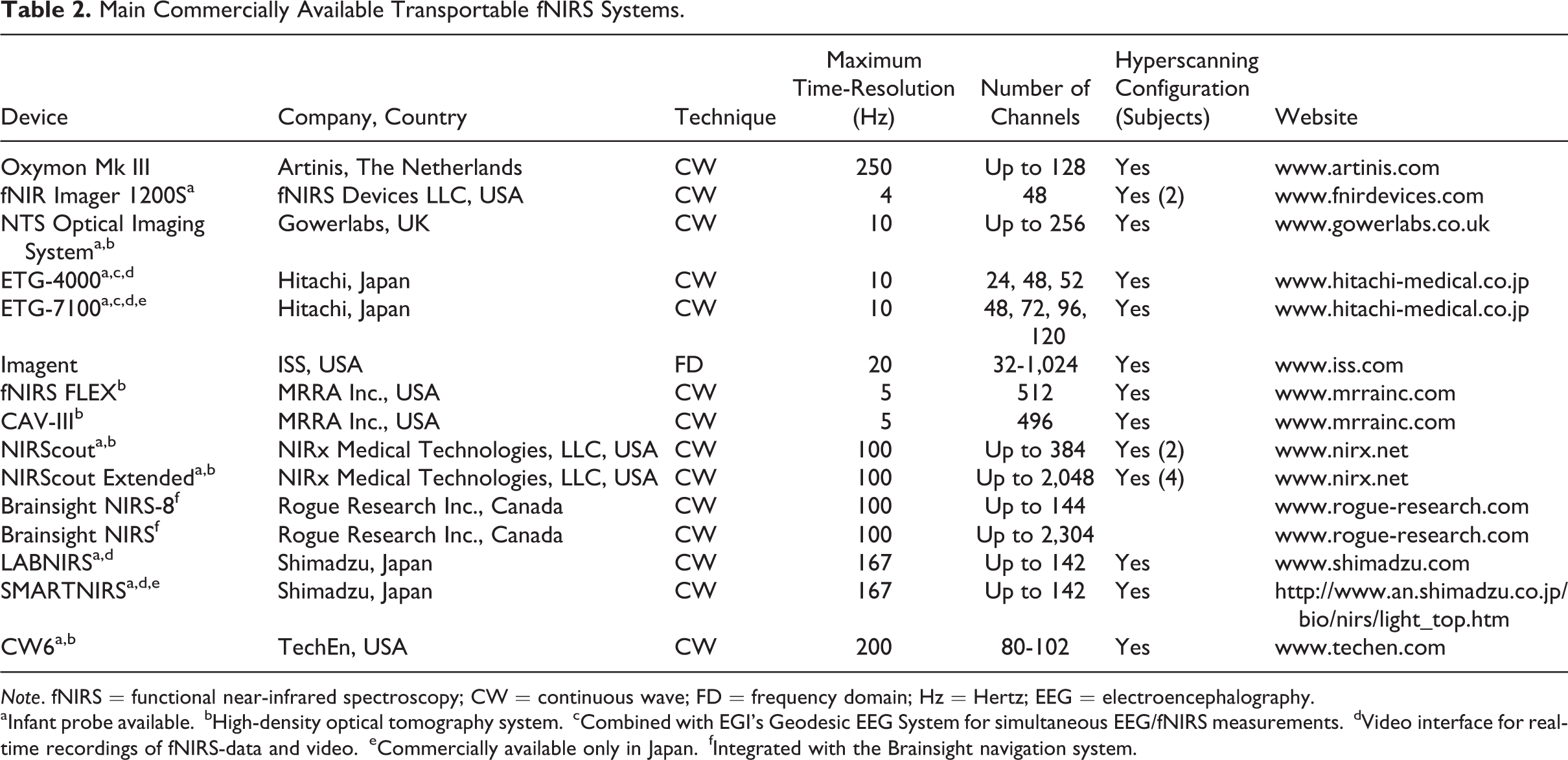
Note. fNIRS = functional near-infrared spectroscopy; CW = continuous wave; FD = frequency domain; Hz = Hertz; EEG = electroencephalography.
aInfant probe available.
bHigh-density optical tomography system.
cCombined with EGI’s Geodesic EEG System for simultaneous EEG/fNIRS measurements.
dVideo interface for real-time recordings of fNIRS-data and video.
eCommercially available only in Japan.
fIntegrated with the Brainsight navigation system.
Main Commercially Available Continuous Wave Portable Wireless fNIRS Systems.
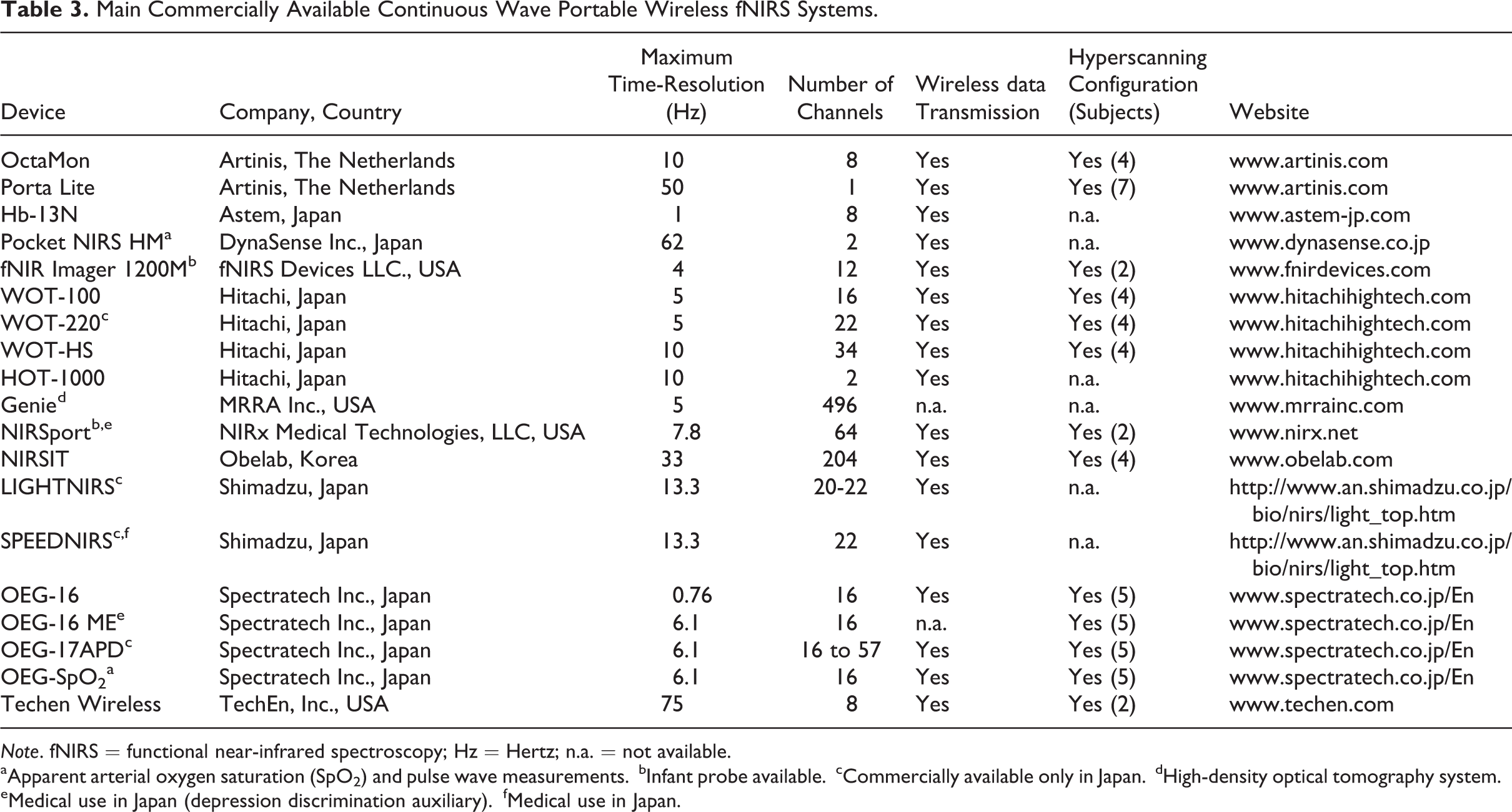
Note. fNIRS = functional near-infrared spectroscopy; Hz = Hertz; n.a. = not available.
aApparent arterial oxygen saturation (SpO2) and pulse wave measurements.
bInfant probe available.
cCommercially available only in Japan.
dHigh-density optical tomography system.
eMedical use in Japan (depression discrimination auxiliary).
fMedical use in Japan.
The fNIRS measurements can be carried out, for example, while a subject is sitting in an armchair (Figure 3) or lying on a table. The fNIRS cap is placed on the top of the head. The NIR light is not harmful and does not provide any sensation. In general, an fNIRS measurement lasts 30 to 60 minutes. The subjects can respond, for example, by pressing a button when they hear or see certain sounds or words during auditory and visual tasks; the subjects may play games or be occupied in other activities such as cognitive tasks on the computer or with an experimenter. A typical activation revealed by fNIRS in a cerebral cortex area is sketched in Figure 4. The increase in O2Hb and the concomitant relatively smaller decrease in HHb reflect an increase in the local arteriolar vasodilatation, with the consequent rise of the local CBF and cerebral blood volume. However, the augmented amount of oxygen transported to the activated cortical area typically exceeds the local neuronal rate of oxygen utilization, resulting in an overabundance of cerebral blood oxygenation in the activated area. The precise neuronal origin of the hemodynamic responses measured by fNIRS and fMRI is still relatively unknown, and it is beyond the scope of this article to cover these important issues.
So far, the fNIRS systems used for monitoring cortical activation vary in complexity from dual channels to “whole-head” arrays of several dozen channels. fNIRS data processing/analysis methods permit topographical assessment of real-time regional cortical O2Hb/HHb changes. Data from multiple simultaneous measurement sites are displayed by fNIRS systems in the form of O2Hb/HHb map or image over a specific cortical area. So far, fNIRS has lacked the combination of spatial resolution and wide field of view sufficient to map in detail the distributed brain functions. The recent emergence of high-density 3-dimensional DOT (using more than 50 fiber optic bundles covering the head) represents the last generation of fNIRS systems, which opens up more potential clinical applications by offering mapping higher-order, distributed brain functions either at rest or in response to interventions (Liao & Culver, 2014). For instance, high-density DOT imaging array has been recently used to map multiple resting-state networks including the dorsal attention and default mode networks and cortical responses during language tasks (Eggebrecht et al., 2014).
The methods of analysis of the fNIRS/DOT/DOI signals have been recently reviewed (Scholkmann et al., 2014). In particular, the methods to separate different components, caused by cerebral and extra-cerebral systemic activity in fNIRS signals—the latter revealed by the shallow light detector (Figure 2), have been reported. Over the years, the complexity of fNIRS systems has grown so that 2- or 3-dimensional topographic images can be obtained. Proprietary reconstruction systems as well as many different open source software products are available. The disadvantage of using fiber optic bundles, whose fNIRS systems are equipped, is that the fibers are often heavy and of limited flexibility and length, perhaps provoking discomfort to the subjects. To overcome these disadvantages, different battery-operated multichannel wearable/wireless systems, suitable for performing fNIRS measurements on adult forehead, have been commercialized since 2009 (Table 3). An example of a 22-channel wearable fNIRS system is shown in Figure 5. This represents the most advanced version of fNIRS systems suitable for evaluating brain activation in response to cognitive tasks performed in normal daily activities (Pinti et al., 2015).
fNIRS to Assess Cerebral Cortex Functions During Active Behavior in Natural and Social Situations
In 2014, the prestigious journal NeuroImage dedicated a special issue to commemorate the first 20 years of fNIRS research (Boas, Elwell, Ferrari, & Taga, 2014). Its 9 review articles and 49 contributed papers offer a comprehensive survey of the exciting advances driving the field forward and of the myriad of applications that will benefit from fNIRS. The main fNIRS applications in some branches of the social sciences, including psychology/education, economics, and linguistics, are reported in Table 4. In addition, the main fNIRS applications in functional neuroimaging basic research and in medicine are included too. So far, fNIRS has been applied successfully to various research domains in healthy populations (newborns, children, and adults) and in patients with neurological and psychiatric disorders. It’s noteworthy to mention that fNIRS and/or EEG rather than fMRI offer the great advantage to understand the infant brain. This would allow a clarification of the early development of human mind including the development of the social brain (Geddes, 2015). Significant fNIRS studies dedicated to the understanding of infant cognition, typical and atypical development, and language and its development are nicely reviewed in recent articles (Table 4). Interestingly, the fNIRS approach has been adopted for addressing some noteworthy developmental issues: object processing (Watanabe, Homae, Nakano, & Taga, 2008), social communication (Minagawa-Kawai et al., 2009), social situations across cultures (Lloyd-Fox et al., 2014), and human action processing (Lloyd-Fox et al., 2015b). More recently, fNIRS has been also used on infants in natural and social situations with live people (Lloyd-Fox et al., 2015a; Urakawa, Takamoto, Ishikawa, Ono, & Nishijo, 2015). Urakawa et al. (2015) performed their study on 7-month-old infants during the social interactive play “peek-a-boo,” in which communicative ostensive signals in both auditory and visual modalities were presented by a real human presenter. A more prominent cortical activation in the dorsomedial prefrontal area was found in response to social play with a partner’s direct gaze compared to an averted gaze, suggesting that a partner’s direct gaze shifts an infant’s attention to the partner’s eyes for interactive communication. Lloyd-Fox et al. (2015a) investigated 6-month-old infants’ sensitivity to ostensive signals for speech and gesture processing during naturalistic communicative interactions. The results of this study have indicated that only the multimodal presentation of combination of ostensive signals (infant-directed gaze and infant-directed speech) is able to enhance the activation of the cortical regions known to be involved in processing auditory and visual aspects of social communication. Both studies have demonstrated how fNIRS can give a contribution in assessing cerebral cortical functions in infants during natural and social situations.
Main fNIRS Applications on Human Beings (Either Infant/Child or Adult) in the Field of Social Sciences, Neuroimaging Basic Research, and Medicine.

Note. fNIRS = functional near-infrared spectroscopy; Pub = publication; A = recent outstanding article; R = review.
aCochlear implant users are not suitable for functional magnetic resonance imaging (fMRI) investigation.
Considering the interest of the journal Organizational Research Methods in promoting a more effective understanding of methodologies and their application in organizational settings, the potential of fNIRS to assess brain functions during active behavior in social environments is nicely documented by some recent studies (related to speech production, neuroeconomic research, neuromarketing, daily cognitive function—virtual shopping—using virtual reality, and human interactions). In fact, overt speech production and its neural correlates cannot be investigated by PET or fMRI due to their high sensitivity to movement artifacts. Suda et al. (2010) investigated face-to-face conversation by a 52-channel fNIRS system. The subjects, while sitting in front of an interviewer, were asked to talk alternately about a certain topic for a predefined amount of time. The conversation periods were accompanied by an activation observed in the superior frontal and superior temporal gyri and not observed in the Broca’s area. Kopton and Kenning (2014) have recently reviewed the fNIRS studies with relevance to (economic) decision making and presented a decision table for neuroeconomic researchers that may enable a better determination of the suitability of fNIRS for studying neuroeconomics. Misawa, Shimokawa, and Hirobayashi (2014) focussed on the price topic (which is a purchasing decision-making factor) and measured the prefrontal cortex activation (16-channel OEG-16, Spectratech Co., Japan) induced by the evaluation of the product price. Their results suggest that prefrontal cortex activation can be used as an indicator of the extent to which consumers feel a product price to be “expensive” or “inexpensive.” Okahashi et al. (2014) applied virtual shopping with three different difficulty levels for investigating in two groups of convalescent brain-damaged patients and healthy young adults, the difference on prefrontal cortex activation, as measured by fNIRS (16-channel OEG-16, Spectratech Co., Japan). They reported that both groups had different task performances and different prefrontal cortex activation patterns as well as different subjective assessment of the rising difficulty of the task. Very recently, Pinti et al. (2015) evaluated the feasibility of using a portable fNIRS 22-channel wireless system (WOT-220, Hitachi, Japan) for monitoring the prefrontal cortex activation during a prospective memory task conducted outside in a typical London street location and mimicking the demands of everyday life. Significant prefrontal cortex activations in response to social and nonsocial prospective memory cues were recorded.
Applying functional neuroimaging modality simultaneously in two or more people, a recent technique known as hyperscanning, it is possible to calculate interbrain neural effects that appear only in interactions between individuals (human interactions) (for reviews, see Babiloni & Astolfi, 2014). The hyperscanning approach in fMRI studies is advantageous for determining precisely the region(s) involved in inter-brain effects. However, it is almost impossible to record inter-brain effects in daily life using fMRI. By contrast, the hyperscanning approach in fNIRS studies, characterized by a reasonable high temporal resolution, captures moment-to-moment interactions in a natural context (for reviews, see Koike et al., 2015; Scholkmann, Holper, Wolf, & Wolf, 2013). An example of hyperscanning fNIRS measurement is shown in Figure 5. In particular, a couple of headsets, which can be used for neuromarketing studies, offers the potential to gauge consumer reactions in relatively normal situations. The first hyperscanning fNIRS study goes back to 2011 (Funane et al., 2011). For that study, a 22-channel wireless system (WOT-220, Hitachi, Japan) was used to monitor cortical responses in two subjects while sitting down face-to-face and synchronously pressing a button (after counting 10 seconds in their mind) when it was requested. Higher prefrontal cortex inter-brain coherence was significantly associated with shorter time intervals between button presses. The authors concluded that the performance of a cooperative behavior is associated with inter-brain coherence.
Face-to-face interaction is one of the most common types of social interaction in everyday life; uncertainties about other people’s intention influence interpersonal interactions. Very recently, Tang et al. (2015) investigated how face-to-face interaction impacts interpersonal brain synchronization during two-person economic exchange by combining an economic exchange game paradigm with fNIRS hyperscanning approach (ETG-4000, Hitachi Medical Company, Japan). In their study, pairs of strangers interacted repeatedly either face-to-face or face-blocked while their cortical activation was simultaneously measured in the right temporo-parietal junction and the control region (right dorsolateral prefrontal cortex). The related fNIRS results have indicated an increased interpersonal brain synchronization during face-to-face interactions in right temporo-parietal junction region with a greater shared intentionality between partners. Gender-related social interactions (two-person game) have been also investigated by Cheng, Li, and Hu (2015). They have observed a coherent brain activity across dyads in relation with the cooperation specifically with opposite-sex partners. The effects of this synchronization were observed roughly in frontopolar, orbitofrontal, and left dorsolateral prefrontal cortex. The authors have suggested the utility of using task-related coherence for differentiating the neural processes that underlie mixed-sex cooperation compared to same-sex cooperation.
Another important social interaction is represented by teaching, which involves a pupil and a teacher. Previously, Holper et al. (2013) recorded prefrontal brain oxygenation during dialog execution simultaneously in 17 teacher-student pairs using a four-channel wireless fNIRS prototype. Their main finding was that in the students who successfully transferred the knowledge, a lower activation (with respect to the students without transfer) was observed. Those results could encourage future studies aimed at investigating brain networks involved in real educational setups where knowledge is acquired in a complex entangled process involving an interaction between a pupil and a teacher.
Although leadership is considered a notable feature of the human society, little is known about the neural basis of leader-follower communication. Very recently using the fNIRS-based hyperscanning approach (ETG-4000, Hitachi Medical Company, Japan) in a realistic three-subject interpersonal-communication context, Jiang et al. (2015) found evidence that human leaders cooperated with their followers to achieve group decision by synchronizing their brain activities with those of the followers through their tactful communication skills and competence. In addition, they found that it was possible to predict leadership based on the interpersonal neural synchronization data as well as communication behaviors early in their interactions. Their findings give an important contribution to the theoretical discussion about the relevance of communications in leader emergence. Finally, the authors have suggested that the fNIRS hyperscanning approach might be used in neuro-feedback or neuro-intervention during leadership training.
In a multiperson interaction environment, the information exchange process is much more complex than that in a two/three-person situation. Therefore, single-brain level analysis and paired-brain level analysis may not support comprehensive study of the characteristics of multi-brain interaction processes. Confronting these challenges, Duan et al. (2015) have recently proposed a general framework for hyperscanning and modeling multiple interacting brains called cluster imaging of multi-brain networks. As a demonstration of this method, the authors have conducted a simple preliminary experiment in which nine participants drummed together and tried their best to make their beats consistent with each other while the oxygenation of the prefrontal cortex and left temporal parietal junction were simultaneously recorded by two fNIRS systems (ETG-4000, Hitachi Medical Company, Japan; LABNIRS, Shimadzu, Japan).
Conclusions
The feasibility and the success of applying fNIRS in some branches of the social sciences, neuroimaging basic research, and medicine have been well documented. The increasing number of publications (about 280 in 2014; source: SCOPUS 2014, Elsevier B.V.) is promising for an expansion of fNIRS in these fields. The development of fNIRS has strongly gained from the advances in microelectronics, computer technology, and optical engineering. With the advent of further miniaturization and integration such as integrated optics, wearable and even disposable fNIRS technology can be envisioned. The ongoing significant developments in time-domain diffuse optics could generate compact and even wearable time-domain fNIRS systems (Dalla Mora et al., 2015). The fNIRS systems that will emerge from these developments would further enlarge the number of fNIRS applications and facilitate the comparison of the fNIRS findings. The prediction of the future directions of fNIRS for assessing brain function during human behavior in natural and social situations is not easy. However, fNIRS will give an outstanding contribution in revealing cortical responses to social stimuli in infant brains and in investigating real social interactions in adult brains.
Footnotes
Acknowledgment
Declaration of Conflicting Interests
The author(s) declared no potential conflicts of interest with respect to the research, authorship, and/or publication of this article.
Funding
The author(s) disclosed receipt of the following financial support for the research, authorship, and/or publication of this article: The research leading to this article was made possible, in part, by the 2014 grant from the “Fondazione Cassa di Risparmio della Provincia dell’Aquila” and the “Abruzzo earthquake relief fund” (Toronto, ON, Canada).