



Abstract
Introduction:
The use of silver (Ag) as an antimicrobial agent has gained attention for its multimodal mechanisms of activity. We have previously demonstrated the efficacy of a 95% titanium dioxide (TiO2) and 5% polydimethylsiloxane (PDMS) silver carboxylate (AgCar)–eluting chemistry, which has been shown to prevent bacterial adherence, proliferation, and biofilm formation on orthopedic implants and prosthetic liners. This project examines the physical and chemical properties of AgCar on polyether ether ketone (PEEK) implant materials and silicone prosthetic liners, which are prone to bacterial colonization.
Methods:
PEEK implants were coated with varying concentrations of matrix, AgCar, or both. To determine physical properties, we used water contact angle for hydrophilicity, atomic force microscopy for surface roughness averages, and graphite furnace atomic absorption spectroscopy for Ag elution rate. For durability studies, silicone was coated and examined for compressive, frictional, and wear rate properties.
Results:
All implants demonstrated hydrophobicity in the presence of the matrix, AgCar, or both; in contrast, uncoated (UNC) PEEK implants were found to be hydrophilic. The 95% 10× matrix exhibited a delayed and extended elution of AgCar into solution compared with other solutions. Implant surface roughness increased as AgCar concentrations increased. Durability studies found no notable differences between UNC and 95% 10× coated implants.
Conclusion:
Consistent with previous work, the 10× AgCar coating demonstrated the best results for inhibitory behavior on two distinct fronts: mechanically, through roughness and increased hydrophobicity, and chemically, through AgCar elution activity. Furthermore, the 10× coating will likely not impair the durability or elasticity of the implant within the body or silicone prosthetic liners. Thus, the 95% TiO2:5% PDMS matrix doped with a 10× AgCar concentration is a prime candidate for clinical application consideration.
As many as 20%–30% of hospital-acquired infections are because of post-operative surgical site infections (SSIs). These infections have a high morbidity rate and often require revision operations.1,2 With the growing prevalence of multidrug-resistant organisms, common antibiotic agents are often inadequate, necessitating the development of alternative therapeutics. 3 The treatment of SSIs is further complicated by the formation of bacterial biofilms and their structural properties.4,5 Biofilm adhesion can be driven by surface characteristics of roughness, hydrophilicity/hydrophobicity, and electrostatic interactions (van der Waal’s forces). 4 Generally, microorganisms are more likely to attach and form biofilms on rough, stiff, hydrophilic surfaces.6,7
Polyether ether ketone (PEEK) is a polyaromatic semi-crystalline polymer, commonly used as an implant material because of its biologically favorable mechanical properties. 8 Its modulus of elasticity and specific strength is similar to that of human bone, and its strong thermal properties make it stable in the body. 9 Silicone is another commonly used medical material, frequently employed for use as a prosthetic liner and for small joint replacements in the hand and foot.10,11 Silicone has low compressive and frictional properties. Prior studies have shown that the wear and durability of both PEEK and silicone may be affected by chemical coatings.10,12–14
In recent years, silver carboxylate (AgCar) has attracted renewed attention as a potential antimicrobial targeted at biofilm formation because of its long-lasting release, broad antibacterial spectrum, and low incidence of antibiotic resistance.15,16 Low resistance is largely because of the multiple intracellular pathways through which silver (Ag) mediates bacterial death, including hydrogen ion leaks that disrupt ATPase functionality, interference with DNA base pairing, disruption of proteins by reaction with thiol groups, and generation of free radical species. 17
Our group has developed and validated a AgCar coating, which utilizes a titanium dioxide (TiO2) and polydimethylsiloxane (PDMS) hybrid matrix to facilitate the controlled release of AgCar particles over time.11,18–20 The coating utilizes the broad antimicrobial activity of Ag, exhibits controllable release pharmacokinetics, and is capable of deep pilosebaceous gland penetration. 19 It has previously been studied as an implant coating on PEEK and liner coating on silicone, in which it demonstrated inhibition of adherence against problematic pathogens, including Cutibacterium acnes, methicillin-resistant Staphylococcus aureus, Staphylococcus epidermidis, and Serratia marcescens.11,18–21 Although its efficacy has been validated, the way in which AgCar alters the surface characteristics of implant materials has not been sufficiently studied.
This study examines the mechanical and surface effects of a 95% TiO2:5% PDMS matrix doped with varying concentrations of AgCar on orthopedic implant materials—PEEK and silicone. Determining roughness and hydrophobicity will clarify whether bacterial adherence to the material is maintained with application of the coating. Elution rate will ensure that the coating maintains a controlled release once applied. Finally, measuring wear and durability is crucial to ensure that implant materials will remain stable and will be unaffected by the coating. We hypothesized that this coating will demonstrate physical characteristics that help to maintain bacterial inhibition, while not impacting durability.
Materials and Methods
AgCar doped TiO2-PDMS coating
The AgCar chemistry used in all experimental trials was developed according to a patented protocol developed by BioIntraface (North Kingstown, RI). It is composed of TiO2, heptane, PDMS, isopropanol, and Ag neodecanoate. The matrix from which the AgCar is eluted has been previously characterized and is a refined version of 95% TiO2:PDMS, which utilizes a 19:1 ratio of heptane to PDMS. 19 Implants were coated with the 95% matrix and contained varying concentrations of Ag neodecanoate. Increasing concentrations of Ag neodecanoate corresponded to greater doping concentrations. The concentrations used are no Ag (0×), small dopage (1×), and large dopage (10×). Implants coated in 100% AgCar and a 1:1 mix of isopropanol and Ag neodecanoate without any matrix were used as positive control group. Uncoated (UNC) implants served as negative control group.
Orthopedic implant material and dip-coating method
Flat PEEK discs measuring 2.5 mm diameter cut via saw were used for hydrophilicity and roughness tests. Elution rate tests were conducted with 2.5 mm-long PEEK semicircular bacilli. Silicone liners were cut into squares for durability tests, which examined compressibility, frictional coefficients, and wear rate. Coating solutions were made according to the ratios in Table 1, and samples were fully submerged into the solution with sterilized tweezers. Implants were then placed into a well plate and allowed to air-dry in a fume hood for 24 hours, after which they were used for property testing.
Composition Ratios of TiO2: PDMS and Silver Carboxylate Dopage Used for Implant Coating Conditions Formulations Used for Implant Coatings Showing the Relative TiO2: PDMS Matrix Ratio and Corresponding Silver Carboxylate (AgCar) Dopage Levels. UNC Denotes Uncoated Control Implants
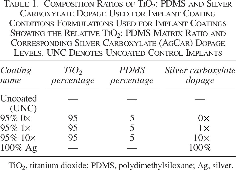
TiO2, titanium dioxide; PDMS, polydimethylsiloxane; Ag, silver.
The implant surfaces of the samples were evaluated using Atomic Force Microscopy (AFM) (MFP-3D-BIO, Asylum Research) using C (contact) mode. AFM data were gathered using a sharp, silicone cantilever (tip diameter <20 nm) with a nominal stiffness value of 40 N/m (Budget Sensors). Topographic height and piezoelectric sensor scans were used to determine surface roughness. Roughness was calculated on the piezoelectric sensor scan of the whole image that had been 2D flattened using AFM software and reported as root mean squared (RMS) values. The scan area was 10 × 10 nm, and 3 spots were scanned per sample for all replicates. To ensure that the data gathered were not imaging artifacts, height and sensor were captured for both directions (trace and retrace). Nine implants were used for each coating condition. Accounting for the trace and retrace measurements per spot on each sample, this yielded a total of 54 data points per coating condition.
Hydrophilicity and contact angle measurements
Nine implants for each coating condition were used for contact angle measurements. At room temperature (20–22°C), 10 mcL water droplets were placed on the surface of PEEK discs in three separate spots for a total of 27 data points per coating condition. To account for variability in coating composition because of dip coating and air-drying, droplets were placed on the left, center, and right portions of the disc. Water droplet magnification, angle calculation, and imaging were done using the DSA100E Drop Shape Analyzer (Krüss Scientific, Hamburg, Germany). After each measurement the implant was pat dried to prevent coating removal. The angle measurements were used to determine the classification and degree of hydrophilicity/hydrophobicity, with a greater contact angling corresponding to increased hydrophobicity.
Elution rate and graphite furnace atomic absorption spectroscopy
After the coated implants were dip coated and allowed to dry, they were added to 10 mL of deionized water in a glass scintillation vial. The implant was then taken out of the vial and moved to a fresh vial of water for another 24 hours. Ten percent nitric acid was added to the previous vial to prevent AgCar from plating onto the glass or precipitating during storage. This process was repeated thrice to establish an elution curve for each coating condition throughout the first 96 hours after coating application.
Before analyzing the samples in the graphite furnace, the solutions being tested were diluted by 20× to achieve concentrations which would not saturate the graphite furnace atomic absorption spectroscopy (GFAAS). GFAAS was done using a PerkinElmer Analyst 600 Graphite Furnace Atomic Absorption Spectrometer at a detection wavelength of 328.1 nm. Absorbance was calibrated using samples of known Ag concentration at 0, 10, 20, 30, 40, and 50 parts per billion (ppb). In addition, NIST 1640 A (Trace Elements in Natural Water) with a known Ag concentration of 8.017 ppb was tested after every five samples to further confirm accuracy. Three implants for each coating condition were each tested three separate times to ensure accurate values, for N = 9 data points per time point per coating condition, N = 45 overall.
Durability tests
Silicone’s compressive and frictional properties have much lower values than PEEK. 22 Therefore, it was used as the coated material to produce detectable values and differences for the compression, frictional, and fatigue tests. A silicone cushion liner was marked with a 20 × 20 mm grid and subdivided with a scissor. As the preferred coating condition, only the 95% 10× condition was examined for these studies, in addition to the UNC control group.
Compression set
Six treated and six untreated samples of silicone (2 × 2 cm each) were placed on flat plates in an Instron E1000 load frame (Instron Corp., Canton, MA). A 6.35 mm diameter steel indenter was lowered to contact the specimen then compressed at a rate of 25.4 mm per minute while collecting force and displacement data at 200 Hz. The test was stopped when the samples were compressed to 75% of starting thickness. Compressive stiffness was sampled from the load displacement between 8 and 9 Newtons. The peak force was the maximum force required during the run to compress samples to 75% of starting thickness and reach the end of the test. Stiffness, peak force, and displacement were reported for each test.
Frictional coefficient
Five treated and unlubricated, five untreated and unlubricated, five treated and lubricated, and five untreated and lubricated silicone samples (1 × 1 cm) were affixed with cyanoacrylate adhesive to the actuator on an Instron E1000 load frame (Instron Corp., Canton, MA). A one-inch by two-inch length of smooth leather (McMaster Carr, Elmhurst, Illinois) was affixed in parallel orientation on a transverse sliding fixture with an outboard pulley where a static weight (17.98 lbs) was suspended. The weight provided a load to the specimen during testing with a resultant calculated surface pressure of 200 kilopascals (kPa). The actuator was lowered at 25.4 mm per minute to 25 mm. The force feedback and displacement data were collected at 200 Hz and used to calculate static and dynamic friction coefficient. Lubricated test groups were moistened with 0.09% physiologic saline solution before and during testing using a saline drip bag (approx. 0.5 Hz drip rate). Photographs were taken before and after testing. Peak static force, displacement at peak static force, minimum dynamic force, and displacement at minimum dynamic force were reported for each test.
Fatigue wear rate
Accelerated wear testing was performed on three 1 × 1 cm silicone implants undergoing the same coating as the treated lubricated group undergoing frictional testing. Samples were loaded into the friction test apparatus, and the actuator was driven with a sinusoidal displacement waveform of 1 Hz at an amplitude of 5 mm. The applied surface pressure was 22.3 kPa. The test was run to a total of 10,100 cycles on each specimen. Lubricant was applied during testing using a sterile saline drip bag (approx. 0.2 Hz drip rate), and the resulting runoff was collected at approximate cycles: 0 to 500, 501 to 1.5k, 1.5k to 2k, 2k to 3k, 3k to 5k, and 5k to 10k, inclusively. The collected liquid samples were sent for dissolved metal analysis (Essco Laboratory, Cranston, RI) to detect elution rates of select coating components. The method reporting limit of the liquid analysis was 1.0 mg/L for the element Ag and 5.0 mg/L for the element titanium.
Results
Average implant roughness
Figure 1 shows roughness values by condition both graphically and numerically. Implant surface roughness decreased by 23% with the application of the matrix alone, as observed from the UNC to 0× comparison. For all implants, roughness increased as Ag dopage increased. The 100% Ag condition exhibited the highest surface roughness, demonstrating a 312% increase from the UNC case. The standard deviations for all coating conditions were similar regardless of the absolute value of roughness. There was statistically significant differences in roughness among the various coating conditions (p < 0.001). Post hoc Tukey analysis indicated that the 10× coating had substantially greater roughness than UNC (335.6 vs. 157.5, p < 0.001), 0× (335.6 vs. 121.2, p = 0.001), and 1× (335.6 vs. 203.9, p < 0.001). The 100% Ag coating also demonstrated substantially greater roughness than UNC (492.0 vs. 157.5, p = 0.001), 0× (492.0 vs. 121.2, p < 0.001), and 1× (492.0 vs. 203.9, p < 0.001). Of note, 100% Ag led to substantially greater roughness compared with the 10× coating (335.6 vs. 492.0, p < 0.001).

PEEK Implant surface roughness on UNC, 0×, 1×, and 10×, 100% Ag coatings. Roughness increased with increasing concentrations of silver applied to the surface. 100% Ag showed significantly greater roughness than all other coating formulations. The matrix decreased surface roughness. Rougher surfaces are more prone to bacterial adherence. Thus, the application of a TiO2-PDMS matrix may decrease bacterial adherence. PEEK, polyether ether ketone; UNC, uncoated; Ag, silver; TiO2, titanium dioxide; PDMS, polydimethylsiloxane.
Coating effect on implant hydrophilicity
Figure 2 shows contact angle values by condition both graphically and numerically. Only the UNC PEEK implants exhibited hydrophilic behavior with contact angles below 90 degrees. The 0×, 1×, 10×, and 100% Ag conditions are all well contained in the hydrophobic regime having final values above 104 degrees, a 32% angle increase from UNC (Fig. 2). One-way Analysis Of Variance (ANOVA) analysis indicated a statistically significant difference in contact angles among the various coating conditions (p < 0.001). Post hoc Tukey analysis suggested that the UNC condition had a substantially lower contact angle compared with all the other groups.
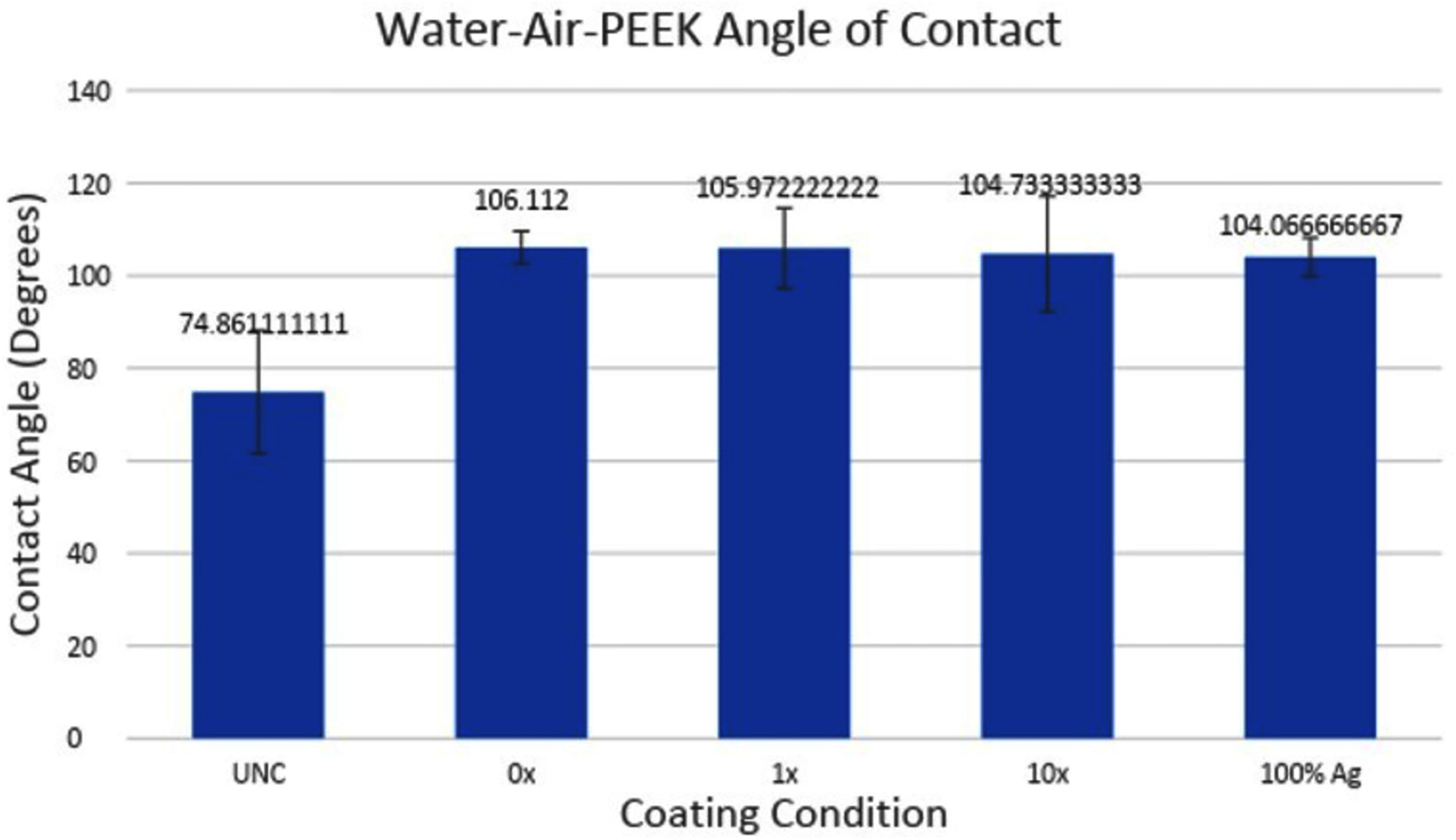
Water-air contact angle on UNC, 0×, 1×, and 10×, 100% Ag coatings. All coating concentrations demonstrated a contact angle of greater than 104, indicating increased hydrophobicity. Bacteria tend to adhere and form biofilms of hydrophilic surfaces, indicating that this change would decrease bacterial adherence. UNC, uncoated; Ag, silver.
AgCar elution rate
Figures 3 and 4 show the Ag concentration in solution after coated PEEK implants were allowed to elute throughout 24-h time periods.

Silver carboxylate elution curve for UNC, 0×, 1×, and 10× PEEK implants. Comparison indicates that only the 10× matrix strongly demonstrates a delayed release through 96 hours. The 100% Ag condition is not included because of scaling factors. UNC, uncoated; PEEK, polyether ether ketone; Ag, silver.
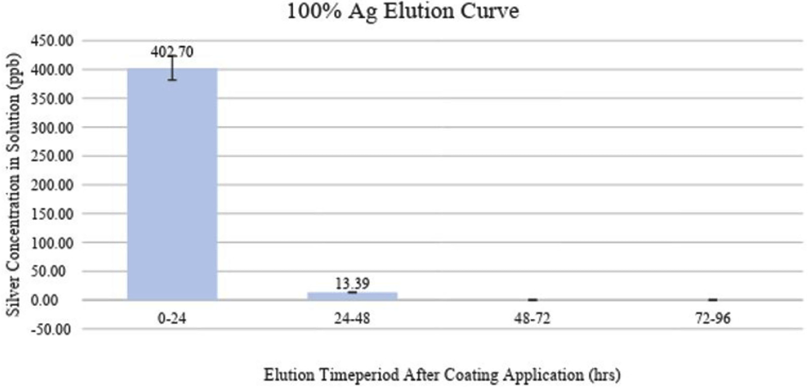
Silver carboxylate elution curve for 100% Ag PEEK implants. Day 1 shows a large silver concentration, whereas days 3 and 4 show no silver concentrations in solution. Ag, silver; PEEK, polyether ether ketone.
The 0× implants demonstrated undetectably low concentrations of AgCar for every day examined (Fig. 3). In contrast, the 1× implant eluted 2.86 ppb the first day and decreased by 35%–50% for each subsequent day (Fig. 3). The 10× condition eluted 1.61 ppb of AgCar during the first 24 hours. Following the first 24 hours, AgCar concentrations rose by 1600% to 27.38 ppb and proceeded to increase slightly through days 3 and 4 (Fig. 3).
The 100% Ag coating demonstrated a high initial elution of 402.70 ppb during the first 24 hours, subsequently dropping by 96.7% to 13.39 ppb after 48 hours (Fig. 4). Days 3 and 4 showed undetectable concentrations of AgCar in solution, suggesting that all particles had been fully eluted in the first 48 hours (Fig. 4).
Coating durability
Compression testing
Table 2 lists the average stiffness, peak force, and peak displacement for UNC and 10× coating conditions on silicone. The peak displacement for all samples was 2.61 mm because of the uniform sample thickness and the 75% thickness end point for each trial. The 10× implant’s peak force was 3.1% larger compared with the UNC condition (17.41 vs. 16.89), resulting in a substantially greater stiffness value for the 10× coated samples (9.32 vs. 8.73, p = 0.001).
Compressive Properties of Silicone Implants With and Without 10x Silver Carboxylate Coating Comparison of Stiffness, Peak Force, and Peak Displacement Between Uncoated (UNC) and 10x AgCar-Coated Silicone Samples During Compression Testing. Values Represent Mean ± Standard Deviation

UNC, uncoated.
Frictional coefficient
Table 3 displays the static and dynamic force as well as the coefficients for the four conditions on silicone. When grouping by coating condition, there was no statistically significant difference in frictional coefficient between the 10× coated and UNC implants (0.117 vs. 0.115, p = 0.352). In contrast, lubricated implants had substantially lower frictional coefficients compared with unlubricated (0.108 vs. 0.123, p < 0.005).
Static and Dynamic Frictional Coefficients of Silicone Implants With and Without 10x Silver Carboxylate Coating Comparison of Static and dynamic Forces and Corresponding Frictional Coefficients for Uncoated (UNC) and 10x AgCar-Coated Silicone Samples Under Lubricated and Unlubricated Conditions. Values Represent Mean ± Standard Deviation

UNC, uncoated.
The 10× unlubricated and UNC lubricated equally had the highest dynamic coefficient of friction, at 0.104 (Table 3). There was no statistical difference in dynamic coefficient between groups (p = 0.102).
Fatigue wear rate
Dissolved metal analysis following accelerated wear testing is displayed in Table 4. Titanium concentrations were below detectable levels for all silicone samples and all wear cycles tested (Table 4). One sample demonstrated wear-induced Ag release within the first interval cycle, slightly above the detectable level at 1.05 mg/L. All other samples demonstrated undetectable wear induced Ag release at any interval cycle.
Dissolved Metal Analysis of Silicone Implants Following Accelerated Wear Testing Measured Concentrations of Silver (Ag) and Titanium (Ti) Released from 10x AgCar-Coated Silicone Samples Across Sequential Wear Cycles. “ND” Indicates Values Below the Detectable Limit. Values Represent Mean ± Standard Deviation
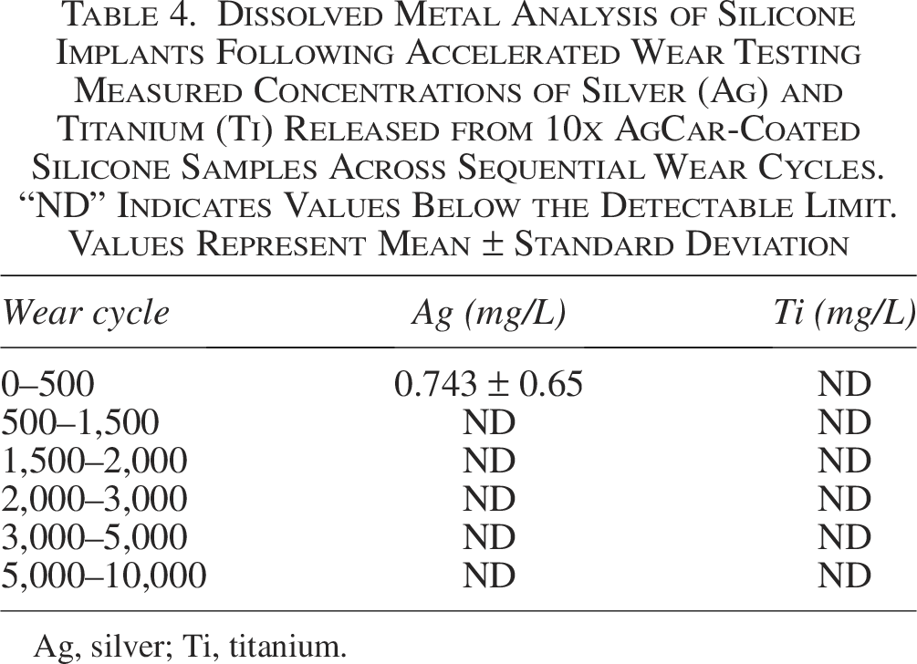
Ag, silver; Ti, titanium.
Discussion
The rising threats of orthopedic device-related infection (ODRI) and antimicrobial resistance both necessitate the development of novel therapeutics and treatment methods. A 95% TiO2:5% PDMS AgCar doped chemistry has previously shown extended efficacy as a coating on orthopedic implants against methicillin-resistant S. aureus, C. acnes, S. epidermidis, and multi-drug resistant S. marcescens.11,18–21 Coating application prevents adherence, proliferation, and biofilm formation of these problematic pathogens, thus serving as a valuable tool against both SSI and ODRI. The biochemical activity of AgCar has been well characterized 18 ; however, little research has been done on the physical properties of the coating following its application on implants. We characterized the coating’s effect on PEEK implant surface roughness and implant hydrophilicity/hydrophobicity, while also studying AgCar elution rate, wear, and durability. Results demonstrate that the addition of a TiO2-PDMS matrix impregnated with AgCar substantially reduces the surface roughness of the material while introducing hydrophobicity to implant materials. Overall, these physical properties help reduce bacterial adhesion on implants. Furthermore, our results demonstrate that the coating maintains extended elution through 96 hours and is durable against wear.
Addition of the TiO2-PDMS matrix acted to reduce roughness compared with the untreated PEEK implants. A rougher surface allows for easier anchoring to an implant and is therefore preferred by bacteria. Consequently, the addition of the matrix alone in the 0× condition may mechanically act to prevent bacterial adherence and subsequent biofilm formation. However, we found that implant surface roughness increases as the amount of AgCar present increases. Even so, the 95% TiO2:5% PDMS matrix substantially mitigates increases in roughness because of Ag. This is most apparent when comparing the 10× coating to 100% Ag, which has comparable amounts of AgCar but substantially greater roughness (492.0 vs. 335.6, p < 0.0001).
Of note, standard deviations of the RMS values used to determine roughness were quite high relative to the average values of roughness. This is likely because of factors creating variability in surface topography. For one, the imprecise nature of dip coating may lead to changes in coating thickness between implants. In addition, it is possible that surface micro-scratches were produced by the tweezers when transferring the implants to and from the coating solutions. Finally, the machining of the implants into their final shape may have caused certain edges to be rougher than others.
Only the UNC PEEK implants exhibited hydrophilic behavior. All coated implants exhibited hydrophobic behavior with substantially lower contact angles. This suggests that the presence of the matrix and Ag, whether independently or together, induces hydrophobicity on the surface. Bacteria prefer to adhere to a more hydrophilic surface, so the presence of the coating acts to mechanically deter bacterial anchoring and biofilm formation.6,7
The elution tests showed consistent and increasing elution from the 10× coating through 96 hours. In contrast, the 100% Ag condition showed a large and immediate release of AgCar in the first 24 hours, followed by a 96.7% decrease by day 2, and no release after 48 hours. This confirms prior work emphasizing the importance of the TiO2-PDMS matrix for controlled release of AgCar. Taken in concert, the 100% Ag coating could be effective at initially eliminating bacteria but does not offer any sort of sustained protection. Furthermore, the greater concentrations of Ag observed with 100% Ag yield increased risk for toxicity, highlighting the importance of the TiO2-PDMS matrix in a clinical setting.
Application of the 10× coating was found to increase silicone liner stiffness by 0.5 N/mm, a change of 6.8%. It is true that bacteria prefer to adhere to stiffer surfaces; however, much larger changes in stiffness are typically required for an appreciable increase in bacterial adherence. 7 Therefore, although there was a statistically significant difference in stiffness following application of the 10× coating, it is unlikely to be clinically significant. By virtue of its greater elasticity, silicone provides a softer surface which must be maintained to be effective in the residual limb-liner interface. AgCar has previously been demonstrated as efficacious on silicone liner. 11 The minimal changes to stiffness following coating application confirm that following coating, the material’s valuable flexibility is maintained.
There was no effect on dynamic friction by either coating or lubrication. Overall, only static friction was substantially changed during frictional testing, and this was because of lubrication and not the coating itself. There were no substantial changes for the dynamic coefficient of friction. Frictional coefficients for all cases were found to be between 0.087 and 0.136, indicating a low resistance to motion both statically and dynamically. Implants are manufactured to minimally impede natural body movement. Our results indicated that application of AgCar would not change this valuable characteristic.
The fatigue wear rate studies did not detect titanium at any point and only detected Ag in one of the 18 sample vials. This detection came at the beginning of the trial and was only 0.05 mg above the detection threshold. These results confirm that the TiO2 matrix is chemically bonded to the material and cannot be abraded off the implant surface by abrasion, thereby keeping the matrix attached. It is unclear on whether the Ag presence came from natural AgCar elution in an aqueous environment or from mechanical abrasion. Still, AgCar is embedded within the matrix, which has proved to be resilient in this situation. In addition, elution occurred within the first 8.5 minutes. Both these points support that the minimal Ag detected is from natural AgCar elution that occurs in an aqueous environment rather than mechanical removal. The human environment is subject to many lateral and torsional forces, and the coating must be able to remain intact. The resistance to abrasion of the coating indicates that it will remain adhered following application.
There are limitations to this study. Fatigue wear testing could not detect levels of AgCar below 0.1 mg/L, although levels this low are likely not of clinical significance. We solely tested AgCar on PEEK and silicone implant materials. Different implant materials have varied surface properties and, thus, unique physical properties when treated with AgCar. We specifically chose PEEK and silicone given the demonstrated antimicrobial efficacy of AgCar on these materials. Furthermore, PEEK, a rough and micro-topographically complex material, and silicone, a softer, more flexible material capture a broad spectrum of implants. We therefore believe that although our results are specific to the materials tested in this study, they may be broadly generalizable to other implant types. Finally, physical properties may be altered following implantation. Further work is required to study if the physical properties of AgCar may change in vivo.
Conclusion
This study characterized the physical and chemical properties of a 95% TiO2:5% PDMS AgCar coating on PEEK and silicone implant materials. Overall, the 10× coating demonstrated the best results for inhibitory behavior on two distinct fronts: mechanically through roughness and increased hydrophobicity and chemically through AgCar elution activity. It was also shown that the 10× coating will not impair physical performance and durability of the implant within the body. Taken in conjunction with the proved biologic efficacy of antimicrobial activity from past studies, the 95% TiO2:5% PDMS matrix doped with a 10× AgCar concentration is a prime candidate for clinical application.
Footnotes
Author Disclosure Statement
D.G. is an equity holder in BI Medical, LLC. C.T.B. holds equity in BI Medical, LLC., and owns stock in Biointraface, Inc. All other authors declare that they have no financial or personal relationships that could inappropriately influence this work.
Funding Information
This study was supported by the Diane N. Weiss Foundation, the Sipprelle Family Foundation, and the National Institute of Allergy and Infectious Diseases of the National Institutes of Health under Award Number R03 AI159776.