



Abstract
Albiflorin (AF), a primary bioactive compound from the roots of Paeonia lactiflora Pall. (Peony), demonstrates anti-inflammatory, antioxidant, and cytoprotective effects. This study aims to investigate the antithrombotic potential of AF and clarify its mechanisms of action. Its anticoagulant effects were assessed via activated partial thromboplastin time, prothrombin time, thrombin, and factor Xa (FXa) activities, as well as fibrin formation and platelet aggregation. Additionally, the effect of plasminogen activator inhibitor type 1 and tissue-type plasminogen activator was evaluated in tumor necrosis factor-α-stimulated human umbilical vein endothelial cells (HUVECs). Our experiments revealed that AF exhibited antithrombotic activity comparable to that of rivaroxaban, a well-established direct FXa inhibitor, particularly in suppressing FXa activity and platelet aggregation induced by adenosine diphosphate (ADP) and U46619, a synthetic thromboxane A2 analog. AF also reduced the surface expression of P-selectin, inhibited phosphorylation of myristoylated alanine-rich C kinase substrate, and decreased PAC-1 activation following ADP or U46619 stimulation. Furthermore, AF enhanced nitric oxide production while preventing excess endothelin-1 release in HUVECs exposed to these agonists. In vivo experiments in mouse models of arterial and pulmonary thrombosis demonstrated that AF is a potent anticoagulant and antithrombotic agent. These findings indicate that AF could serve as a promising lead compound for developing innovative anti-FXa and antiplatelet therapeutics.
INTRODUCTION
Disruptions in intrinsic and extrinsic coagulation pathways, along with platelet activation processes—including adhesion, aggregation, and granular release—are central to thrombus formation, a leading cause of global mortality. 1 In recent years, a new group of anticoagulants—known as direct oral anticoagulants (DOACs)—has emerged for the management of thromboembolic disorders. Several preclinical and clinical studies show that DOACs surpass older agents such as warfarin, offering more predictable pharmacokinetics, faster therapeutic onset, and a reduced risk of drug–drug interactions.2,3 DOACs demonstrate significant efficacy in preventing and managing venous thromboembolism and in reducing the incidence of stroke and systemic embolism in patients with nonvalvular atrial fibrillation.4,5 They also offer a more favorable safety profile, including a lower risk of major bleeding than that of more traditional anticoagulant therapies. Consequently, international consensus guidelines now recommend DOACs for controlling and treating thromboembolic disease.6–8 Direct thrombin inhibitors and factor Xa (FXa) inhibitors have largely replaced vitamin K antagonists and partly replaced low-molecular-weight heparins across many cardiovascular indications. These agents act through distinct mechanisms and generally avoid the paradoxical thrombotic effects occasionally observed with conventional anticoagulants. Nevertheless, some evidence indicates that dabigatran may be associated with a higher risk of myocardial infarction, a concern that requires careful clinical consideration.9–12
Robust clinical research is essential for clarifying the interactions between anticoagulant and antiplatelet therapies. The AGUSTUS trial shows that while initiating therapy with a combination of a yclooxygenase-1 (COX-1) inhibitor, a P2Y12 blocker, and a FXa inhibitor offers short-term benefits, this regimen ultimately increases the risk of major bleeding. 13 Therefore, a comprehensive assessment of individual patient profiles and personalized therapeutic strategies is critical for optimizing outcomes when using combination regimens. 14
Platelet aggregation is central to many thromboembolic disorders, as platelets are essential not only for maintaining hemostasis but also for initiating and amplifying thrombus formation. 15 Comprehensive clinical investigations demonstrate that the early administration of antiplatelet therapy enhances patient prognosis in conditions including heart attacks, coronary stent procedures, and severe stomach bleeding. Nonetheless, the ongoing need for developing novel antiplatelet agents remains owing to bleeding complications and treatment resistance associated with current therapies.16,17
In traditional Chinese medicine, thrombotic diseases are commonly attributed to blood stasis syndrome. 18 While aspirin remains a cornerstone for preventing thrombosis, growing evidence suggests that certain patients exhibit reduced responsiveness to its antithrombotic effects, manifesting as aspirin resistance. This has intensified interest in identifying alternative or complementary treatments from traditional Chinese medicine to address blood stasis, especially in patients with reduced aspirin responsiveness. 19
Albiflorin (AF; Fig. 1A), a major glycoside isolated from the roots of peony (Ranunculaceae), has drawn attention for its anti-inflammatory, antioxidant, and neuroprotective effects. 20 AF also alleviates neuropathic pain and anxiety-like behaviors resulting from chronic sciatic nerve compression via inhibiting NLRP3 inflammasome activation. 21 Additionally, AF mitigates inflammatory damage and oxidative stress via modulating the Nuclear Factor kappa B/NOD-, LRR-, and pyrin domain-containing protein 3 (NF-κB/NLRP3) pathway in models of methotrexate-induced enteritis 20 and influences neurological inflammation and metabolic dysfunction. 21 Despite these diverse biological activities, the potential antithrombotic effects of AF—particularly on FXa and platelet function—remain unexplored. Therefore, this study aims to determine whether AF exerts antithrombotic activity by assessing its effects on FXa function, coagulation parameters, and platelet aggregation. We further evaluated its efficacy in vivo using murine thrombosis models. Our findings could provide the first evidence that AF exerts antithrombotic effects by inhibiting FXa and platelet function.

MATERIALS AND METHODS
Cell culture and reagents
Human umbilical vein endothelial cells (HUVECs) were obtained from Cambrex Bio Science (Charles City, IA, USA) and cultured following established protocols.22,23 Tumor necrosis factor (TNF) and rivaroxaban were purchased from Abnova (Taipei, Taiwan) and Bayer HealthCare (Leverkusen, Germany), respectively. Collagen and AF were sourced from Sigma-Aldrich (St. Louis, MO, USA), and U46619 was provided by Calbiochem-Novabiochem Corp. (San Diego, CA, USA). A collection of coagulation-related proteins—including plasmin, FVIIa, FX, FXa, activated protein C, tPA, trypsin, and thrombin—was obtained from Haematologic Technologies (Essex Junction, VT, USA). Reagents for thromboplastin assays, including activated partial thromboplastin time (aPTT) and prothrombin time (PT), were purchased from Fisher Diagnostics (Middletown, VA, USA). Chromogenic substrates specific for various enzymes—S-2222 for trypsin, S-2228 for tPA, S-2238 for thrombin, S-2251 for plasmin, S-2366 for activated protein C, and S-2765 for FXa—were obtained from Chromogenix AB (Mölndal, Sweden). Flow cytometry antibodies, including anti-CD61-Fluorescein isothiocyanate (FITC), anti-CD62P-PE, anti-PAC-1, and anti-CD61-PE, were sourced from BD Pharmingen (BD Biosciences, San Diego, CA, USA), while anti-tissue factor (TF) antibodies were purchased from Santa Cruz Biotechnology (Santa Cruz, CA, USA).
Animal care and blood correction
Seven-week-old male C57BL/6 mice (average weight 27 g) were obtained from Orient Bio Co. (Sungnam, South Korea) and acclimated for 12 days before experimentation.22,23 All animal handling procedures followed the Guidelines for the Care and Use of Laboratory Animals and were approved by the Institutional Animal Care and Use Committee of Kyungpook National University (IRB No. KNU 2021-107). Experimental compounds were administered intravenously in a 200 μL volume containing 10% (v/v) solution. Subsequently, blood samples (800 μL) were collected via cardiac puncture of each mouse, centrifuged at 1500×g for 15 min to isolate plasma, and promptly stored at −80°C. Plasma proteins remained stable under these storage conditions, allowing their immediate use in subsequent analyses.
Human plasma and platelets
Fasting venous blood was obtained from ten normotensive, nonsmoking volunteers (6 female, 4 male; aged 24–28 years) with no documented history of allergy, cardiometabolic disease, or nutritional supplement use. Following immediate centrifugation of citrate-anticoagulated samples at 1300×g for 15 min, platelet-rich plasma (PRP) was prepared and adjusted to a standardized concentration of 1 × 10⁹ platelets/mL. Washed platelets were resuspended in (2-hydroxyethyl)-1-piperazineethanesulfonic acid (HEPES) buffer with 1 mM CaCl2, equilibrated for 30 min at room temperature, and subsequently used in 10 µL aliquots for coagulation assays. The study was conducted in accordance with the ethical guidelines approved by the Kyungpook National University Hospital Institutional Review Board (approval number KNUH 2012-01-010).
In vitro coagulation assay
Plasma for PT and aPTT analyses were obtained from blood drawn 1 h postadministration of dimethyl sulfoxide (DMSO) or AF, followed by centrifugation at 2000×g for 10 min. Coagulation assays were performed on a thrombotimer (Behnk Elektronik, Germany) according to established methods. 24 In the aPTT procedure, 100 µL of platelet-poor plasma was incubated with an equal volume of silica-based activator reagent at 37°C for 1 min prior to the addition of 100 µL of 20 mM calcium chloride to initiate clotting. For PT determination, 100 µL of plasma was added to 200 µL of prewarmed thromboplastin reagent and incubated at 37°C for 3 min before measuring the clot formation time.
In vitro platelet aggregation assay
PRP was incubated with AF dissolved in DMSO for predetermined intervals of 1, 3, 5, or 10 min. Platelet activation was then triggered by adding one of the following agonists: collagen (1 mg/mL), adenosine diphosphate (ADP) (10 µM), U46619 (6 µM), or thrombin (3 U/mL). Platelet aggregation responses were then quantitatively measured using a Chronolog aggregometer (Havertown, PA, USA).
Inhibition of FXa amidolytic activity
AF was dissolved in a 50 mM Tris buffer (pH 7.4) and then combined with 150 µL of FXa solution at 1 U/mL. The mixture was incubated at 37°C for 1 min. Subsequently, 150 µL of 1.5 mM S-2222 substrate was added, and the change in absorbance of the reaction at 405 nm was monitored over 20 min using a spectrophotometer. Enzymatic activity was determined from the initial reaction velocity (Vi), calculated from the slope of the absorbance versus time plot. The percentage inhibition of AF was calculated using the formula:

Inhibitory constant for FXa
Kinetic inhibition constants (Ki) for AF were derived from chromogenic assays performed with at least seven compound concentrations in duplicate. Reactions were initiated by adding enzyme solution to a mixture containing 50 µL of substrate and 25 µL of either AF or control buffer in a microplate (Cergy Pontoise, France). Continuous spectrophotometric monitoring at 405 nm over a 60-min incubation at 37°C quantified the release of p-nitroaniline, with initial velocities expressed in mOD/min. Ki values were subsequently determined through Dixon plot analysis, graphing the inverse of the maximal velocity against the corresponding inhibitor concentration.
Production of FXa on the surface of HUVECs
HUVEC monolayers in 96-well plates were exposed to AF for 10 min prior to a 6-h stimulation with 10 ng/mL TNF-α. Following this, cells were incubated with 10 nM FVIIa in HEPES-buffered saline (pH 7.45) containing CaCl2 and BSA for 5 min, with or without a TF-neutralizing antibody. Factor X was then introduced, and the reaction proceeded for 15 min before termination with EDTA. FXa activity was subsequently measured by cleavage of the chromogenic substrate S-2765, with absorbance at 405 nm used to calculate enzyme concentration from a purified FXa standard curve.
Cell viability assay
Cell viability was assessed using 3-(4,5-dimethylthiazol-2-yl)-2,5-diphenyltetrazoliumbromide (MTT) assay, following the method described by Hahn et al. 25 Cells were seeded in 96-well plates at a density of 5000 cells per well and allowed to adhere for 24 h. After washing with fresh culture medium, the cells were treated with AF. Subsequently, the medium was replaced with 100 µL of MTT solution (1 mg/mL), and cells were incubated for an additional 48 h. Formazan crystals were solubilized by adding 150 µL of DMSO to each well, and absorbance was recorded at 540 nm using a spectrophotometer to quantify cell viability.
Western blotting
Equal protein amounts from HUVEC lysates were separated with Sodium dodecyl sulfate–polyacrylamide gel electrophoresis (SDS-PAGE) and transferred onto Immobilon membranes (Millipore, Billerica, MA, USA) via electroblotting. The membranes were blocked and incubated for 1.5 h at room temperature with an anti-phospho-MARCKS antibody (Santa Cruz, CA, USA), followed by HRP-linked secondary antibody treatment. Chemiluminescent signals were detected using Enhanced chemiluminescence (ECL) reagents. To confirm equal protein loading, membranes were probed with anti-β-Actin antibody (1:1000, Santa Cruz, CA, USA). Densitometric quantification was performed using ImageJ Gel Analysis software (NIH, Bethesda, MD, USA).
Measurement of intracellular calcium mobilization
The quantification of platelet intracellular calcium ([Ca2+]i) was conducted as described by Wu et al. 26 Briefly, Fura 2-AM-loaded platelets were washed, resuspended in a calcium-free buffer, and adjusted to 5 × 10⁷ cells/mL. After reintroducing extracellular calcium, agonist stimulation was performed for 1 min. [Ca2+]i was then derived from the 339/500 nm fluorescence excitation/emission ratio using the standard calibration equation of Grynkiewicz et al. 27
Measurement of PAC-1 and P-selectin expression
PAC-1 and P-selectin expression levels were determined using a modified version of established protocols. 28 Washed platelets (0.5 mL; 2 × 10⁸ platelets/mL) were incubated with 20 µL of either DMSO or AF (10–20 µM) for 3 min. Platelets were then activated with either 10 µM ADP or 6 µM U46619 for 6 min at 37°C, and then cooled to 4°C. For PAC-1 detection, 50 µL of the treated platelet suspension was stained with saturating amounts of activation-specific anti-PAC-1 antibody and anti-CD61-PE, followed by incubation in the dark for 25 min. Similarly, P-selectin expression was evaluated by incubating 50 µL of the platelet suspension with anti-CD62P-PE and anti-CD61-FITC antibodies under the same conditions. The samples were then diluted <4-fold and analyzed by flow cytometry (BD Biosciences, San Diego, CA, USA). Platelet populations were identified based on forward and side scatter characteristics and gated for CD61 positivity to exclude background. A minimum of 5000 events per sample was recorded, and mean fluorescence intensity was quantified using the BD Accuri C6 software. Data were obtained from five independent donor samples.
Quantification of nitric oxide and endothelin-1
NO and ET-1 levels in the cell culture medium were measured using a commercially available enzyme-linked immunosorbent assay kit (R&D Systems, Minneapolis, MN, USA).
Arterial thrombosis animal model
An arterial thrombosis model was established in male C57BL/6 mice using ferric chloride (FeCl3). The mice fasted overnight prior to intravenous administration of the test compound, dissolved in 0.2 mL of DMSO, and were then allowed to rest. The carotid artery was surgically exposed, and a 200 µm diameter cotton thread soaked in 0.25 M FeCl3 was applied to the adventitial surface of the vessel for 5 min. Following removal of the FeCl3-soaked thread, the site was rinsed with saline. Thrombus formation was monitored in real time at 35°C using three-dimensional imaging techniques. Thrombi were categorized based on their size: small (50–75 µm), medium (100–150 µm), large (200–300 µm), or multiple when more than three thrombi were observed. Additionally, the time required for a significant thrombus to fully occlude the carotid artery after FeCl3-induced endothelial injury was recorded.
Acute pulmonary thrombosis induced via combined treatment of collagen and epinephrine in an animal model
Following an overnight fast, mice were divided into groups of 10. AF dissolved in DMSO was administered intravenously to each animal. One hour later, acute thrombosis was induced via injecting a mixture of collagen and epinephrine (500 µg/kg each). The mice were monitored for 15 min to assess recovery, onset of paralysis, or mortality. For histological assessment of pulmonary thrombus formation, lung sections were stained with hematoxylin and eosin and randomly selected from five mice per group. Within each left lung lobe section, five fields were randomly chosen to quantify the arteries larger than 20 µm in diameter containing thrombi, totaling 25 fields per group. Approximately 60–80 vessels per lung segment were imaged using a Leica imaging system (Germany). Thrombus burden was quantified as the number of thrombi per 25 mm2 lung tissue, following established criteria. 29
Statistical analyses
Data are presented as the means ± standard deviations from five independent experiments, each performed in duplicate. After a significant one-way analysis of variance, pairwise comparisons between groups were conducted using Tukey’s post hoc test. Differences were considered statistically significant at P < .05.
RESULTS
Effects of AF on cellular viability, clotting time in vitro and ex vivo
The cytotoxic effects of AF on HUVECs were evaluated using the MTT assay. Figure 1B shows that the exposure to AF at concentrations ranging from 5 to 50 µM does not adversely affect cell viability. The effects of AF on coagulation were subsequently examined by measuring aPTT and PT, key indicators of blood clotting. Rivaroxaban, a direct FXa inhibitor, served as a positive control. AF treatment significantly prolonged aPTT at 5–20 µM (Fig. 2A). Notably, AF and rivaroxaban at 8.17 and 7.19 µM, respectively, each doubled the clotting time during aPTT assessments. For rivaroxaban, aPTT values increased from 20.1 s (baseline) to 31.5 s (2.5 µM), 42 s (5 µM), 53 s (10 µM), and 57 s (20 µM), while AF produced values of 20.9 s (baseline), 21.1 s (2.5 µM), 37.1 s (5 µM), 57 s (10 µM), and 65 s (20 µM). To corroborate these in vitro findings, mice (n = 5 per group) received intravenous injections of AF once daily for 4 days in ex vivo experiments. These results demonstrated that AF dose-dependently extended clotting times (Fig. 2B), with doses of 0.34 and 0.38 mg/kg for AF and rivaroxaban doubling the aPTT values, respectively. Circulating blood volume in mice was estimated at ∼2 mL per animal, based on an average body weight of 27 g and a blood volume of 72 mL/kg. 30 Consequently, after AF administration at 0.09, 0.18, 0.36, or 0.71 mg/kg, the corresponding estimated plasma concentrations were 2.5, 5, 10, or 20 µM, respectively.
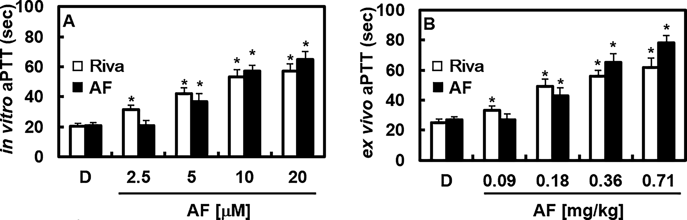
Effects of AF on clotting time.
Effects of AF on platelet aggregation in vitro
The antiplatelet activity of AF was evaluated against multiple agonists in PRP. In contrast to rivaroxaban, which does not inhibit platelet aggregation,31,32 AF produced a concentration-dependent suppression of aggregation induced by ADP, collagen, thrombin, and the thromboxane A2 analog U46619, as shown in Figure 3.
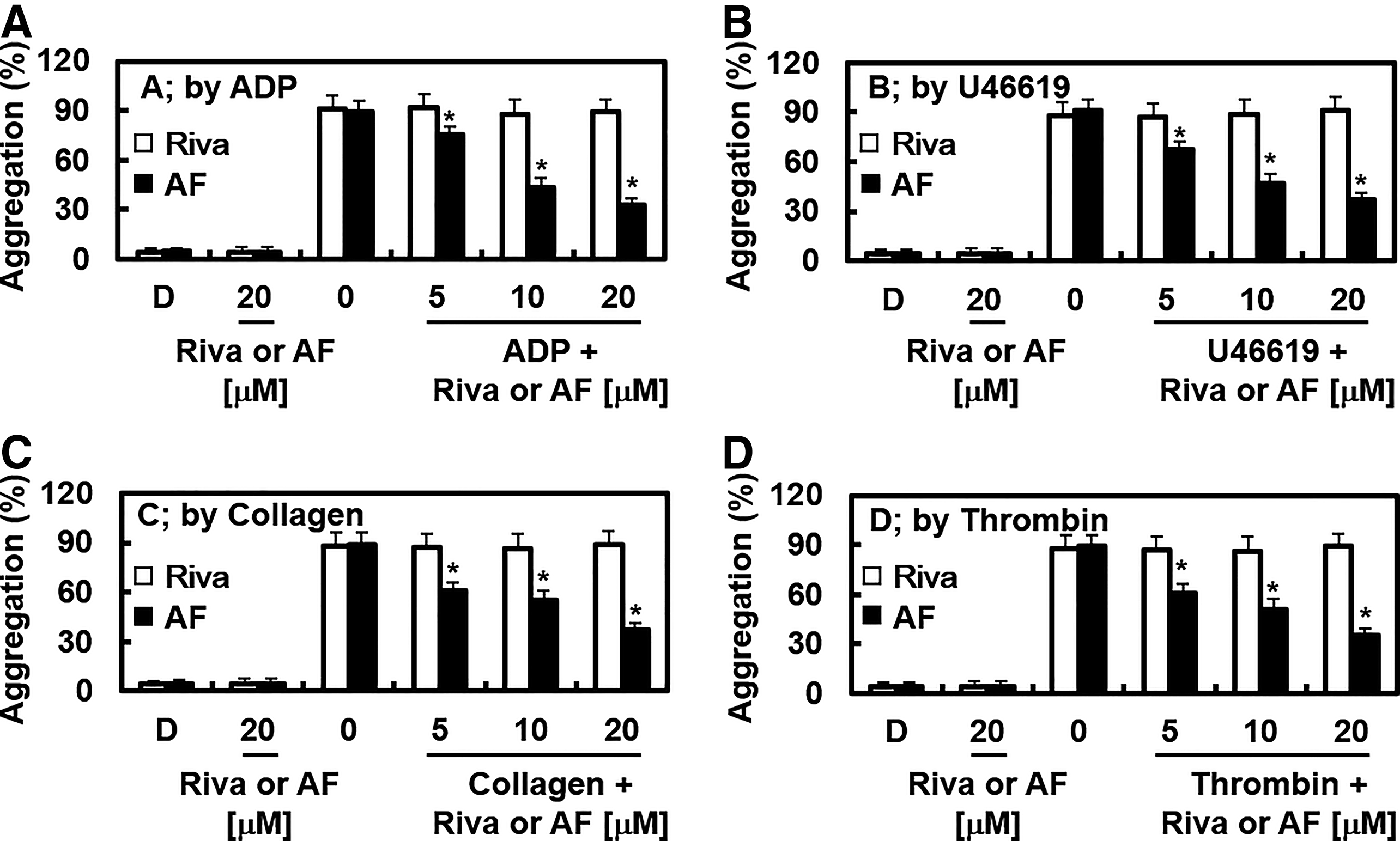
Effects of AF on agonist-induced platelet aggregation. Platelet-rich plasma (PRP) samples were incubated for 5 min with varying doses of AF, Riva, or the vehicle control. Platelet aggregation was then triggered using ADP (10 µM, panel A), U46619 (6 µM, panel B), collagen (1 µg/mL, panel C), or thrombin (3 U/mL, panel D), and the extent of aggregation was measured with an aggregometer. Results represent the means ± SD from five independent experiments. Asterisks indicate statistical significance (*P < .05) compared with the vehicle-treated group
Effects of AF on animal models of arterial and pulmonary thrombosis
To investigate the antithrombotic effects of AF in vivo, we employed a FeCl3-induced carotid artery thrombosis model based on the protocol of Izuhara et al. 33 Table 1 summarizes the effects of AF on the onset and extent of FeCl3-induced thrombus formation. While FeCl3 induced rapid formation of sizable thrombi through endothelial injury, AF treatment markedly delayed thrombus growth. Furthermore, AF demonstrated a protective effect in an in vivo pulmonary thrombosis model in which mice injected with a collagen–epinephrine cocktail developed severe lung thrombosis and immediate paralysis. AF administration significantly reduced mortality and thrombus burden in lung tissues compared with mice treated with only the collagen–epinephrine mixture (Fig. 4; Table 1).

Effects of AF on microvascular thrombosis. Representative H&E sections of lungs from mice treated as labeled. C + E: Treatment of collagen with epinephrine. Magnification: × 200. Scale bar: 100 µm.
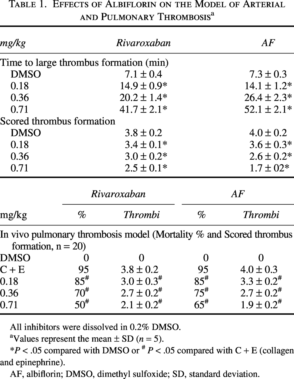
All inhibitors were dissolved in 0.2% DMSO.
aValues represent the mean ± SD (n = 5).
*P < .05 compared with DMSO or # P < .05 compared with C + E (collagen and epinephrine).
AF, albiflorin; DMSO, dimethyl sulfoxide; SD, standard deviation.
Effects of AF on the catalytic activity and production of FXa in vitro
To elucidate how AF modulates coagulation and platelet aggregation, we investigated its effects on FXa activity and production. AF inhibited the enzymatic activity of FXa on the chromogenic substrate S-2222 in a dose-dependent manner. Michaelis–Menten analysis revealed that AF reduced reaction velocity, yielding slopes of 0.585 and 0.846 observed at 10 and 20 µM concentrations, respectively, compared with a slope of 0.458 in untreated controls (Fig. 5A). Lineweaver–Burk plots (Fig. 5A inset) showed that AF acted as a noncompetitive FXa inhibitor, reducing Vmax without altering the Km for S-2222. The calculated Ki for AF against FXa was 3.71 µM (Table 2). AF showed remarkable selectivity for FXa over other coagulation enzymes, with a selectivity ratio >300 based on Ki values (Table 2). Given that TF expression is essential for FVIIa-mediated activation of FX in TNF-α-stimulated HUVECs.32,34 We also assessed the effect of AF on this pathway. TNF-α treatment increased FVIIa-mediated FX activation ∼11.4-fold (105.1 ± 7.1 nM vs. 9.2 ± 0.7 nM; Fig. 5B), an effect significantly inhibited by anti-TF Immunoglobulin G (IgG) (12.2 ± 1.9 nM). Preincubation with AF reduced FX activation in a dose-dependent manner (Fig. 5B). Based on recent evidence that FXa directly promotes platelet aggregation, 35 we evaluated the ability of AF to inhibit FXa-induced platelet aggregation. AF significantly suppressed FXa-stimulated platelet aggregation (Fig. 5C).
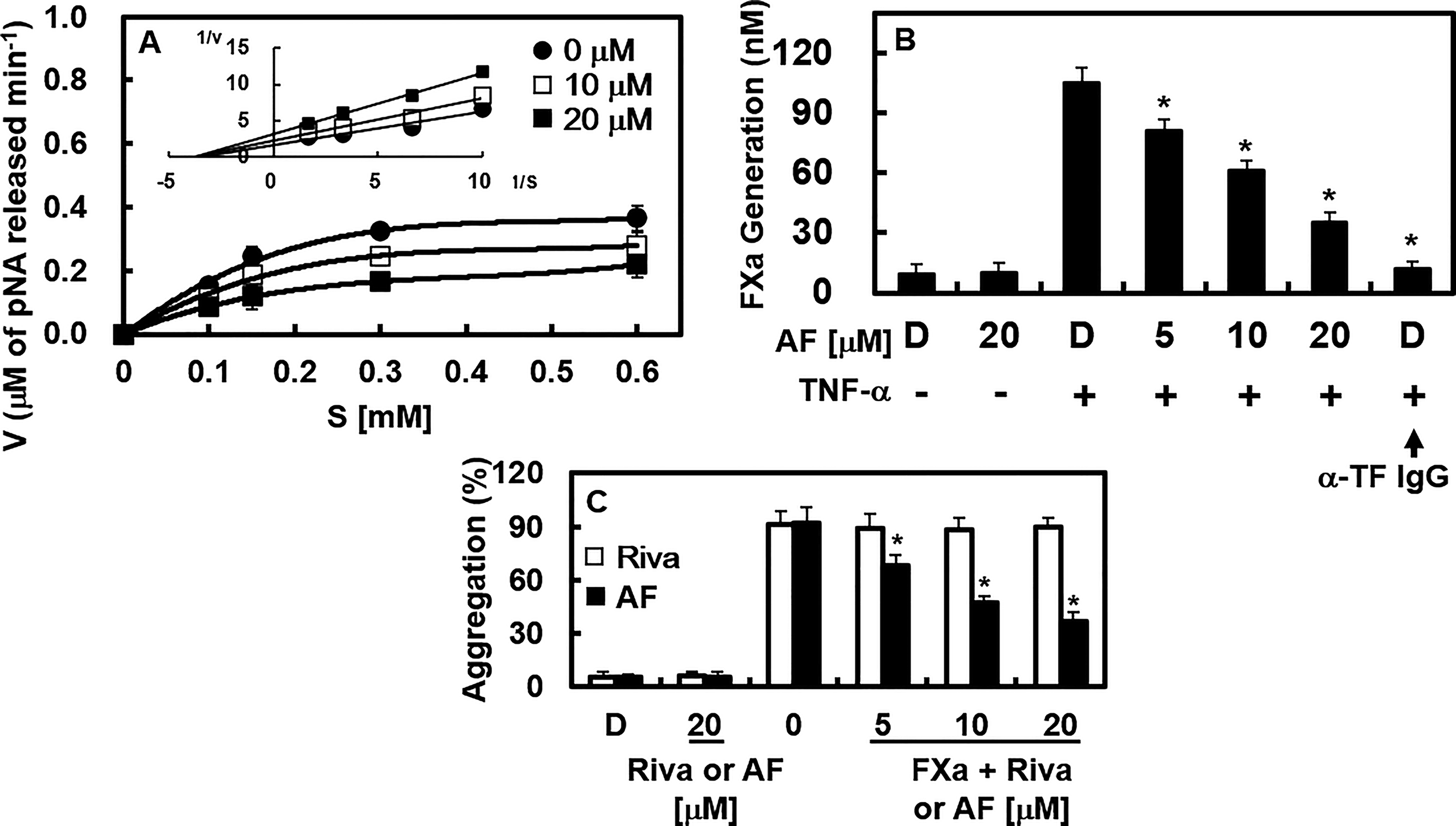
Effects of AF on the catalytic activity and production of FXa.
Enzyme Kinetics and Selectivity of Albiflorin Against Different Human Enzymes

tPA, tissue plasminogen activator.
aKi is represented by the mean ± SD (n = 5), µM.
bRatio = Ki enzyme/Ki Factor Xa.
Effects of AF on the activation of protein kinase C and mobilization of intracellular calcium
We then investigated the molecular basis through which AF modulates platelet aggregation. We examined the effect of AF on protein kinase C (PKC) activity by assessing MARCKS phosphorylation, a key PKC target in human platelets. Since PKC activation and [Ca2+]i elevation are key triggers of platelet aggregation, 36 we examined these pathways. AF treatment inhibited ADP- and U46619-induced MARCKS phosphorylation, indicating suppression of PKC signaling (Fig. 6A). Agonist-induced platelet activation triggers phospholipase C to cleave phosphatidylinositol 4,5-bisphosphate into inositol 1,4,5-trisphosphate and diacylglycerol, which elevate cytosolic calcium and activate PKC, respectively. 37 Ca2+ and PKC synergize to promote granule secretion and activate PAC-1 (GPIIb/IIIa), the key integrin in platelet aggregation. 37 Consistent with these mechanisms, AF reduced ADP and U46619-induced increases in intracellular Ca2+ (Fig. 6B and 6C). Collectively, these findings indicate that AF impairs platelet aggregation via inhibiting PKC activation and dampening calcium signaling.

Effects of AF on activation of protein kinase C (PKC) and mobilization of intracellular calcium.
Effects of AF on the expression of P-selectin and PAC-1
Upon platelet activation, P-selectin moves to the platelet surface, and fibrinogen bridges PAC-1 receptors on adjacent platelets, promoting aggregation. 38 AF treatment significantly reduced surface P-selectin and PAC-1 in ADP or U46619-stimulated platelets (Fig. 7A and 7B). The downregulation of key adhesion molecules via AF contributes to its capacity to suppress platelet aggregation.

Effects of AF on the expression of P-selectin and PAC-1 and the production of NO and ET-1.
Effects of AF on nitrogen monoxide and endothelin-1
NO—a key endogenous vasodilator—critically inhibits platelet aggregation and maintains arterial integrity. Conversely, ET-1, a potent vasoconstrictor synthesized via vascular endothelial cells, regulates NO release.39–41 Examining the effect of AF on NO and ET-1 levels clarified its mechanism of action in suppressing platelet aggregation. A proper balance between NO and ET-1 production is essential for regulating vascular tone and maintaining vascular homeostasis.42,43 Our findings indicate that AF normalized the elevation of ET-1 induced by ADP or U46619 and restored NO secretion to baseline (Fig. 7C and 7D).
DISCUSSION
Various antiplatelet agents, alone or in combination with other therapies, are currently employed to manage thrombotic conditions. Despite their widespread use, prolonged treatment with these drugs often causes adverse effects.44,45 Our findings indicate that AF effectively inhibits platelet aggregation induced by ADP, U46619, collagen, and thrombin, highlighting its potential as a novel antithrombotic therapy with reduced side effects. Given the pivotal role of FXa in the coagulation cascade, 46 we hypothesized that its selective inhibition could provide a targeted and effective strategy for developing new antithrombotic drugs, as FXa bridges the intrinsic and extrinsic pathways. Current thrombin inhibitors have a narrow therapeutic window.47,48 Moreover, the relatively low-molecular-weight of AF (480.46 Da) compared with conventional FXa inhibitors may offer advantages, including simpler manufacturing and reduced immunogenicity.
We evaluated the anticoagulant effect of AF by measuring clotting times via the intrinsic (aPTT) and extrinsic (PT) pathways. 49 AF administration selectively and dose-dependently prolonged aPTT in mice without affecting PT, suggesting a preferential inhibition of the intrinsic coagulation cascade, potentially through factors such as XIa, XIIa, or the cofactor VIIIa. In parallel ex vivo platelet function tests, AF demonstrated targeted antiplatelet activity by concentration-dependently inhibiting aggregation triggered by ADP, collagen, U46619, and thrombin.
Our findings indicate that AF exerts antithrombotic effects comparable to those of rivaroxaban, effectively reducing FXa production and enzymatic activity, prolonging clotting times, and delaying thrombus formation and onset. While rivaroxaban had no effect on ADP or U46619-induced platelet aggregation, AF suppressed platelet aggregation in a dose-dependent manner. Mechanistic investigations showed that the antiplatelet effects of AF involve modulation of PKC activation, intracellular calcium mobilization, and downregulation of P-selectin and PAC-1 expression. Consequently, AF exhibits comprehensive antiplatelet activity by inhibiting platelet aggregation, prolonging coagulation, reducing cytosolic Ca2+ flux, suppressing FXa activation, reducing MARCKS phosphorylation via the PKC pathway, and downregulating P-selectin and PAC-1 expressions. Additionally, AF modulates vascular tone by reducing ET-1 synthesis, a potent vasoconstrictor, and increasing NO, a vasodilator. Platelet activation, adhesion, and aggregation are critical for initiating coagulation.46,50 Vascular injury disrupts the endothelium, exposing blood to subendothelial tissue and triggering platelet activation, TF-factor VII interaction, and fibrin clot formation. 46 The growing thrombus activates the intrinsic pathway via factors VIII and IX, thereby recruiting additional platelets and amplifying the coagulation cascade. 46 A key aspect of this amplification is the thrombogenic surface provided by activated platelets and endothelial cells. Thus, platelet activation and functional changes are key molecular targets for controlling coagulation and aggregation, highlighting their central role in antithrombotic and antiplatelet therapies.
The influence of rivaroxaban on platelet aggregation appears context-dependent.31,32,51–53 While in vitro studies, including our own, show no direct effect of rivaroxaban on aggregation induced by standard agonists, clinical observations by Petzold et al. note reduced aggregation in patients on therapy. 35 This discrepancy may arise from metabolic alterations or drug interactions in patients with comorbid cardiovascular and metabolic diseases, potentially unveiling indirect modulatory effects. Crucially, our data align with the premise that FXa is a potent platelet activator. AF, by effectively suppressing FXa-induced aggregation, functions as an antiplatelet agent through targeted interference with FXa-mediated signaling pathways.
The anticoagulant profile of AF raises questions about its potential effect on bleeding risk and hemostatic balance, alone or combined with other anticoagulant therapies. While this study shows that AF prolongs aPTT and inhibits FXa inhibition comparable to rivaroxaban, we did not assess clinically relevant safety endpoints, including spontaneous bleeding, delayed hemostasis after vascular injury, or interactions with standard antithrombotics. Given that most anticoagulant agents have a narrow therapeutic window, even modest additive or synergistic effects on coagulation or platelet function may increase the risk of hemorrhagic complications, particularly in patients on multiple medications or with hemostatic disorders. Therefore, future studies should systematically evaluate bleeding time, blood loss in surgical or trauma models, and recovery of hemostasis after AF exposure, and its pharmacodynamic interactions with other anticoagulants and antiplatelet drugs. Additionally, detailed toxicological and pharmacokinetic evaluations will be essential to define a safe AF dosing range and determine whether its dual effects on FXa and platelet function can be therapeutically harnessed without increasing bleeding risk.
Once in the circulatory system, rivaroxaban may interact with other drugs, potentially altering its properties. These modifications may influence platelet aggregation by stimulating or suppressing unidentified signaling pathways involved in platelet activation. Petzold et al. also highlight FXa as a powerful stimulator of platelet aggregation. 35 Our findings indicate that AF effectively inhibits FXa-induced platelet aggregation, suggesting its antiplatelet effect is mediated via the FXa signaling pathway.
In conclusion, this study identifies AF as a novel agent that targets the intrinsic coagulation cascade, acting as an FXa inhibitor and a suppressor of platelet aggregation. These insights may aid the development of novel treatments to prevent thrombotic disorders caused by abnormal coagulation.
AUTHORS’ CONTRIBUTIONS
Conceptualization, methodology, investigation: Y.H.J. and X.-X.D. Investigation: G.H. Conceptualization, methodology, supervision, resources, writing—reviewing and editing: J.-S.B.
Footnotes
AUTHOR DISCLOSURE STATEMENT
No competing financial interests exist.
FUNDING INFORMATION
This research was supported by the National Research Foundation of Korea (NRF) grant funded by the Korea government (MSIT) (NRF-RS-2025-00555195).