
Abstract
Lactoferrin (Lf), an iron-binding glycoprotein present in various exocrine secretions, exhibits a wide range of biological activities, including cell proliferation, immune modulation, and antimicrobial effects. This study investigates the bioactivities of commercially available Lf derived from bovine milk. Commercial Lf, with a purity exceeding 95%, was analyzed using chemical and cell-based assays. Fourier-transform infrared spectroscopy confirmed the protein’s identity through characteristic peaks. Cell viability assays demonstrated noncytotoxic effects, while cell morphology analysis and proliferation assays revealed that Lf stimulated human keratinocyte (HaCaT) proliferation in a concentration-dependent manner. Wound healing assays showed that Lf promotes wound closure, with optimal effects observed at lower concentrations. However, antimicrobial assays revealed that Lf alone did not exhibit activity against Staphylococcus aureus and Pseudomonas aeruginosa. These findings highlight the importance of purity and concentration in determining the biological activity of Lf. Further studies are warranted to investigate the molecular mechanisms underlying these effects and the potential applications of bovine Lf in therapeutic contexts.
INTRODUCTION
Lactoferrin (Lf) is an iron-binding glycoprotein an 80 kDa primarily found in milk and other human exocrine secretions, including saliva, tears, and gastrointestinal fluids. Lf exists in two forms: apo-Lf, the iron-unsaturated form predominantly present in breast milk, and holo-Lf, the iron-saturated form. 1 Both in vitro and in vivo studies have demonstrated the diverse biological activities of Lf, including cell proliferation, immune modulation, and antimicrobial and antiviral effects.2,3
Bovine lactoferrin (bLf) and human lactoferrin (hLf) share approximately 70% amino acid sequence homology and have similar tertiary structures. Commercially available purified bLf from bovine milk exhibits biological activities comparable with those of hLf. 4 Due to its numerous health-promoting and therapeutic potentials, Lf has been widely explored for nutraceutical applications. Lf acts as a key modulator of the innate immune system, influencing processes such as granulation tissue formation, reepithelialization, and the stimulation of fibroblast and keratinocyte proliferation and migration.5,6
In the context of wound healing, Lf’s multifunctional properties, including antimicrobial, antioxidant, and anti-inflammatory activities, play a central role. Evidence suggests that Lf promotes wound contraction and enhances the synthesis of extracellular matrix (ECM) components, including collagen and hyaluronan. 7 Its therapeutic potential is further supported by studies in skin conditions such as psoriasis and atopic dermatitis, where its immunomodulatory effects have shown promise.1,8
Although the benefits of Lf are well established, its biological activity can vary depending on factors such as source, glycosylation patterns, and extraction methods. Despite the high homology between hLf and bLf, variations in receptor-binding regions and glycosylation may lead to distinct bioactivities. For instance, bLf demonstrates broad-spectrum antimicrobial effects, while hLf exhibits more specialized immunological functions. 9 These distinctions underscore the necessity of evaluating Lf within specific biological and therapeutic contexts.
Considering these factors, this study aims to evaluate the bioactivities of commercially available bLf, focusing on its influence on keratinocyte proliferation, migration and antimicrobial activity against microorganisms most frequently associated with skin infections.
MATERIALS AND METHODS
Lactoferrin
The Lf assessed in the following tests was commercially obtained from Lactoferrin Company (Byron Bay, AUS). It was extracted from bovine-derived milk, gently spray-dried, and has a high purity level (>95%) with a pH value ranging between 5.2 and 7.2.
Chemical analysis
Fourier transformed infrared spectroscopy in the Attenuated Total Reflectance mode (ATR-FTIR, Nicolet iS50, Thermo Fisher Scientific Inc., Waltham MA, USA) was performed on the bLf powder. The FTIR spectra were baseline-corrected and normalized for analysis. The iron content of the commercial (bLf sample was determined by energy dispersive X-ray spectroscopy (EDX). The analysis was performed using an EDX-720 spectrometer under vacuum conditions, allowing qualitative and quantitative elemental characterization. The measurements were carried out in triplicate, and elemental quantification was obtained using the fundamental parameters method. The results were expressed as percentage composition, enabling the assessment of iron presence and relative abundance in the bLf sample.
Cell-based assays
Biological assays were performed using human skin keratinocyte lineage (HaCaT; Cell Lines Service, 300493). These cells were cultured in complete DMEM media (Dulbecco’s Modified Eagle’s Medium; Gibco, Carlsbad, CA, USA) containing 1% of antibiotic solution (Penicillin-Streptomycin 10,000 U/mL; Gibco) and 10% of fetal bovine serum (FBS; Gibco) in a humidified incubator (37°C, 5% CO2; Thermo Fisher Scientific). Lactoferrin was dissolved directly in DMEM, which served as the vehicle for its preparation.
Cell viability by alamarBlue™ assay
To evaluate cell viability, 3 × 10³ cells per well were seeded onto each of three different 96-well plates in complete DMEM for 24 h. After the cells attached to the microplate bottom, the supernatant was removed, and then the cells were treated with different concentrations of bLf (5 µg/mL, 10 µg/mL, and 50 µg/mL) and incubated for 24 h; this time point was denoted as day 1. Afterward, cells were incubated in DMEM without FBS containing 10% alamarBlue™ reagent (Invitrogen, Thermo Fisher Scientific Inc., Waltham, MA, USA) for 3 h. The same procedure was repeated at time points 3 and 7 days, with media refreshed every other day. Cell viability was determined at 560 nm (excitation range: 540–570 nm) and an emission of 590 nm (emission range: 580–610 nm) using a fluorescence-based plate reader (SpectraMax iD3, Molecular Devices, LLC, San Jose, CA, USA).
Cell proliferation by CyQUANT™ assay
Quantitative and qualitative CyQUANT™ cell proliferation assay (Invitrogen; Carlsbad, CA, USA) was conducted following the manufacturer’s instructions; this test evaluated total cellular DNA content by binding a fluorescent dye. Cells (2 × 10⁴) were seeded on 24-well plates (Corning—Costar, New York, NY, USA) in complete DMEM for 24 h. After this period, cells were exposed to different treatments: FBS-free media (negative control); complete media (with FBS) (positive control); FBS-free media containing 5 µg/mL of bLf (Lac 5+); and complete media containing 5 µg/mL of bLf (Lac 5−) for 1, 3, and 7 days. The treatments were refreshed every other day. At the end of each time-point, treatments were removed, the fluorescent CyQUANT™ reagent was added to the cells, and the plates were incubated for 30 min in the dark at 37°C and 5% CO2. Subsequently, for quantitative analysis, fluorescence intensity was measured at 480 nm excitation and 520 nm emission using a microplate reader (Spectra iD3; Molecular Devices LLC, San Jose, CA, USA); for qualitative analysis, photomicrographs of the wells were performed via a fluorescence microscope at 4× magnification (scale bar 460 µm; ECHO Revolve Microscope; Discover Echo Inc., San Diego, CA, USA).
Cell morphology
To evaluate cell morphology, 2 × 10⁴ cells were cultured in 24-well plates as previously described for 24 h. After this period, cells were exposed to different treatments according to each experimental group (negative control, positive control, Lac 5+, and Lac 5−) for 12, 24, and 48 h. At each time point, cells were fixed with 70% ethanol for 1 h at 4°C, followed by washing with phosphate-buffered saline (PBS; Gibco) and F-actin staining using red fluorescent dye for actin filaments (1:20 in PBS; ActinRed 555 ReadyProbes reagent; Invitrogen) for 30 min. After this period, cell nuclei were stained in blue with DAPI (1:5000 in PBS; Thermo Fisher Scientific). Photomicrographs of each well were obtained using a fluorescence microscope (ECHO Revolve Microscope) at 4× (scale bar 460 µm) and 10× (scale bar 180 µm) magnifications.
Wound healing by scratch assay
Cells were cultured in 24-well plates at a seeding density of 2 × 10⁵ cells per well in complete culture media and allowed to grow until a 100% confluent monolayer was formed. A scratch was created in the cell monolayer using a P200 tip, and cellular debris was then washed with sterile PBS. Next, cells were treated with 5 µg/mL of bLf in complete DMEM, supplemented with 1% FBS or without FBS. The closure of the scratched area was evaluated at time points 0, 6, 12, 24, and 48 h using the EVOS FL Auto 2.0 imaging platform combined with automated image analyses, and the cell migration rate was quantified by ImageJ software (Wayne Rasband, National Institute of Mental Health, NIH, Bethesda, MD, USA). The medium of the different groups was refreshed before analyzing each time point.
Antimicrobial assay by MIC
The reference strains used in this study were selected based on the most common bacteria isolated from chronic wounds. S. aureus (LAC, USA300; MRSA strain) and P. aeruginosa (PAO1) were generously provided by the laboratory of Dr. Christopher Fenno at the University of Michigan. The minimum inhibitory concentration (MIC) was determined in eightfold by the broth microdilution technique using a modified version of the reference documents M27-A320 and M7-A721. To standardize the inoculum, culture suspensions were prepared and diluted in 0.9% saline using a 0.5 McFarland scale to achieve approximately 1.5 × 10⁸ Colony Forming Units (CFU/mL) of bacteria. The susceptibility test was conducted on 96-well microplates. bLf was dissolved in sterile PBS, and the solution was added to the 96-well plates containing BHI broth at concentrations ranging from 6.25 to 75 mg/mL. Furthermore, the bacteria were incubated for 24 h. Microplate wells containing only the BHI broth served as the negative control, whereas the positive control was established with wells containing bacterial suspensions plus the antibiotic (1 mg/mL ciprofloxacin). The inhibition of bacterial growth was determined by measuring the turbidity of the broth culture measured at an absorbance of 625 nm in a spectrophometer (Spectra iD3; Molecular Devices LLC, San Jose, CA, USA).
RESULTS
Connection between experimental design and study objectives
To investigate the bioactivities of bLf, a multifaceted experimental approach was employed. The alamarBlue™ assay was chosen to assess cell viability, providing insights into the potential cytotoxicity of bLf at various concentrations. Complementing this, the CyQUANT™ assay quantified DNA content to evaluate cell proliferation, elucidating how bLf influences keratinocyte growth under different conditions. The wound healing assay was conducted to directly observe the migratory behavior of keratinocytes in response to bLf, simulating a critical aspect of the wound repair process. In addition, the analysis of cell morphology using fluorescence imaging offered qualitative data on cytoskeletal organization and cellular responses to bLf, shedding light on underlying mechanisms such as migration and cytoskeletal remodeling. Together, these assays provided a comprehensive assessment of bLf’s bioactivity, addressing key aspects of keratinocyte behavior that are central to its proposed therapeutic applications in wound healing.
Fourier transformed infrared spectroscopy
Figure 1 shows the spectra observed in the sample of commercially available bLf. The first absorbance peak observed at 3278 cm−1 represents the O–H bond, indicating the presence of water in the sample. 10 Peaks at 1637 cm−1 and 1529 cm−1 correspond to amide groups I and II, respectively, as previously described. 11 The presence of amide I at 1637 cm−1 is due to C = O stretching vibration of the peptide group, and the amide II at 1529 cm−1 corresponds to N–H bending with a contribution of C–N stretching vibrations. 12 Lastly, the peak at 1451 cm−1 probably indicates the presence of amide III, although this group is usually observed between 1225 and 1425 cm−1. Considering the origin and method of obtaining Lf, these may interfere with the absorbance of this group. 13
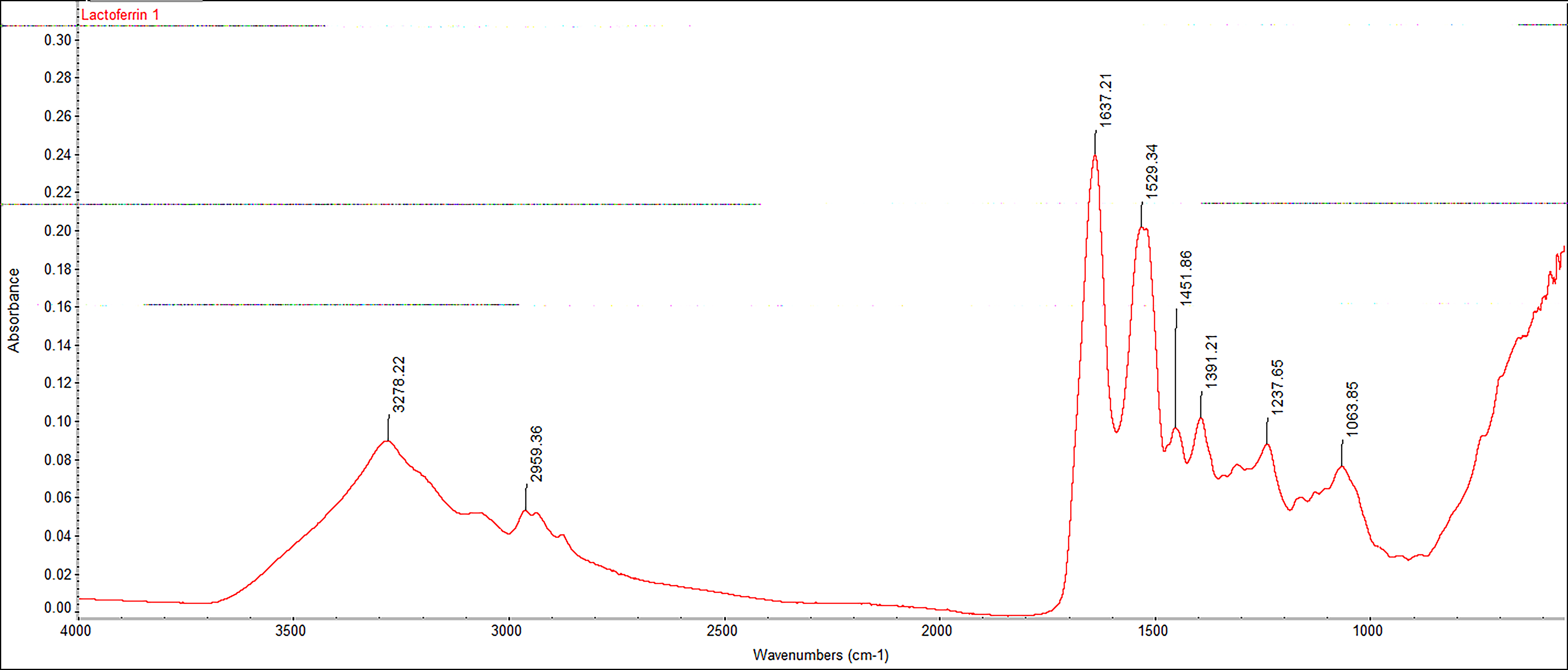
FTIR spectra for commercially available bLf derived from bovine-derived milk. bLf, bovine lactoferrin; FTIR, Fourier transformed infrared spectroscopy.
Iron content of bovine lactoferrin
Elemental analysis by EDX confirmed the presence of iron in the commercial bLf sample. Quantitative results revealed an iron content of approximately 1.94% (±0.07), indicating that the protein is not predominantly in its iron-free (apo) form. The detection of iron suggests that the Lf used in this study presents partial or predominant iron saturation (Supplementary Fig. S1).
Cell viability
Cell viability of HaCaT cells exposed to bLf at concentrations of 5, 10, and 50 µg/mL was assessed using the alamarBlue™ assay (Fig. 2). No cytotoxic effects were observed at 5 and 10 µg/mL, with cell viability exceeding 95% relative to the untreated control. A slight increase in proliferation (∼12%) was observed at 10 µg/mL after 24 h. Time-course analysis showed stable viability across 24–72 h, indicating sustained biocompatibility of bLf at the tested concentrations. At the highest tested concentration (50 µg/mL), a significant reduction in cell viability was observed compared with lower concentrations, suggesting that higher bLf concentrations may affect keratinocyte metabolic activity. Based on comparable efficacy and material economy, 5 µg/mL was selected for subsequent experiments. Statistical differences between groups and time points are indicated by distinct lowercase and uppercase letters, respectively, as described in the figure legend.
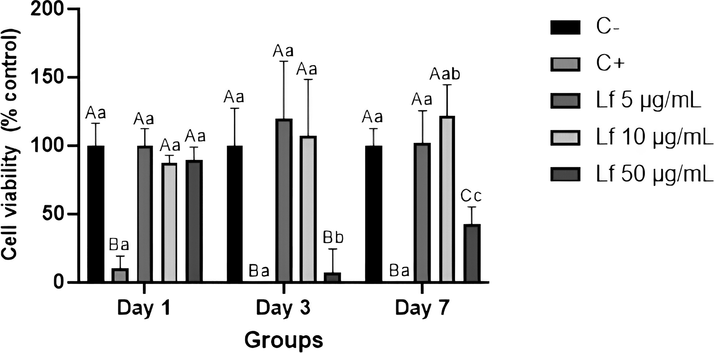
Graphic representation of the mean ± standard deviation of cell viability (%) of HaCaT cells after treatment with bLf on days 1, 3, and 7 at the following concentrations: 5 µg/mL, 10 µg/mL, and 50 µg/mL. Different lowercase letters indicate statistically significant differences between experimental groups within the same time point, while different uppercase letters indicate statistically significant differences between time points within the same group (P < .05). bLf, bovine lactoferrin.
Cell proliferation
Cell proliferation increased progressively from day 1 to day 3 in all experimental groups (Figs. 3 and 4). On day 7, most groups maintained elevated proliferation levels; however, the 5 µg/mL FBS-free group showed a decline (∼15% decrease), likely due to cells reaching confluence and detaching from the microplate. These results highlight that bLf supports keratinocyte proliferation over short- and medium-term intervals under the tested conditions. Comparative analysis between FBS-containing and FBS-free groups revealed higher proliferation rates in the presence of FBS, reflecting the combined effects of serum and bLf on cell growth. 14 Quantitative data are presented in Figure 3 as mean ± 95% confidence intervals, while representative fluorescence microscopy images illustrating cell proliferation patterns over time are shown in Figure 4.
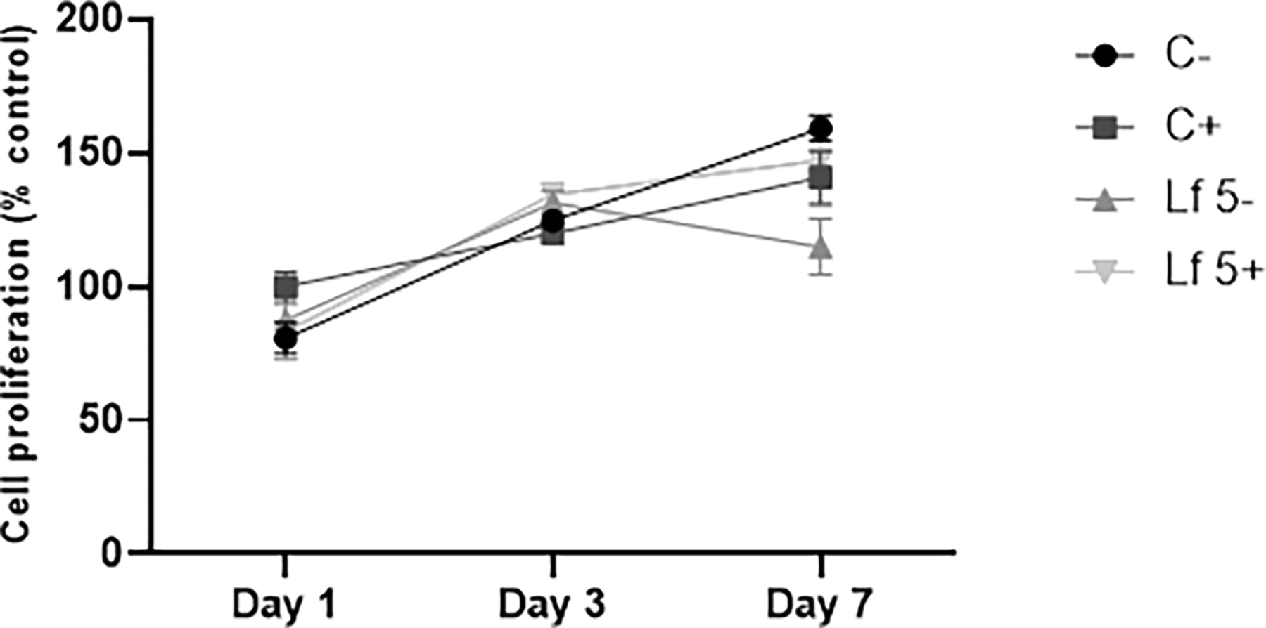
Graphic representation of mean and standard deviation of cell proliferation (%) (CyQUANT™ Cell Proliferation Assay) of HaCaT cells after 1, 3, and 7 days of contact to different treatments: culture media FBS-free (C−); culture media with FBS (C+); lactoferrin (5 µg/mL) solution in culture media FBS-free (Lf 5−); Lactoferrin (5 µg/mL) solution in culture media with FBS (Lf 5+). Geometric symbols are means and error bars represent 95% confidence intervals (α = 5%). FBS, fetal bovine serum.

Representative images of fluorescence microscopy for qualitatively analysis of HaCaT proliferation (CyQUANT™ Cell Proliferation Assay) after 1, 3, and 7 days of contact to different treatments: culture media FBS-free (C−); culture media with FBS (C+); lactoferrin (5 µg/mL) solution in culture media FBS-free (Lf 5−); lactoferrin (5 µg/mL) solution in culture media with FBS (Lf 5+). Images were acquired at 4× and 10× magnifications for qualitative morphological assessment. Scale bar = 460 µm (4× magnification) and 180 µm (10× magnification). FBS, fetal bovine serum.
Cell morphology
Fluorescence microscopy analysis was performed to qualitatively assess the morphology of HaCaT cells following treatment with bLf (5 µg/mL). Images acquired at 4× and 10× magnifications (Figs. 4 and 5) revealed preserved cell morphology, with keratinocytes exhibiting a typical polygonal shape, intact nuclear staining, and organized actin filament distribution across all experimental groups. No evident morphological alterations, such as cell rounding, detachment, or cytoskeletal disruption, were observed under the experimental conditions evaluated. Morphological evaluation was conducted using DAPI staining for nuclei and ActinRed555 for actin filaments (Fig. 5). In FBS-containing conditions (C+ and Lf 5+), cells exhibited well-organized actin filaments and intact nuclei. In contrast, FBS-free groups (C– and Lf 5–) displayed less intense actin staining and a higher proportion of rounded cells, indicative of cytoskeletal stress. In addition, nuclear staining appeared less intense in FBS-free conditions, whereas FBS-containing groups showed more pronounced DAPI fluorescence, consistent with enhanced cellular organization and metabolic support provided by serum. 15 These findings highlight the influence of FBS on cytoskeletal organization and overall cell morphology.
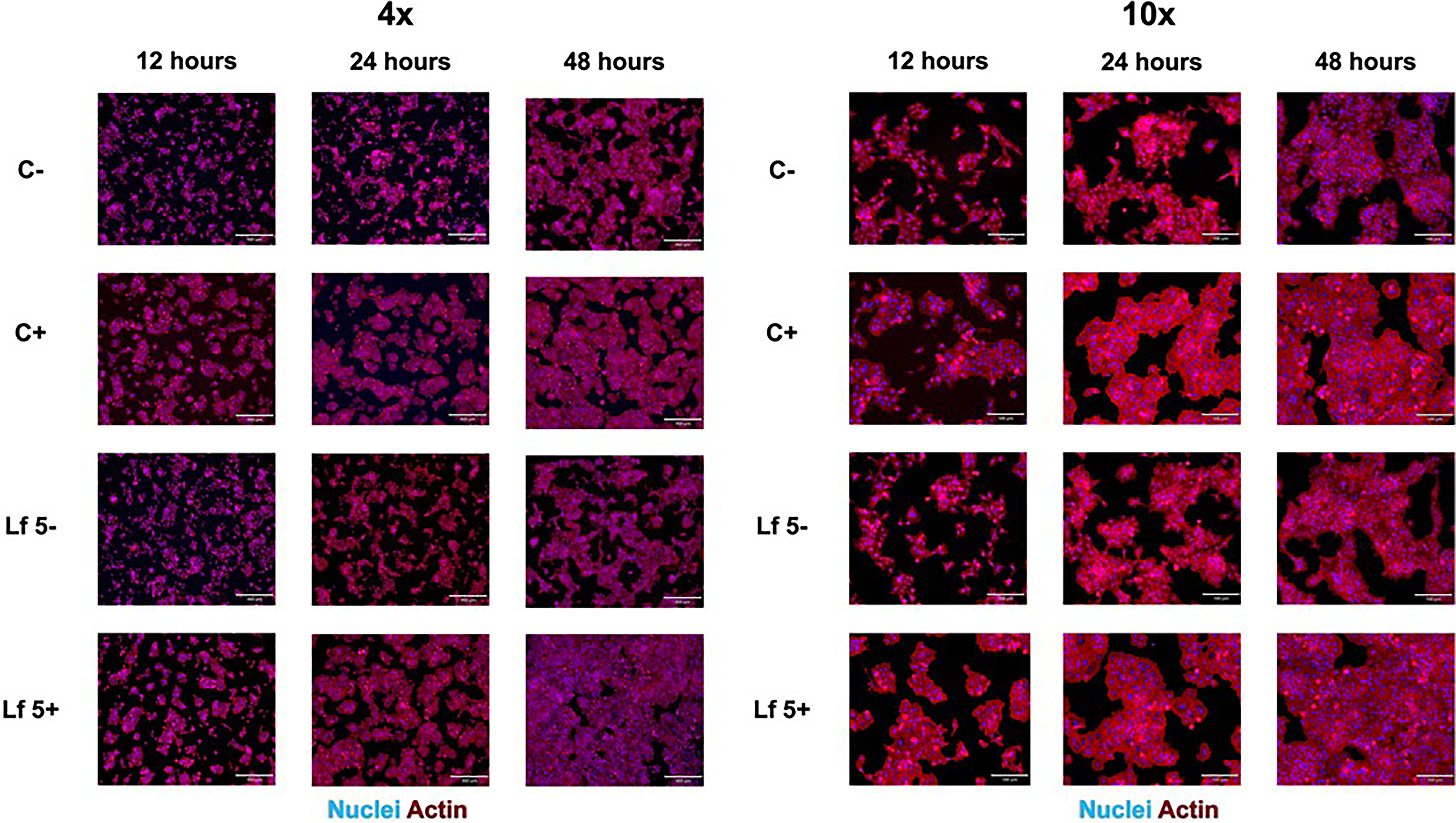
Representative images of fluorescence microscopy of HaCaT cells morphology after 12, 24, and 48 h of contact to different treatments: culture media FBS-free (C−); culture media with FBS (C+); Lactoferrin (5 µg/mL) solution in culture media FBS-free (Lf 5−); Lactoferrin (5 µg/mL) solution in culture media with FBS (Lf 5+). Cell nuclei are stained in blue (DAPI), and actin filaments are stained in red (ActinRed 555). Scale bar represents 460 µm (4× magnification) and 180 µm (10× magnification). FBS, fetal bovine serum.
Normalized metabolic activity analysis
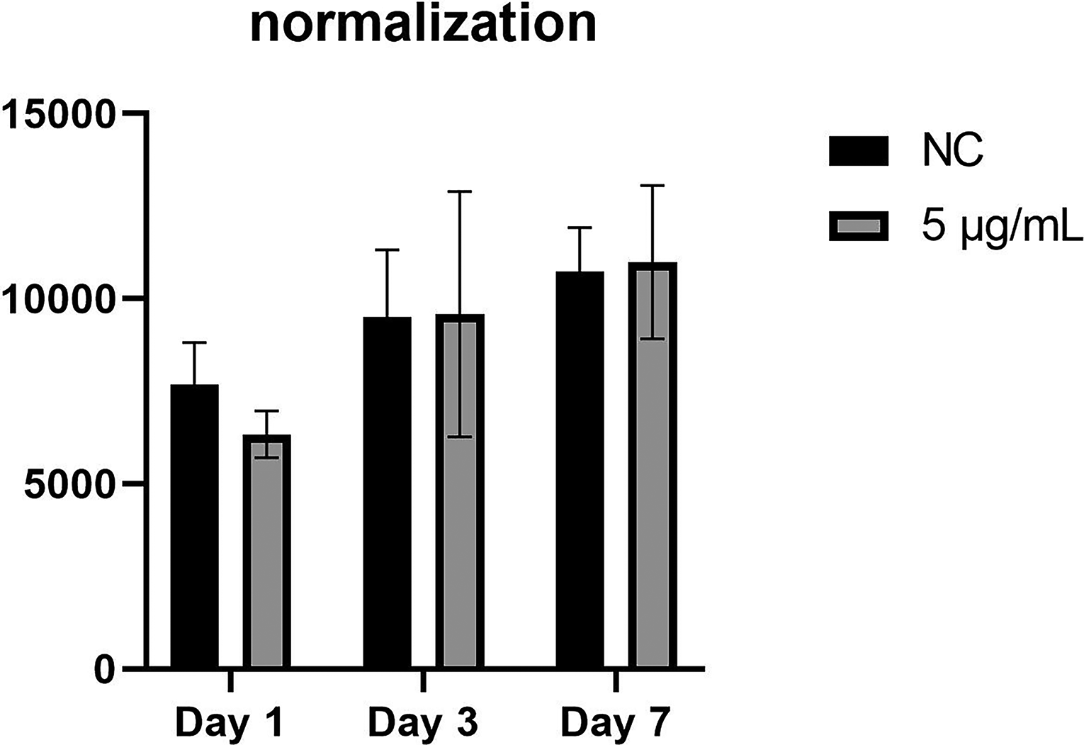
Metabolic activity normalized to DNA content (alamarBlue™/CyQUANT™) revealed no statistically significant differences between control and 5 µg/mL bLf groups at days 1, 3, and 7 (P > .05, Fig. 6). A slight increase (∼5%) in metabolic activity per cell was observed at day 3 in the treated group. Overall, these findings indicate that bLf exposure maintains cellular functionality and is not cytotoxic under the tested conditions.
Normalized metabolic activity of HaCaT cells relative to DNA content. Data represent the ratio of metabolic activity (alamarBlue™ assay) to DNA content (CyQUANT™ assay) for the control group (NC) and 5 µg/mL lactoferrin group across three time points (days 1, 3, and 7). Bars represent mean ± 95% confidence interval. No significant differences were observed between the groups (P > .05).
Wound healing
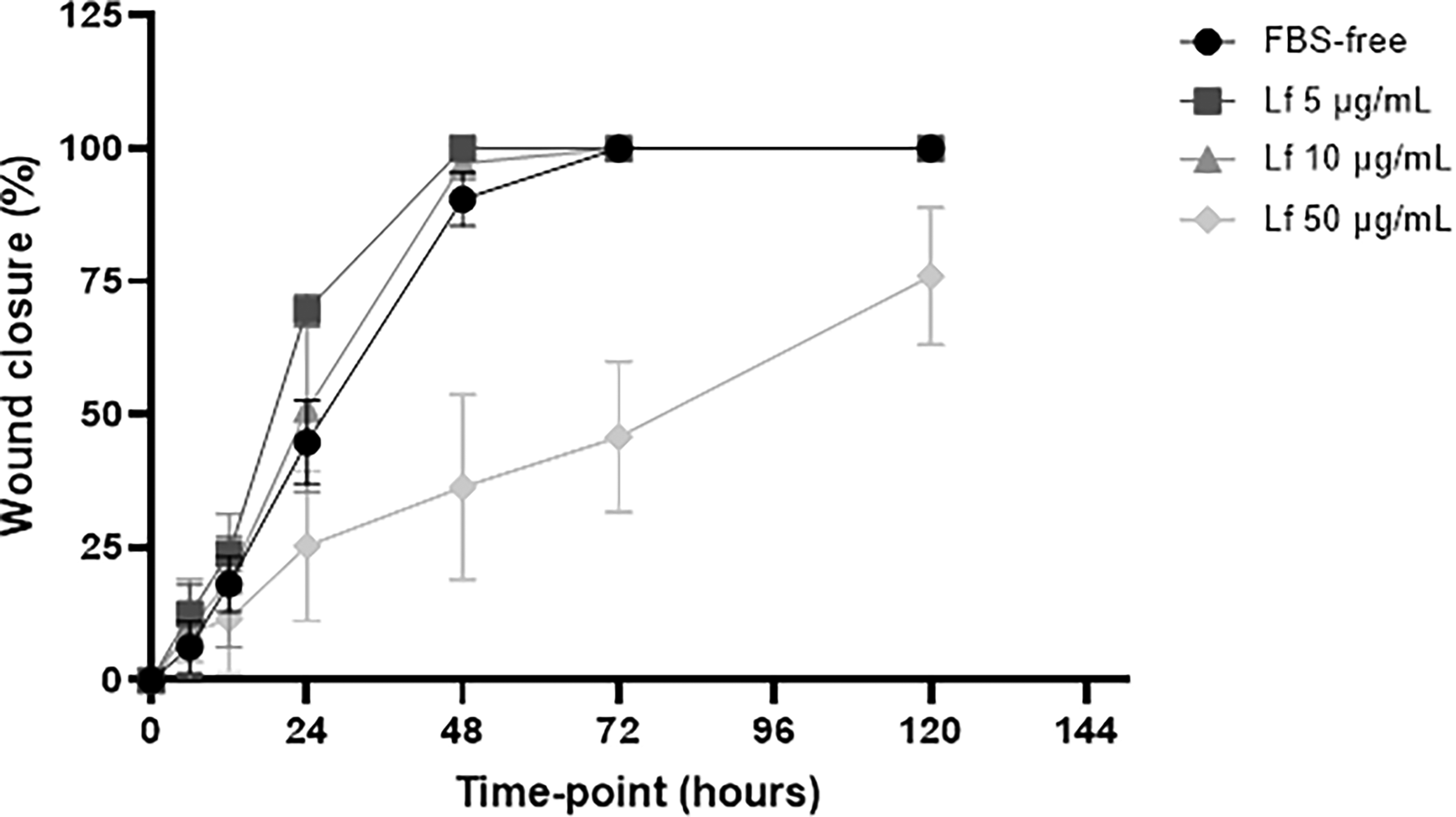
Wound closure assays demonstrated a concentration-dependent effect of bLf on HaCaT cell wound healing (Fig. 7). Cells treated with lower bLf concentrations exhibited accelerated wound closure during the first 24 h, with the 5 µg/mL group achieving approximately 60% closure, followed by the 10 µg/mL group (∼45%). In contrast, treatment with 50 µg/mL resulted in a markedly delayed response, with only ∼30% wound closure at 24 h. By 48 h, complete wound closure was observed in all experimental groups except the 50 µg/mL group, which maintained an open wound area. These findings indicate that lower concentrations of bLf optimally support keratinocyte wound closure, whereas higher concentrations negatively affect this process. The reduced closure rate observed at 50 µg/mL is consistent with the decreased cell viability and metabolic activity detected at this concentration in the alamarBlue™ assay, suggesting a concentration-dependent impairment of cellular fitness. As the scratch assay was conducted without mitomycin C, wound closure reflects the combined contribution of keratinocyte migration and proliferation, providing a physiologically relevant model of the wound healing process. Therefore, the delayed closure at higher bLf concentration is likely attributable to reduced cellular performance rather than a direct inhibitory effect on migratory capacity.
Graphic representation of mean and standard deviation of wound closure (%) of HaCaT cells after treatment with Lf of contact to different concentrations: 5 µg/mL, 10 µg/mL, and 50 µg/mL. Lf, lactoferrin.
Antimicrobial activity
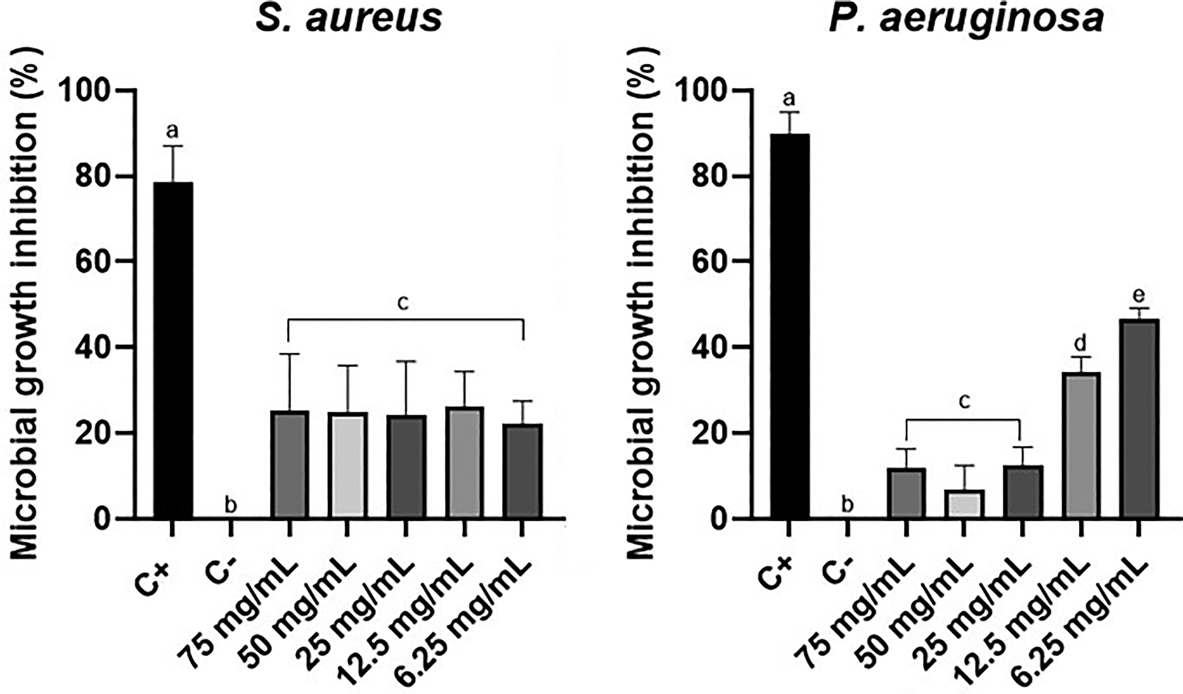
Lf alone did not exhibit antimicrobial activity against S. aureus or P. aeruginosa at any tested concentration, including 50 µg/mL (Fig. 8). No inhibition zones were observed, suggesting that under these experimental conditions, bLf does not directly suppress growth of these bacterial strains.
Graphic representation of mean and standard deviation of microbial growth (%) of S. aureus and P. aeruginosa after the treatment with different Lf concentrations. Lf, lactoferrin.
DISCUSSION
The present study investigated the bioactivities of commercially available bLf on keratinocyte viability, proliferation, migration, and antimicrobial properties, integrating complementary assays to provide a comprehensive analysis of its potential in wound healing and regenerative medicine.
The absence of cytotoxicity at low concentrations (5–10 µg/mL) and the observed stimulatory effect on proliferation at 10 µg/mL align with previous studies reporting safe therapeutic windows for Lf.2,3 In a study conducted by UCHIDA 16 and colleagues in 2017, they explored the impact of bLf on HaCaT cells, examining its role in both promoting proliferation and fostering differentiation. Their investigation revealed an elevation in the expression of TER, a recognized marker of epithelial barrier function. However, at its highest concentration (50 µg/mL), bLf exhibited a reduction in cell viability levels, a result that contradicts some findings in the current literature. Notably, FATHIL 17 et al. reported that hLf, even at a concentration of 125 µg/mL, did not induce cytotoxicity in HaCaT cells over a 72-h incubation period. Similarly, ABDALLA 2 et al. found no cytotoxic activity of hLf on human dermal fibroblasts, even at concentrations ranging from 32 µg/mL to 125 µg/mL. The reduced viability observed at 50 µg/mL may be associated with concentration-dependent cellular stress, potentially related to increased iron availability or saturation of Lf receptors. 16 Similar effects have been reported for bioactive proteins at higher concentrations, where excessive ligand exposure may disrupt cellular homeostasis.2,10
The observed disparity in cytotoxicity responses across different concentrations can be attributed to two primary factors: (1) variations in the origin and methods employed for obtaining Lf, and (2) the inclusion or exclusion of Lf in combination with another molecule. In both studies, Lf activity was assessed using a hydrogel containing Ag nanoparticles conjugated to Lf. In the current investigation, the partial results presented stem from initial data with the aim of evaluating the isolated activity of the target molecule. This underscores the significance of considering not only the origin of Lf but also its association, or lack thereof, with other molecules—whether biological, chemical, or nanoparticles—as these factors can significantly influence cellular activity.
Cell proliferation and metabolic activity demonstrated progressive growth from day 1 to day 3, with stabilization or slight decline by day 7, particularly in the serum-free 5 µg/mL group, likely due to confluence-related detachment. 14 These intervals were chosen to capture both immediate and sustained responses, showing that bLf supports keratinocyte viability and proliferation without inducing apoptosis, in agreement with prior studies.12,13,18 The normalization of metabolic activity (alamarBlue™ assay) to DNA content (CyQUANT™ assay) further confirmed that bLf treatment did not alter per-cell metabolic activity significantly, underscoring its noncytotoxic nature at therapeutic doses. Importantly, the influence of FBS must be considered, as proliferation rates were consistently higher in serum-containing conditions, suggesting synergistic effects between FBS and bLf. Future work should refine experimental conditions to better isolate the specific contribution of bLf to keratinocyte proliferation.
In all groups, cell proliferation levels increase between days 1 and 3. On day 7, the pattern is repeated, except for group 5 µg/mL (−), where proliferation has decreased. Comparison between time points indicates that Lf does not induce cell apoptosis, as has already been established, 16 and under certain conditions and/or depending on the concentration, it can stimulate proliferation. 19 The data presented in this study corroborate what is known about Lf not inducing cell apoptosis. However, to affirm that the Lf used in these assays is also capable of stimulating cell proliferation, it must be considered whether the response obtained is not being influenced by the presence of FBS in the culture medium. Furthermore, the concentration of Lf, along with its association with other molecules, can be crucial factors influencing cell proliferation. 20 While the qualitative results corroborate the quantitative findings, it is imperative to thoroughly analyze these aspects to complete the presented data.
Fluorescence microscopy revealed that cytoskeletal remodeling was strongly influenced by serum, with FBS-containing groups showing more pronounced actin filament organization and nuclear activity. These findings are consistent with reports linking serum components to enhanced cytoskeletal reorganization and cell motility.8,21 While Lf has been reported to independently affect actin dynamics and migration,22,23 its specific impact was less distinguishable in the presence of FBS, suggesting that serum effects may mask Lf-driven responses. This emphasizes the need for serum-depleted models or complementary approaches to delineate Lf-specific pathways. Moreover, differences in nuclear morphology observed between groups point to potential variations in proliferation and differentiation states, reinforcing the role of microenvironmental factors in modulating keratinocyte responses. 20
The analysis of HaCaT cell migration was performed after the cells were exposed to treatment containing Lf in different concentrations (Fig. 7). As expected, the rate of wound closure varied according to the Lf concentration. The group with the lower concentration (5 µg/mL) showed a higher rate of wound closure in the first 24 h. However, 48 h after treatment, all groups achieved complete closure of the wound, except for the group with the highest concentration, 50 µg/mL, in agreement with the data obtained regarding cytotoxicity and higher concentrations of Lf. The scratch assay was performed without the use of mitomycin C, a compound typically employed to inhibit cell proliferation and isolate migratory behavior. While the standard protocol suggests its inclusion, we chose not to use mitomycin C to maintain a physiological environment more representative of the wound healing process. This decision aligns with the objective of evaluating both the migratory and proliferative responses of HaCaT cells to Lf treatment, as both processes are critical components of reepithelialization during wound healing. Nonetheless, the lack of mitomycin C may result in overlapping effects of proliferation and migration in the observed wound closure rates. To address this, the results were interpreted with caution, correlating them with independent proliferation assays and cytotoxicity data to minimize confounding effects. Moreover, the complete closure of wounds by 48 h, except for the highest concentration group (50 µg/mL), supports the conclusion that the observed effects were consistent with the biological activity of Lf rather than being solely proliferation-driven. Future studies could incorporate additional controls to further delineate these processes, enhancing the robustness of the findings. The delayed wound closure observed at the highest bLf concentration (50 µg/mL) further supports the concentration-dependent effects of lactoferrin on keratinocyte behavior. While lower concentrations promoted wound closure, higher concentrations negatively affected cell viability, which likely compromised both proliferative and migratory responses. These findings underscore the importance of optimizing bLf concentration for wound healing applications, as excessive doses may impair keratinocyte function rather than enhance tissue repair.
Recombinant Lf has also been observed to significantly stimulate the proliferation of human HaCaT, in addition to increasing the rate of reepithelialization of second-degree burns in pigs.16,20 However, it is noted that, in addition to the origin, the association of the protein with other molecules or particles is a crucial factor that can influence its activity in cell migration, as reported by ABDALLA 10 et al.. In their study, Lf, when conjugated to Ag nanoparticles, did not disturb the normal cell migration, showing that the risk of this hydrogel to cause adverse effects on the cells was low during the healing process. As well, according to BELVEDERE 3 et al., the combination of Lf and mesoglycan enhances the individual effects of each substance on the migration and invasion rates of keratinocytes, as well as other cell types, including endothelial cells and fibroblasts. These effects play a critical role in the cellular response during the wound healing process, and their enhancement highlights the potential beneficial impact of the tested combination. Lastly, in the same study, the authors evaluated markers using confocal analysis, which confirmed keratinocyte differentiation, fibroblast conversion into myofibroblasts, and epithelial-mesenchymal transition, all influenced by the combination of Lf and mesoglycan, thus validating the effect of this combination on cell activation. It is well established that during the reepithelialization phase, keratinocytes initially migrate in a directed manner and then differentiate to facilitate the proper formation of the skin layers.22,23
As well, in a study conducted by Mouritzen and Jenssen 24 , bLf modulated the expression of MMP-2 and MMP-9, enzymes critical for the remodeling of the ECM during healing. Not only that, a decrease in the expression of pro-inflammatory cytokines (such as IL-6 and TNF-α) was observed, which may contribute to a more favorable environment for tissue repair. Although the referenced study similarly utilized HaCaT cells and a scratch assay to compare human and bLf, the present study extends this groundwork by investigating previously unexplored facets of lactoferrin’s bioactivity. A primary distinction lies in its focus on commercially available bLf, which enhances its translational relevance for therapeutic development. Furthermore, this work rigorously examines concentration-dependent effects, systematically evaluating how varying bLf concentrations modulate keratinocyte proliferation, migration, and cytoskeletal organization, thereby identifying optimal therapeutic dosage ranges. To capture a big picture of bLf’s bioactivity, the study integrates complementary assays (alamarBlue™, CyQUANT™, and morphological analyses), offering insights beyond the scratch assay’s scope. Crucially, by framing results within the context of wound healing and tissue repair, this research bridges fundamental findings with practical applications, setting it apart from purely comparative studies. Collectively, these advances position the current work as both a novel contribution to lactoferrin research and a catalyst for its clinical translation in regenerative medicine.
In vitro and in vivo assays have already demonstrated the antimicrobial activity of Lf against the main pathogens that can impede the wound healing process through microbial infection.10,17 Similarly, in addition to its antimicrobial activity, Lf conjugated to mesoglycan—an association of glycosaminoglycans that present potential antimicrobial activity—demonstrates that these two substances can mutually contribute to antimicrobial effects. This is different from when they are isolated, which is another factor to be considered regarding the discrepancy in results found.
The determination of iron content in the commercial bLf sample provides critical insight into the biological responses observed in this study. The presence of iron (∼1.94%) indicates that the protein is likely in a partially or predominantly iron-saturated (holo or intermediate) form. This characteristic is consistent with the maintenance of keratinocyte viability and the stimulation of cell proliferation observed at lower concentrations, as iron-bound lactoferrin has been reported to support cellular growth. Conversely, the absence of bactericidal activity against S. aureus and P. aeruginosa may be explained by the reduced antimicrobial potential of iron-saturated lactoferrin compared with its apo form.5,9 Furthermore, at higher concentrations (50 µg/mL), the presence of iron may contribute to cellular stress or altered receptor interactions, potentially explaining the reduced viability and migration observed under these conditions. 9 Collectively, these findings reinforce the importance of considering iron saturation when evaluating lactoferrin bioactivity and interpreting its effects on wound healing–related cellular processes.
A key variable that was not specifically controlled in this study is the iron saturation state of Lf, whether in its apo (iron-free) or holo (iron-saturated) form. The iron saturation state of Lf is known to influence cellular responses, including its antimicrobial, anti-inflammatory, and pro-healing properties. However, due to time constraints and the exploratory nature of this study, no specific analyses were conducted to determine or control for the iron saturation levels of the Lf used. This represents a limitation of the current investigation, as variations in iron saturation may partly explain the observed divergence with certain findings in the literature. Notably, the preparation methods for Lf and the conditions under which it was used are consistent with those reported in similar studies, suggesting that the form used here likely represents a mixed or predominantly apo state. While the absence of this analysis does not invalidate our findings, it highlights the need for further research to dissect the precise contributions of Lf’s iron-binding state to its biological activity. Future studies will benefit from incorporating iron saturation analyses, potentially through spectroscopic methods or specific assays, to refine our understanding of Lf’s mechanistic roles in wound healing and cellular behavior.
The current study focused exclusively on the experimental evaluation of bLf, with comparative references to hLf drawn solely from existing literature.
Despite the lack of side-by-side testing under identical conditions, the discussion of hLf was included to provide broader context and highlight potential parallels and divergences in the biological activity of these two forms of lactoferrin. This speculative comparison aims to offer a foundation for future studies rather than to assert definitive conclusions. Future research comparing bLf and hLf under identical experimental conditions would be critical to substantiate claims of comparative efficacy and better understand their respective roles in wound healing and cellular responses.
CONCLUSION
The results of this study highlight the importance of variables related to the purity and method of obtaining commercial Lf in determining its bioactivities. Although the Lf evaluated showed bioactive properties, some discrepancies observed in relation to the literature indicate the need for further investigations. Future studies should seek to elucidate the molecular mechanisms involved in the bioactive activities of Lf, as well as to evaluate in more detail the impacts of structural characteristics and extraction conditions.
AUTHORS’ CONTRIBUTIONS
The authors’ contributions are as follows: M.L.A., L.M.C., and C.A. conducted the experimental assays. M.L.A. was responsible for drafting and writing the article. R.G.L. provided leadership in coordinating the research group and supervising the project.
Footnotes
ACKNOWLEDGMENTS
The authors would like to express their sincere gratitude to the University of Michigan, particularly to the members of the Bottino Lab and the Castilho Lab, for their invaluable support, collaboration, and for providing an enriching environment during the development of this study. We also extend our thanks to the funding agency CAPES/PRINT for their financial support through scholarships and research grants, which were essential for the completion of this work. In addition, we acknowledge the National Council for Scientific and Technological Development (CNPq) for the researcher fellowship (DT-1D CNPq Grant #306225/2021-2) of one of the authors (RGL), as well as the Coordination for the Improvement of Higher Education Personnel, Brazil (CAPES), for a PhD fellowship (Finance Code 001) awarded to one of the authors (MLA).
AUTHOR DISCLOSURE STATEMENT
There are no conflicts to declare.
FUNDING INFORMATION
This work was supported by Coordination for the Improvement of Higher Education Personnel, Brazil (CAPES), Finance Code 001.
Supplemental Material
References
Supplementary Material
Please find the following supplemental material available below.
For Open Access articles published under a Creative Commons License, all supplemental material carries the same license as the article it is associated with.
For non-Open Access articles published, all supplemental material carries a non-exclusive license, and permission requests for re-use of supplemental material or any part of supplemental material shall be sent directly to the copyright owner as specified in the copyright notice associated with the article.