



Abstract
We examined the contribution of dermatoscopy to the reliability of the diagnosis and management of non-melanocytic skin tumours using store-and-forward teledermatology. A total of 150 patients with non-melanocytic skin tumours were enrolled into the study. The reliability of the diagnoses and management plans was measured by comparing teledermatology with face-to-face examination; the effect of adding dermatoscopy images was also analysed. The accuracy of the diagnoses was measured by comparing teledermatology with histology; the effect of adding dermatoscopy images was also analysed. Diagnostic reliability (kappa) for teledermatology without dermatoscopy was 0.75 and 0.77 for two different dermatologists, A and B. The reliability increased significantly when dermatoscopy was added, to 0.86 and 0.88 respectively (P < 0.05). The reliability of management plans without dermatoscopy was 0.67 and 0.70, but it did not increase significantly when dermatoscopy was added. The accuracy of the diagnoses was significantly increased by the addition of dermatoscopic images, from 85% to 94% for dermatologist A and from 88% to 95% for dermatologist B. Teledermatology is a reliable technique for the diagnosis and management of non-melanocytic skin tumours and the addition of dermatoscopic images increases the reliability and the accuracy of teledermatology.
Introduction
Dermatoscopy is a tool for the diagnosis of pigmented and non-pigmented skin lesions, which allows the clinician to visualize features that are not discernible by naked-eye examination. 1 It is regarded as the most helpful technique in the diagnosis and management of pigmented skin lesions.1,2
Many studies have shown that teledermatoscopy has high reliability compared with face-to-face examination, ranging from 85 to 91%.3–6 However, the majority of studies have investigated only melanocytic skin lesions. To the best of our knowledge, no study has specifically investigated non-melanocytic lesions. Furthermore, there have only been a few studies which included non-melanocytic skin lesions and performed store-and-forward teledermatology. In one of these studies, store-and-forward teledermatology was demonstrated to be an effective, accurate, reliable and valid approach for the routine management of patient referrals in skin cancer and pigmented lesion clinics. 7 In another study, store-and-forward teledermatology was shown to be effective and accurate as a preoperative tool for non-melanoma skin cancer, avoiding unnecessary visits to the hospital and shortening the waiting intervals to the surgical treatment. 8
We hypothesized that the reliability of diagnosis and management in non-melanocytic skin tumours would be increased by the addition of dermatoscopic images to store-and-forward teledermatology. For this purpose, we compared teledermatology and face-to-face evaluation, and examined the contribution of dermatoscopy.
Methods
One hundred and fifty patients who presented to the dermatology outpatient clinic at the Ankara hospital between April 2009 and September 2009 and had a non-melanocytic skin tumour on clinical examination were enrolled in the study. Patients were not excluded in terms of age, gender or skin phototype. The study was approved by the appropriate ethics committee.
Lesions which had been diagnosed as a non-melanocytic skin tumour in face-to-face examination were evaluated first with store-and-forward teledermatology based on clinical images and a standard information sheet (Table 1) and then they were re-evaluated with store-and-forward teledermatology containing additional dermatoscopic images. The reliability of teledermatology for diagnosis and management was established by comparison with face-to-face examination. The accuracy of teledermatology for diagnosis was established by comparison with histopathological examination.
The standard information form
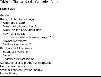
Two dermatologists (Teledermatologists A and B) from the Department of Dermatology at Adana Hospital conducted the teledermatology assessments. Two dermatologists from the Department of Dermatology at Ankara Hospital performed the face-to-face assessments to establish the reference values. The same technician took clinical and dermatoscopic images of the patients and stored them electronically.
Teledermatology
Clinical and dermatoscopic images of the lesions were obtained for each lesion by the technician and saved to a database on a computer. Standard guidelines and previous studies were followed for digital imaging.3,9–11 Two macro images (distance and close-up) at 7.2 megapixel resolution (3072 × 2304 pixels, JPEG format) were obtained for each lesion using a digital camera (Cyber-shot DSC-W70, Sony). One dermatoscopic image was also taken for each lesion with the same digital camera and a lens attachment (Dermlite II Pro HR, 3Gen Inc).
Following face-to-face evaluation, the images and the standard information form for each patient were stored by the technician on the separate web areas for teledermatologists A and B. The teledermatologists evaluated the clinical information and images of 150 cases sorted randomly and recorded a diagnosis for each case. The teledermatologists also chose one of three management plans: lesion destruction (destruction or removal of the lesion); medical treatment (antibacterial, antiviral or anti-fungal therapy) or follow-up.
Two months later, dermatoscopic images of the lesions of the same cases were added to the web data and the teledermatologists were asked to establish a diagnosis and recommend a management plan for each lesion again. The cases were presented in random order.
There was a two-month-interval between the first (without dermatoscopic images) and the second teledermatology evaluation (with dermatoscopic images). The second assessment was carried out in a different lesion order from the first one and the teledermatologists were prevented from seeing the results of their first evaluations.
Reference values
Each case had a single management plan after face-to-face examination which was used to establish the reliability of teledermatology. Each case could have two reference diagnoses: the face-to-face diagnosis, which was used to establish the reliability of teledermatology, and the histopathology diagnosis, which was used to establish the accuracy of teledermatology.
All of the patients were examined by a dermatologist face-to-face. Histopathological examinations of the lesions were performed only in the cases in which a diagnosis could not be established with clinical evaluation.
Histopathological diagnoses were accepted as the gold standard.
Reliability
The reliability of teledermatology was evaluated by comparing diagnoses and management plans with the in-person evaluation. If the diagnoses and management plans of the teledermatologist were the same as those made in face-to-face examination they were accepted as being in agreement. Reliability was measured by the kappa coefficient. The McNemar test was used to analyse the contribution of dermatoscopy to the reliability of teledermatology.
Accuracy
The accuracy of teledermatology was calculated as the percentage of correct diagnoses in the cases diagnosed by histopathological examination (the gold standard). If the teledermatology diagnoses were the same as those of the histopathological examination they were accepted as correct. Fisher's exact test was used to analyse the contribution of dermatoscopy to the accuracy of teledermatology. Standard packages (SPSS 16.0, SSS Inc. Illinois and MedCalc 10.4, MedCalc Software, Mariakerke, Belgium) were used for statistical analysis.
Results
Seventy six (51%) of 150 patients enrolled in the study were male. The mean age of the patients was 55 years (61 years for males and 50 years for females). None of the patients had a melanoma diagnosis in their medical history. However, nine of the patients (6%) had a non-melanocytic skin cancer history. The mean duration of the lesions was 1.7 years, SD 0.3. Only five lesions had appeared in the previous three months. Most of the patients (53%) did not have any symptoms worthy of note. The most frequent recorded symptom was a change in the lesion size.
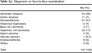
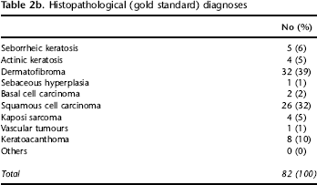
In 150 cases, the most common face-to-face diagnosis was dermatofibroma (31%), followed by seborrheic keratosis (30%), basal cell carcinoma (17%), actinic keratosis (7%), vascular tumor (6%) and squamous cell carcinoma (3%) (Table 2a). The diagnoses of 82 cases were based on histopathological examinations. Dermatofibroma was also the most common histopathological diagnosis (39%) and it was followed by squamous cell carcinoma (32%) and keratoacanthoma (10%) (Table 2b).
Diagnoses on face-to-face examination
Histopathological (gold standard) diagnoses
The most common management plan on face-to-face examination was follow-up (n = 94, 63%) followed by destruction of the lesion (n = 56, 37%). Medical treatment was suggested for none of the patients.
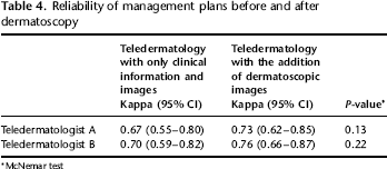
The reliability of the diagnoses made by the teledermatologists was significantly increased by the addition of dermatoscopic images, from 0.77 to 0.85 for teledermatologist A (P = 0.03) and from 0.75 to 0.86 for teledermatologist B (P < 0.001), see Table 3. The reliability of the management plans made by the teledermatologists was also increased by the addition of dermatoscopic images, but these increases were not significant [from 0.67 to 0.73 for teledermatologist A (P = 0.13) and from 0.70 to 0.76 for teledermatologist B (P = 0.22)] (Table 4). Some malignant lesions were under-managed by teledermatologists based on under-diagnoses made by teledermatology, with and without dermatoscopy images (Table 5).
Reliability of diagnoses before and after dermatoscopy
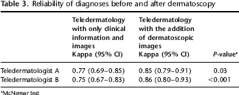
McNemar test
Reliability of management plans before and after dermatoscopy
McNemar test
Malignant lesions which were under-managed by the teledermatologists

SCC = Squamous cell carcinoma, KS = Kaposi sarcoma, BCC = Basal cell carcinoma
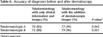
The accuracy of the diagnoses made by the teledermatologists was significantly increased by the addition of dermatoscopic images, from 85% to 94% for teledermatologist A (P = 0.04) and from 88% to 95% for teledermatologist B (P < 0.03), see Table 6.
Accuracy of diagnoses before and after dermatoscopy
Fisher's exact test
Discussion
In the present study of non-melanocytic skin tumours the diagnostic accuracy and reliability of both teledermatologists was significantly increased by dermatoscopy. In addition, the reliability of the management plans was also increased, although not significantly.
To the best of our knowledge this is the first study investigating the contribution of dermatoscopy to teledermatology in the diagnosis and management of non-melanocytic skin tumours. There are many studies of the efficacy of teledermatoscopy in the diagnosis and management of melanocytic lesions. 12 However, there have been few studies examining efficacy of teledermatology in both melanocytic and non-melanocytic lesions. Efficacies of store-and-forward teledermatoscopy and face-to-face evaluation were compared in three of these studies.3,13,14 Fabbrocini et al. found that face-to-face evaluation had a higher diagnostic reliability than teledermatoscopy although there was excellent agreement between teledermatoscopy and face-to-face evaluation in the studies of Tan et al. and Piccolo et al.4,13,14 The low diagnostic accuracy of teledermatoscopy of the last study in contrast to the other studies was attributed to inclusion of the lesions with atypical dermatoscopic patterns. 14 As in our study, two studies in the literature have specifically investigated the contribution of dermatoscopy to store-and-forward teledermatology6,12 Moreno-Ramirez et al. compared diagnostic efficacies of clinical (clinical information and pictures) and dermatoscopic (clinical information and pictures with dermatoscopic images) teleconsultations in order to triage melanocytic lesions. 12 In this study the agreement with gold standard was 0.91 (95% CI 0.82–1.00) for clinical teleconsultation and 0.94 (95% CI 0.88–1.00) for teledermatoscopy (P > 0.05). However, teledermatoscopy has improved the diagnostic confidence level, specificity and referral rates of a teledermatology-based screening system for pigmented lesions. 12 In a similar study including both melanocytic and non-melanocytic skin tumours Kroemer et al. reported that clinical and dermatoscopic evaluations demonstrated strong concordance with the gold standard (kappa = 0.4 for each). 15 In the study carried out by Warshaw et al., which most closely resembles our study because it contained only non-pigmented lesions, the diagnostic reliability of teledermatology was significantly increased with the addition of polarized light dermatoscopy but reliability of the management plans was not improved as in our study 6
A finding of our study is that the increase in the reliability of management plans was not significant. We think that that teledermatologists might have chosen the biopsy/removal option to rule out a possible malignancy even though they could make a diagnosis for a suspected lesion. For example, teledermatologists offered the removal option for six seborrheic keratoses which were correctly diagnosed by teledermatology. However, they never suggested follow-up for malignant tumours (basal cell carcinomas and squamous cell carcinomas). Also, offering only two management options to the teledermatologist might have been a contributing factor.
There are two important limitations of the study. First, the same dermatologists carried out teledermatology, with and without dermatoscopic images. Second, since we did not have a large number of lesions we could not evaluate diagnostic and management reliability separately for each disease.
The results confirm that teledermatology is a reliable technique for the diagnosis and management of non-melanocytic skin tumours and that the addition of dermatoscopic images increases the reliability and the accuracy of teledermatology. Further studies with a larger number of patients are now needed.