



Abstract
In this study, we compared the assessment of remote smartphone photographs to in-office exams in the diagnosis of two groups of external eye diseases, red-eye pathology and post-operative eyelid surgery complications. Participants were examined and received an in-office diagnosis by either a corneal or oculoplastic specialist. After viewing an educational video on smartphone photography, the patient’s companion then took a series of standardized photographs. Two additional corresponding specialists then made a separate diagnosis via the interpretation of only smartphone images and the patient’s history. ‘Remote’ and in-office diagnoses were compared using a kappa test for agreement. The remote and in-office diagnoses were in agreement for 27 of 28 eyes, representing a chance-corrected Kappa agreement rate of 93% (95% confidence interval: 79–99%). Among the 16 red eyes, the diagnoses were in agreement for 15 of 16 red eyes, representing a chance-corrected Kappa agreement rate of 92% (95% confidence interval: 77–99%). Among the 12 eyes with post-operative eyelid surgery complications, the diagnoses were in perfect agreement. Our results suggest that the diagnosis of 1) red-eye pathology and 2) post-operative eyelid surgery complications based on smartphone images may be comparable to in-office exams.
Introduction and background
Telemedicine is emerging as a viable platform across healthcare subspecialties and settings, upon which remote patient assessments can be conducted. Remote patient interactions have the potential to enhance patients’ access to care across the entire healthcare spectrum, especially in rural settings.1,2 They also have the potential to decrease patients’ travel burden.3,4 To date, the majority of tele-ophthalmology initiatives have focused on posterior segment eye pathology such as diabetic retinopathy and utilize technology unavailable to the average American.1,5–9 An improvement in telemedicine initiatives would utilize technology that most patients and clinicians already possess, such as smartphones, rather than rely on novel technology that could impose prohibitive expenses and prolonged learning curves.
In their January 2017 commentary on the evolution of technology, the Pew Research Center reported that 77% of Americans own a smartphone. 10 A recent study by Woodward and associates evaluated the diagnostic accuracy of detecting certain types of corneal disease via photographs using smartphone technology, but they used certified ophthalmic photographers in order to maximize image quality. 11 Our study used smartphone images obtained by patients’ companions (e.g. family, friends, etc.) instead of certified ophthalmic photographers to more closely simulate a real-life scenario. We also introduced two previously unstudied pathologies in the context of tele-ophthalmology – 1) red-eye pathology and 2) post-operative eyelid surgery complications. By analysing the diagnostic accuracy of these unpublished pathologies via smartphone images taken by a patient’s companion and interpreted by ophthalmologists, this study design is both novel and practical.
Red-eye is a common anterior segment pathologic presentation. In fact, red-eye is the most common ocular disorder seen by primary care physicians. 12 Stagg and associates reported that 23% of patients who presented to the emergency department with an eye problem had a non-urgent ocular condition of common ‘red-eye’ origin. 13 Red-eye is a non-specific finding associated with multiple aetiologies, including subconjunctival haemorrhage, pterygium, iritis, corneal ulceration, post-trauma inflammation, scleritis/episcleritis, and conjunctivitis. In this study, we assessed the diagnostic accuracy of these aetiologies.
In their March 2016 educational resource for patients, the American Academy of Ophthalmology listed blepharoptosis and dermatochalasis as two of the most common indications for eyelid surgery. 14 Surgical approaches to correct these entities share common post-operative complications that are the focus of in-office post-operative evaluations. Such post-operative complications include superficial ecchymosis or haematoma, lagophthalmos and wound dehiscence. 15 Prompt recognition is critical to proper management and successful outcomes. Fortunately, these clinical findings are readily visible to external photography. 15
A variety of studies have shown high patient satisfaction with tele-ophthalmology services.16–19 Our study evaluates a novel approach to tele-ophthalmology by leveraging smartphone photography, a familiar and common technology. Overall, the ability to accurately diagnose both red-eye pathology and post-operative eyelid surgery complications via smartphone images has the potential to address four important and growing patient concerns: 1) access to care, 2) patient travel burden, 3) time to intervention/treatment and 4) patient satisfaction.
We hypothesize that the diagnostic accuracy of 1) red-eye pathology and 2) post-operative eyelid surgery complications based on interpreting smartphone images taken by a patient’s companion is not inferior to that based on in-office examinations by a board-certified ophthalmologist with expertise in cornea or oculoplastics.
Methods
For this prospective study, we recruited patients at the Loyola Outpatient Center from May 2018 until May 2019. Individuals eligible for inclusion were those over the age of 18 years who presented to the ophthalmology clinic with a red-eye or having undergone surgical repair of blepharoptosis or dermatochalasis within the past 30 days. Eligibility was limited to those accompanied by a companion or caregiver willing and able to take photos of the involved tissue with the study smartphone.
At the time of enrolment, participants were issued the study smartphone and shown an instructional video outlining a standardized technique for external eye photography in order to minimize image variability among participants. Standardized measures included: 1) location of patient and companion within the examination room, 2) positions of eye gaze, 3) distance between smartphone and patient, 4) room lighting, 5) study smartphone flash set ‘ON’ and 6) study smartphone camera magnification set at 50%. The patient’s companion was then asked to take a series of photographs as described in the instructional video. Enrolled participants were also asked to complete a medical history form in order to provide the interpreting ophthalmologist with historical data, including descriptions of symptoms (i.e. redness, swelling, pain), duration and time course, exacerbating and alleviating factors, responses to therapy, changes in appearance and precipitating events.
We used one dedicated ‘study smartphone’ to enrol red-eye patients, and a separate dedicated study smartphone to enrol post-operative eyelid surgery patients. Both smartphones were an iPhone 6 (Apple, Cupertino, California, USA). The iPhone 6 has a 1334 × 750-pixel resolution display screen and an 8-megapixel camera with 1.5µ pixels. Images of study eyes were both captured and interpreted on the same study smartphone. The study smartphones did not have an activated cellular data plan or a wireless internet connection. Thus, no images were electronically transmitted from patient to clinician or from clinician to clinician, ensuring Health Insurance Portability and Accountability Act (HIPAA) compliance.
This study included two independent patient groups (i.e. a red-eye cohort and post-operative eyelid surgery cohort). All enrolled participants were seen and examined in-person by a corresponding sub-specialist. Red-eye patients were seen by a cornea specialist, while post-operative eyelid surgery patients were seen by an oculoplastics specialist. All enrolled participants then had their smartphone images and historical data reviewed by a second and separate corresponding sub-specialist on a different date. In all cases, diagnostic impressions based on in-person examinations served as the gold standard against which the diagnostic impressions based on remote assessments were compared. In this study, an exact version of the Kappa test was used to estimate the chance-corrected diagnostic agreement rate between paired remote and in-office diagnoses using SAS version 9.4 (Cary, North Carolina, USA).
Results
Red-eye patients
Twenty patients were screened, and 14 patients with red-eye pathology were enrolled, providing 16 affected eyes available for analysis. Twelve of the 14 patients presented with one affected eye, and two of the 14 patients presented with both eyes affected. The remote and in-office diagnoses were in agreement for 15 of these 16 red eyes, representing an agreement rate of 94%. The chance-corrected Kappa agreement rate was 92% (95% confidence interval: 77–99%). Subconjunctival haemorrhage and pterygium were the most common red-eye pathologies observed. Five eyes (31%) presented with subconjunctival haemorrhage and another five eyes (31%) presented with pterygium. The remaining eyes were diagnosed as corneal ulceration (n = 1, or 6.3%), episcleritis (n = 1, or 6.3%), uveitis (n = 1, or 6.3%), blepharitis (n = 1, or 6.3%) or other non-specific acute inflammation (n = 2, or 13%). Figure 1(a) depicts an image series of a patient who presented with subconjunctival haemorrhage in the right eye. As demonstrated in Table 1, the red-eye presentations that were less common were still accurately diagnosed. The single missed remote diagnosis was a patient presenting with a red eye as a result of non-specific inflammation from an occluded nasolacrimal duct, and the remote diagnosing physician interpreted the presentation to be episcleritis. Upon later retrospective review of this missed diagnosis, the interpreting ophthalmologist did not believe image quality played a role, simply that the diagnosis required closer inspection than the available smartphone photographs could provide.

(a) Smartphone image series of a ‘red-eye’ patient presenting with sub-conjunctival haemorrhage in the right eye. (b) Smartphone image series of a post-operative eyelid surgery patient presenting seven days after surgery with mild, expected ecchymosis.
Agreement between in-office and remote diagnoses of red-eye pathologies.

Shaded cells represent perfect agreement. Agreement = 94%. Chance corrected agreement (Kappa) = 92% (95% confidence interval: 77–99%).
aOther = non-specific acute inflammation.
SH: subconjunctival haemorrhage.
Post-operative eyelid surgery patients
Nine patients were screened and six patients with bilateral post-operative eyelid surgery complications were enrolled in the second group, providing 12 post-operative eyes available for analysis. The remote and in-office diagnosis were in perfect agreement for all eyes (100%). Table 2 shows that nine of these post-operative eyes (75%) presented with mild expected ecchymosis, all of which were recognized via smartphone photo interpretation. Thus, both the sensitivity and specificity for mild ecchymosis was 100% in this sample of eyes. Figure 1(b) depicts an image series from a patient presenting with mild, expected ecchymosis.
Agreement between in-office and remote diagnoses of post-operative eyelid surgery complications.
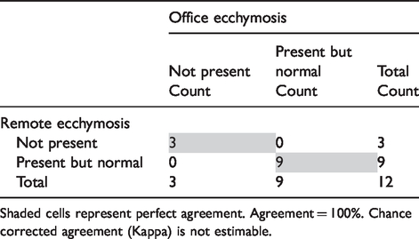
Shaded cells represent perfect agreement. Agreement = 100%. Chance corrected agreement (Kappa) is not estimable.
No patient presented with severe ecchymosis to the point of requiring further intervention and, therefore, the sensitivity of a remote diagnosis of severe ecchymosis was inestimable. No patients presented to the office with lagophthalmos or wound dehiscence, and there were no false positive remote diagnoses of these pathologies. Therefore, the sensitivity of a remote diagnosis of lagophthalmos was inestimable as was the sensitivity of a remote diagnosis of wound dehiscence.
Discussion
Our study assessed the ability to establish accurate diagnoses of anterior segment ocular pathology based on smartphone photographs taken by a patient’s companion. In particular, we wanted to assess the ability to recognize specific ‘red-eye’ pathologies and recognize the post-operative status of patients following eyelid surgery. Following a thorough review of the literature, we were unable to identify any tele-ophthalmology studies to date evaluating these specific pathologies via smartphone technology. Thus, this represents a novel application of tele-ophthalmology practices. Our findings suggest that certain ‘red-eye’ pathologies are diagnosable via smartphone photographs with relative consistency. In particular, subconjunctival haemorrhage and pterygium were readily recognizable via smartphone photos. Given that subconjunctival haemorrhage can often present with an alarming appearance to patients and healthcare providers inexperienced with eye care, smartphone-enabled diagnosis and follow-up would enhance patient care.
Certain red-eye pathologies such as corneal ulceration, episcleritis, uveitis and blepharitis were not as commonly encountered in our study. With the use of smartphone images and a detailed history, these conditions were able to be successfully diagnosed remotely in a small sample of patients. Corneal ulceration has already been shown to be readily diagnosable via the use of smartphone images taken by a trained photographer. 11 Further studies are warranted to evaluate whether smartphone images can be used to consistently diagnose these other red-eye pathologies such as uveitis, episcleritis and blepharitis.
Our study suggests that one’s post-operative eyelid surgery status can be accurately assessed via smartphone photographs. Post-operative ecchymosis was diagnosed with 100% accuracy. This presents a promising new method for following eyelid surgery patients post-operatively. Telemedicine for post-operative care in other fields of surgery has already shown to have: 1) comparable clinical outcomes as clinical visits, 2) cost savings for both patients and healthcare systems, 3) enhanced patient satisfaction and 4) increased accessibility. 20 Therefore, it is reasonable to believe that tele-ophthalmology for post-operative eyelid surgery patients has the potential to yield these benefits as well.
In order to mitigate issues with image quality, our study design incorporated an instructional video for the patient and companion which standardized multiple factors that influence photograph quality. Such factors standardized in our study included patient distance from the camera, position of gaze, camera magnification at 50%, camera flash set ‘ON’ and room lighting. In future studies, it may be worthwhile to incorporate an image grading system in order to more fully assess the impact of image quality.
Several tele-ophthalmology models of care have already established feasibility for screening and triaging a variety of posterior ocular segment conditions.22–25 These models mostly involve local primary care clinicians capturing and transmitting images to ophthalmologists for triage and consultation. Many of these models involve smartphones but require specialized attachments and software applications. 26 While these models offer tremendous benefit, they are limited in that patients do not possess this technology at home and must still present to their nearest primary care physician. Our model is novel and offers enhanced value in that the majority of medical providers and patients already own a smartphone, and no costly attachments or software applications are required. Thus, our model has the potential to reduce the number of emergency room and primary care physician visits since patients are able to send smartphone photographs directly from home. Our study demonstrates feasibility of concept specifically in the diagnosis of several external and anterior segment ocular pathologies. However, if the presenting ocular condition ends up being posterior segment in nature, external photographs of the eye may be of limited value in and of themselves. When used in combination with a clinical history, however, standardized external photographs may still help primary care providers and ophthalmologists to triage the patient. Further studies are warranted to evaluate these concepts more fully.
Limitations of the study
During the course of our study we found that patients with extreme light sensitivity may not be ideal candidates for this type of smartphone-enabled follow-up. Patients with light sensitivity had difficulty keeping their eyes open during photography. Based on our pre-enrolment testing with study equipment, we determined that the iPhone flash would optimize lighting for the purposes of image interpretation. In order to provide standardized conditions, we had every patient take photographs with the flash set ‘ON’. Otero and associates showed that different modes of illumination may affect the colour quality of images produced, but that subjective evaluation of the conjunctiva amongst ophthalmologists was still remarkably consistent. 21 Further research and experimentation will be required to assess the best lighting conditions for smartphone-enabled tele-ophthalmology follow-up. Should flash be necessary for accurate image interpretation, light-sensitive patients may not benefit from this type of follow-up. Another significant limitation of the study is sample size. Unfortunately, due to constraints with funding, our study enrolment period was truncated. As a result, we are limited in the definitive conclusions we are able to draw. However, we believe our study includes enough participants to demonstrate feasibility of concept and to warrant future study.
Conclusion
Overall, the findings of our study present potential for a novel means of patient follow-up. An increasing percentage of individuals in the United States and abroad own smartphones. Thus, follow-up based on smartphone photographs taken at home has the potential to enhance access to care, reduce patient travel burden, expedite the patient’s treatment process and increase patient satisfaction. Furthermore, this approach is patient-centred and promotes patient engagement irrespective of patient location. Unfortunately, our patient enrolment was limited due to constraints with funding. Given our sample size, further studies would be beneficial to validate the results of our study. Further work is also necessary to design a HIPAA compliant platform for patient-physician media transfer. When this occurs, there is potential for patients to benefit greatly from smartphone-enabled tele-ophthalmology follow-up.
Footnotes
Declaration of conflicting interests
The author(s) declared no potential conflicts of interest with respect to the research, authorship, and/or publication of this article.
Funding
The author(s) disclosed receipt of the following financial support for the research, authorship, and/or publication of this article: Research Grant, Illinois Society for the Prevention of Blindness (519350).
