



Abstract
We evaluated the accuracy of tele-ophthalmology in diagnosing the major causes of chronic blurring of vision. Thirty consecutive patients attending a primary eye-care facility in Singapore (the Ang Mo Kio Polyclinic, AMKP) with the symptom of chronic blurred vision were recruited. An ophthalmic technician was trained to perform Snellen acuity; auto-refraction; intraocular pressure measurement; red-colour perimetry; video recordings of extraocular movement, cover tests and pupillary reactions; and anterior segment and fundus photography. Digital information was transmitted to a tertiary hospital in Singapore (the Tan Tock Seng Hospital) via a tele-ophthalmology system for teleconsultation with an ophthalmologist. The diagnoses were compared with face-to-face consultation by another ophthalmologist at the AMKP. A user experience questionnaire was administered at the end of the consultation. Using face-to-face consultation as the gold standard, tele-ophthalmology achieved 100% sensitivity and specificity in diagnosing media opacity (n = 29), maculopathy (n = 23) and keratopathy (n = 30) of any type; and 100% sensitivity and 92% specificity in diagnosing optic neuropathy of any type (n = 24). The majority of the patients (97%) were satisfied with the tele-ophthalmology workflow and consultation. The tele-ophthalmology system was able to detect causes of chronic blurred vision accurately. It has the potential to deliver high-accuracy diagnostic eye support to remote areas if suitably trained ophthalmic technicians are available.
Introduction
Most tele-ophthalmology systems are diagnosis-specific, such as screening for diabetic retinopathy1–3 and glaucoma.4,5 However, there has not been a validated and professional application of tele-ophthalmology in diagnosing various pathologies caused by a single symptom.
Telemedicine in ophthalmology in Singapore has been limited to photographic assessment of the fundus for diabetic retinopathy changes. Previously patients with ophthalmological conditions requiring consultation had to make an appointment with the ophthalmologist in a tertiary hospital and travel there for a face-to-face consultation. This inconvenienced both the care-givers and patients, especially those with limited mobility.
We have therefore studied the accuracy of tele-ophthalmology in diagnosing the major causes of chronic blurring of vision.
Methods
We conducted a prospective study of 30 consecutive patients with chronic blurred vision who attended a primary eye-care facility from July 2008 to September 2008. The study was approved by the appropriate ethics committee.
The tele-ophthalmology software was developed and validated by Wei et al., 6 in collaboration with the National Healthcare Group at the Tan Tock Seng Hospital (TTSH), a tertiary hospital in Singapore. The system not only allowed offline data retrieval, but also online, real-time sampling of patient data. It captured images and videos from the cameras attached to the slit lamp, and stored them in the appropriate format. These data were transferred securely from the primary eye-care facility in Singapore (the Ang Mo Kio Polyclinic, AMKP) to the TTSH via the hospital network. After transmission of data, videoconferencing between the patient at the AMKP and an ophthalmologist at the TTSH was performed with a shared electronic white-board. During the conferencing, the ophthalmologist could manipulate both images and video data on both consultation sides. Any image operation (such as drawing, zooming in or out) or video operation (such as fast forward, pause) at the ophthalmologist's end at the TTSH was displayed on the computer screen at the patient's end at the AMKP.
Workflow
The general physician at the AMKP obtained the history of chronic blurred vision from the patients who attended the general outpatient clinics. If the patients met the inclusion and exclusion criteria, they were referred to the tele-ophthalmology clinic, which was co-located at the AMKP. If patients refused, they were given an appointment to the ophthalmology clinic at the tertiary hospital. The inclusion criteria were: aged 21-75 years old and with chronic blurring of vision of more than six months’ duration. The exclusion criteria were: younger than 21 or older than 75 years; acute blurring of vision of less than one month's duration; red, swollen or painful eyes; symptoms of floaters or photopsia; ocular trauma or surgery within the preceding three months; visual hallucinations; pregnant or nursing mothers; poorly communicative patients; and non ambulant patients.
A qualified ophthalmic technician was trained to use the tele-ophthalmology system. He had many years of experience in ophthalmic imaging and photography in the department prior to this study. The same technician performed the tests for all the recruited patients.
At the tele-ophthalmology clinic at the AMKP, the telemedicine workflow was:
Registration. On arrival at the tele-ophthalmology clinic, the ophthalmic technician registered the patient using the tele-ophthalmology software. This was reflected in the system at both AMKP and TTSH sites.
Data capture. The same ophthalmic technician took a brief, guided history, including the duration and quality of blurring of vision, associated symptoms, past ophthalmic history, medical history such as diabetes mellitus, and family history. Distant and near Snellen visual acuity, auto-refraction (Canon RK-F1, Canon Inc., Japan) and non-contact tonometey were taken. Visual field was assessed with the Red Colour Perimetry Chart. 7
Infrared video-clips of pupillary examination under low ambient lighting were recorded with a HD camcorder (HDR-HC9E, Sony Corporation, Japan). Extraocular movements and cover/uncover tests were also video-recorded.
Anterior segment photographs were captured with a photo slit-lamp (Righton Zoom Photo Slit Lamp RS-1000, Right Mfg. Co., Japan): low magnification (7.5x) diffuse illumination of the cornea and the bulbar conjunctiva, parallelepiped section of the corneal stroma and central anterior chamber, optical slit of the peripheral anterior chamber depth (similar to the Van Herick test), and high magnification (32.3x) specular microscopy photograph of the corneal endothelium.
Transmission of information. The history, data, images and videos were stored by the tele-ophthalmology software and transmitted to the TTSH.
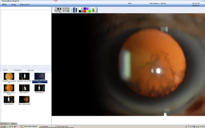
Review of information. The information was downloaded and reviewed by an ophthalmologist at the TTSH. If the Van Herick test showed a grading of three or above, the ophthalmologist would call the tele-ophthalmology clinic and ask for mydriasis. After pupillary dilation, photographs of the lens, red reflex and fundus were taken (CR6-45NM, Canon Inc., Japan), and were transmitted to the TTSH (Figures 1 and 2).
(A) Screenshot of the desktop images that were transmitted from the AMKP to the TTSH. (B) Screenshot of an infrared video recording of pupillary examination Red reflex taken with retroillumination demonstrating posterior subcapsular cataracts and cortical cataracts
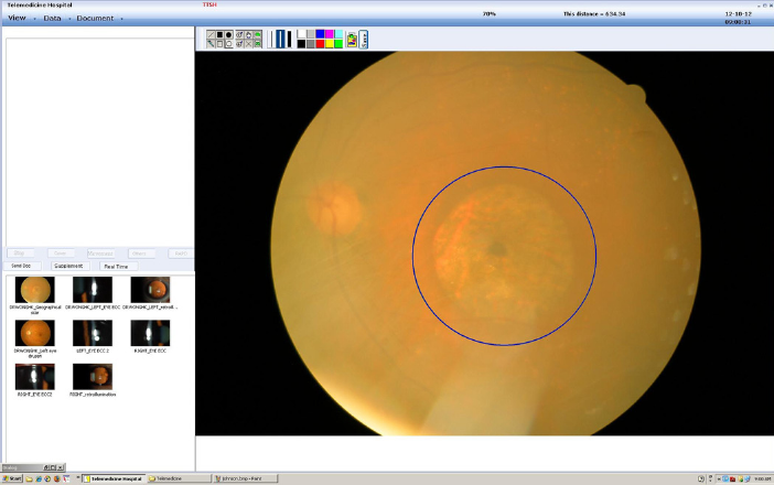
Videoconferencing. Videoconferencing (Figure 3) between the patient at the AMKP and the ophthalmologist at the TTSH allowed both parties to interact with the aid of the electronic white-board feature of the proprietary software (Figure 4).
6
(A) the ophthalmologist (left) videoconferencing with the patient. (b) the patient (right) at the other end of the videoconference Large geographic macular degeneration. The circle drawn over the macula by the ophthalmologist at the TTSH with the electronic white-board feature of the software was seen simultaneously by the patient at the AMKP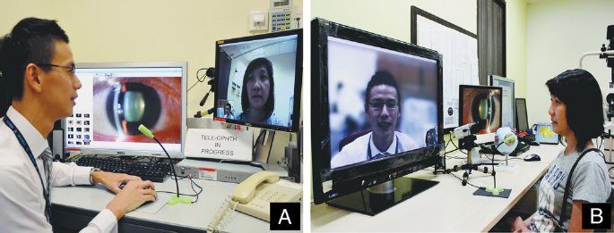
Online transfer of instructions back to the AMKP. At the end of the consultation, the ophthalmologist at the TTSH completed an online electronic medical record with the appropriate diagnoses and dispositions. This was transmitted back to the technician at the AMKP for further action, including scheduling of appointments to the hospital or the tele-ophthalmology clinics at the AMKP, or referrals to the optometrist for fitting spectacles.
The patients underwent a face-to face ophthalmic examination by another ophthalmologist at the AMKP; this served as the reference or gold standard. The AMKP ophthalmologist had no prior information from the TTSH ophthalmologist, and the patients were instructed not to divulge any information given to them by the TTSH ophthalmologist during the tele-consultation.
If both eyes of a patient were equally affected, one eye was randomly selected. Four diagnostic groups were captured using dichotomous variables: media opacity, maculopathy, neuropathy and keratopathy. Sensitivity and specificity for each was analysed against the gold standard. A questionnaire, previously validated with ten patients in an internal trial of the system, was administered. Dichotomous variables (Yes/No) were used in the questionnaire to survey the patients’ experience with tele-ophthalmology workflow and consultation compared to face-to-face consultation, and a 5-point response (from very satisfied to very dissatisfied) to the overall experience with the telemedicine workflow.
Results
Thirteen males and 17 females participated in the study. There were 24 Chinese, 4 Malays and 2 Indians. The mean age was 62 years (range 34-75). Eighteen affected eyes were right, and 12 were left.
The sensitivity and specificity of the tele-ophthalomology system in diagnosing chronic blurred vision due to media opacity, maculopathy optic neuropathy and keratopathy is shown in Table 1. One eye had previous cataract surgery, and was excluded in the analysis of media opacity. Seven and six eyes were also excluded from the categories of maculopathy and optic neuropathy respectively, due to poor image quality of the fundi from media opacity. There were no cases of exudative age-related macular degeneration, retinal artery occlusion, retinal vein occlusion, sectoral disc pallor, disc swelling or corneal oedema.
Sensitivity and specificity of the tele-ophthalmology system in diagnosing causes of chronic blurred vision
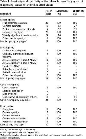
AREDS, Age-Related Eye Disease Study
ARMD, Age-Related Macular Degeneration
Reflects the number of cases used in the analysis of each category, and includes negative cases in that category
All patients participated in the patient experience survey (Table 2). The majority (97%) were satisfied with the process.
Patients’ experience with tele-ophthalmology (n = 30)

Discussion
There was high sensitivity of 96% and specificity of 100% in detecting visually significant media opacity. While cortical cataracts had a sensitivity of 63% and specificity of 77%, and posterior subcapsular cataracts with sensitivity of 67%, all other categories achieved more than 90% sensitivity and specificity. The face-to-face doctor was able to diagnose cortical and posterior subcapsular cataracts with dynamic slit lamp examination, while the reviewer tended to miss these with still images. However, these were not visually significant.
With clear media, the fundus photographs taken were of a sufficiently high resolution and quality to accurately identify the presence or absence of hard exudates, microaneurysms or haemorrahges in the macula. The 100% sensitivity and specificity in the category of maculopathy in this study was not unexpected.
The lack of stereopsis and different exposure of the fundus photographs taken (due to varying amount of media opacity) posed a difficulty in determining the cup-disc ratio, hence, there was a lower sensitivity and specificity in identifying “optic nerve abnormality, others”. Corneal pathologies were easily diagnosed due to ease of image capture for ocular surface pathology.
Photography took longer than examination as the technician had to adjust the focus and flash exposure. Despite that, there was a high satisfaction rate (97%) among the patients surveyed.
Validation of the tele-ophthalmology system encompassed equipment, workflow and reader-accuracy The software had been previously validated to confirm negligible image degradation during transmission. 6 The transfer of data through the hospital intranet was smooth, and the sequence of workflow was logical. The present study validated the accuracy in diagnosing causes of chronic blurred vision using the tele-ophthalmology system. Use of the electronic white-board feature aided in patients’ understanding of their ophthalmic conditions.
The main limitation of the present study was the small sample size. A larger sample size could capture more cases of common pathologies that could further validate the robustness of our system. Moreover, there were seven eyes (23%) with significant media opacity that degraded the quality of the fundus images, while the ophthalmologist at the AMKP was able to visualise the fundi.
Future developments include videos of slit lamp examination of the anterior segment and stereoscopic fundus photographs to detect macula oedema. Possible applications of the tele-ophthalmology system and workflow include patients presenting with acute red eyes and post-cataract extraction surgery follow-up.
In conclusion, most tele-ophthalmology systems are diagnosis-specific, rather than symptom-targeted. We were able to employ tele-ophthalmology in a novel clinical consultation workflow, and widen its application to the realm of diagnosis. The system was able to detect causes of chronic blurred vision accurately. It has the potential to deliver high-accuracy diagnostic eye support to remote areas if suitably trained ophthalmic technicians are available.
Footnotes
Acknowledgements
We thank Siew Lian Yau, Jonah Nehemiah Chung Khai Huang, Kucy Ping Ng, Sing Yong Lim and Mui Leng Tan for their contributions to this study. We are grateful for support from the National Healthcare Group Right Siting Fund.