



Abstract

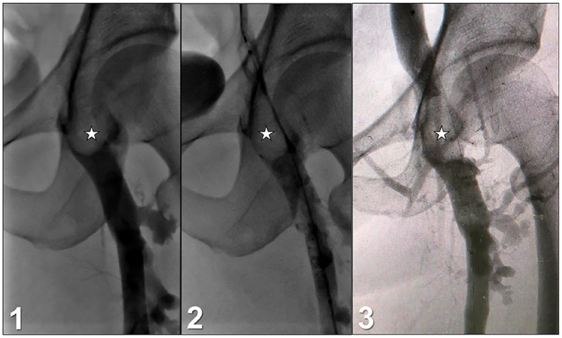
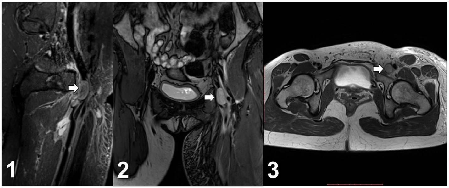
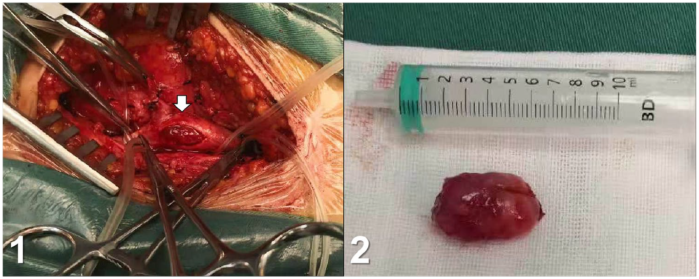
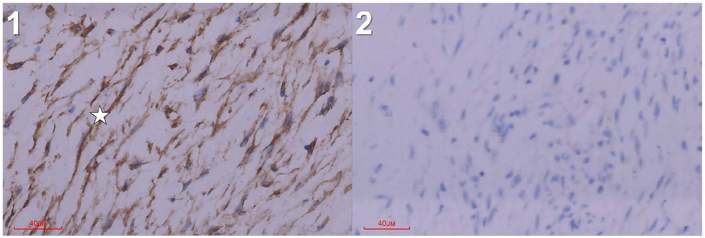
A 39-year-old woman was admitted with presentation of left leg pain and swelling for 2 weeks. She had no history of recent trauma or surgery and was not on any medication. Her past medical history was unremarkable. D-dimer assay was positive with a level of 0.98 μg/ml (reference range: 0.00–0.50 μg/ml) and venous ultrasound revealed focal thrombosis of the left common femoral vein. An aggressive ‘thrombus-removal’ strategy with multiple modalities including pharmacomechanical thrombectomy (PMT), percutaneous transluminal angioplasty (PTA), and additional catheter-directed thrombolysis (CDT) was performed with the aim of early restoration of venous patency, preservation of venous valve function, and reducing the risk of post-thrombotic syndrome. The treatment for deep venous thrombosis (DVT) was not as effective as expected. The venographic assessments, including a pre-PMT image (Panel A-1), a post-PMT and pre-CDT image (Panel A-2), and a post-CDT image (Panel A-3), demonstrated an ovoid-shaped intraluminal lesion in the left common femoral vein (Panel A, star), remaining unchanged during the treatment. Magnetic resonance imaging (MRI) for further evaluation showed a longitudinal soft tissue mass lesion at the same location (Panel B, arrow), occupying the lumen and leading to total occlusion of the left common femoral vein. Finally, a surgical excision was performed, and the patient was found to have a soft, ovoid mass with a clear boundary, which was located at the distal part of the common femoral vein (Panel C-1, arrow). The mass was resected without venous reconstruction. Pathologic examination reported that the mass was a fibromyxoid tissue comprising spindle-shaped cells which were fascicular or irregularly arranged. Immunohistochemical studies showed the spindle-shaped cells were positive for smooth muscle actin (Panel D-1, star) and negative for S-100, desmin, FLI-1, ALK, and CD34 (Panel D-2). The results were consistent with intravascular fasciitis.
Intravascular fasciitis is a rare, benign, intravascular tumor-like lesion characterized by the proliferation of myofibroblasts arising from the wall of small- and medium-sized blood vessels. 1 Histopathology is required to obtain the diagnosis; however, a high index of suspicion is required. Intravascular fasciitis may be mistaken for DVT when it obliterates the vessel lumen, as was the case with our patient. 2
Clues to nonthrombotic intravascular lesions include: (1) suspected DVT in unexpected locations, including an uncommon distribution or pattern of the thrombus; (2) young patient age or low risk for thrombosis; and (3) lack of treatment response to anticoagulation or more invasive therapies for venous thrombosis. MRI may provide important information for diagnosis and treatment, given its excellent soft-tissue contrast and multiplanar reconstruction capabilities.
‘Images in vascular medicine’ is a regular feature of Vascular Medicine. Readers may submit original, unpublished images related to clinical vascular medicine. Submissions may be sent to: Heather Gornik, Editor in Chief, Vascular Medicine, via the web-based submission system at http://mc.manuscriptcentral.com/vascular-medicine
