



Abstract

A 55-year-old man with a past medical history of renal transplantation and residual stenosed upper-extremity arteriovenous fistula (AVF) presented to the emergency department with chest pain of 1-day duration. The pain was localized to the right chest wall, associated with redness and a palpable mass. He did not recall any trauma to his chest and had not noticed any discharge from the nipple. Physical examination revealed a well-defined erythema following a linear pattern across the anterior chest wall with underlying firmness to palpation following the same linear pattern (Panel A).
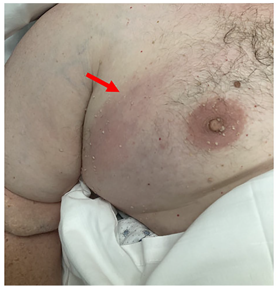
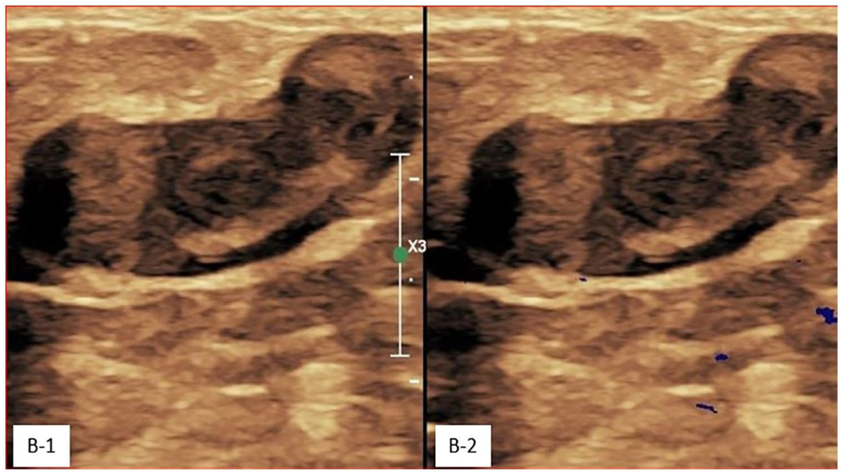
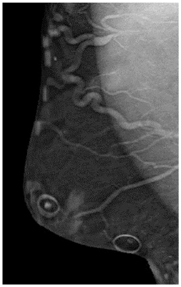
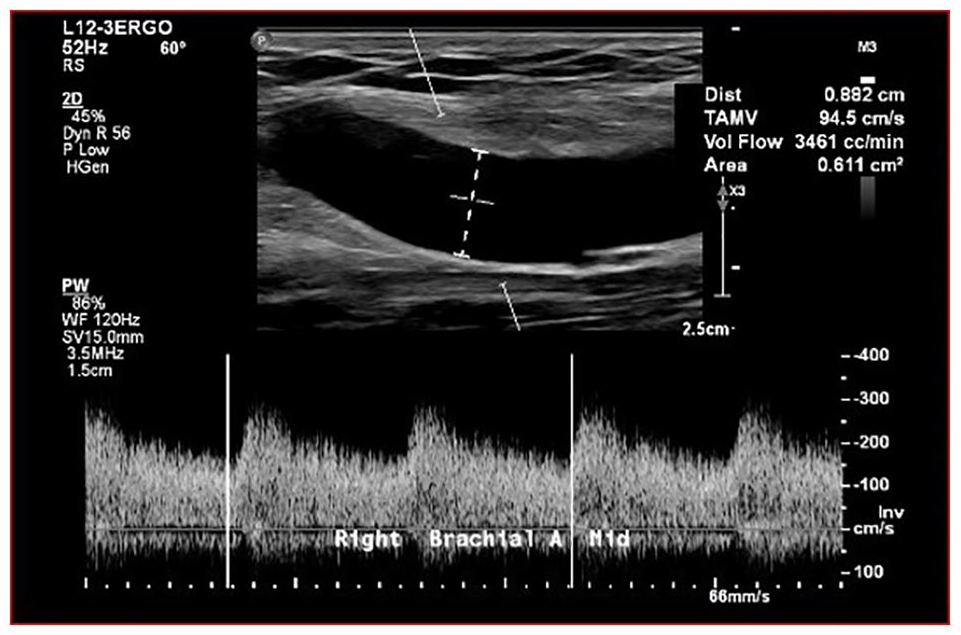
A venous Doppler ultrasound examination was performed, and he was found to have acute superficial thrombophlebitis in the basilic vein extending from the mid-upper arm to the distal-upper arm. There was also acute superficial thrombophlebitis in a superficial vein of the right chest, extending deeper into the thoracic cavity (Panel B). The vascular medicine team was consulted, and based on the history, clinical examination, and venous duplex, the diagnosis of Mondor’s disease was made. He underwent bilateral diagnostic mammogram that revealed multiple varicosities in the upper outer aspect of the right breast but was negative for mass or calcification (Panel C). He subsequently had AVF ultrasound that demonstrated high-output AVF > 3000 mL/min (Panel D). The patient was treated with 6 weeks of anticoagulation with a prophylactic dose (10 mg) of rivaroxiban. His symptoms completely resolved. A follow-up venous duplex at 2 months showed resolution of thrombophlebitis.
Mondor’s is a rare disease, resulting from sclerosing superficial thrombophlebitis that can affect several different veinous drainage systems. It predominantly affects the anterolateral thoracoabdominal wall, and thoracoepigastric, superficial epigastric, and lateral thoracic veins are the most common sites of involvement. 1 In a minority of cases, the axilla and penis can be affected. 2 The pathogenesis of Mondor’s is poorly understood. Though considered to be an inflammatory condition involving superficial phlebitis, it has occasionally been associated with lymphangitis. 3 The common etiopathogenic factors include traumatic events, excessive physical exercise, infections, concomitant oral contraceptive use, pregnancy, and breast surgery, but breast cancer is an important consideration in a newly diagnosed case. In our case, high-output AVF most likely led to venous hypertension and varicose veins of the upper extremity, ultimately resulting in symptomatic thrombophlebitis. Mondor’s can be diagnosed clinically, but if further investigation is warranted, venous duplex ultrasound is the best test. 4 Mammographic evaluation is often indicated to rule out malignancy. In general, Mondor’s is a self-limiting disease that spontaneously resolves within 6–8 weeks without any special treatment. 1 There are studies reporting the effectiveness of anticoagulation in the acute phase of Mondor’s, and some have adopted the anticoagulation strategy of using a prophylactic dose to treat the condition. 5
Footnotes
Note – Figures are in color online.
Declaration of conflicting interests
The authors declared no potential conflicts of interest with respect to the research, authorship, and/or publication of this article.
Funding
The authors received no financial support for the research, authorship, and/or publication of this article.
