
Abstract
The early detection of Alzheimer's disease, before symptoms have appeared, is integral to the development of effective treatments. Dynamic light scattering spectroscopy measures the Brownian movement of proteins at the molecular level. This technique may facilitate early Alzheimer's disease diagnosis and the discovery of pharmaceuticals that may prevent symptom development.
Introduction
The term “early diagnosis” is dependent upon the sensitivity of the diagnostic method employed. Biomarkers have differing sensitivities and specificities. As science advances, “early diagnosis” is increasingly becoming “earlier.”
The ultimate treatment goal is to prevent symptom occurrence. Alzheimer's disease offers a time spectrum for therapeutic interventions with many opportunities to intervene years before the development of permanent symptoms. But that opportunity depends on the earliest diagnosis of the condition.
At the present time, Alzheimer's disease is diagnosed after symptoms have occurred. A preclinical biomarker would be a valuable addition to the development of more effective pharmaceuticals that prevent, not just slow or ameliorate symptoms.
The retina of the eye is a neural tissue, originating from an outpouching of the diencephalon. For this reason, much efforts have been directed at determining whether changes in retinal imaging may be utilized as a biomarker for diagnosing Alzheimer's disease. 1
Ocular coherence tomography (OCT), an imaging test, has demonstrated retinal nerve fiber thinning in patients diagnosed with Alzheimer's disease.2–7 There are multiple issues with the use of this technology. Imaging changes are a relatively late finding reflecting long standing changes in molecular synthesis and cell development. There is an age-related decrease in the thickness of the retinal nerve fiber layer, yet age-matched OCT studies are generally not performed. Other confounding variables include the health of the retina, specifically, the presence of glaucoma, age-related macular degeneration, and diabetic retinopathy. While these conditions have been associated with the development of Alzheimer's disease, they can also affect OCT measurements, making this test, in the absence of a retinal examination, subject to error. 8
What is needed is a sensitive, quantitative, inexpensive test that is independent of the patient's age and retinal condition.
Methods
Dynamic light scattering (DLS) spectroscopy is an optical technique that measures the Brownian movement of molecules. 9 DLS has demonstrated the ability to make reproducible measurements from the retina of the human eye and offers a predictive capability.10–12
It is self-evident that a change in a molecule will affect its Brownian movement. Conceptually, the change in Brownian movement velocity should be a much earlier indicator of a pathological process than the release of a substance in the cerebrospinal fluid, or blood, or an imaging change such as the development of amyloid plaques. The same criticism also applies to recent efforts using AI to analyze retinal OCT images.
Ethics statement
The study was Institutional Review Board approved and was performed in accordance to the Declaration of Helsinki. All participants provided written informed consent.
Results
The reproducibility of DLS testing was determined over several months. Seventeen patients with no history of ocular or neurologic disease were tested. There was no significant difference in measurements obtained between the left, or right eyes, between men or women, or at different times of the day. Patient age was not a confounding variable.
The DLS device is interfaced and parfocal with a clinical ophthalmic fundus camera. The patient is instructed to stare at a 14 μW light. The DLS operator confirms the location of the light on the retina by looking through the camera's oculars. The measurement duration is 5 s. Experimentation has determined the use of a 12.5 ns sampling time. An autocorrelation curve is automatically generated by the embedded software.
Fifteen patients with mild cognitive impairment and a normal retinal examination underwent DLS testing. Four patients with mild cognitive impairment, in the absence of an Alzheimer's disease diagnosis at the time of DLS measurement, demonstrated a slowing in Brownian motion by DLS testing. These patients were later diagnosed with Alzheimer's disease from 6 months to 1 year after the DLS measurement.
Seventeen patients with presumed Alzheimer's disease underwent DLS testing. Eleven patients were confirmed to have Alzheimer's disease by positron emission tomography (PET) amyloid nuclide scans (Figure 1), 5 were correctly identified as having negative nuclide scans, and one patient with a negative PET amyloid nuclide scan exhibited a borderline DLS Alzheimer's measurement predictive of a future diagnosis. Figure 2 is the DLS measurement of an age-matched control patient without neurologic symptoms.
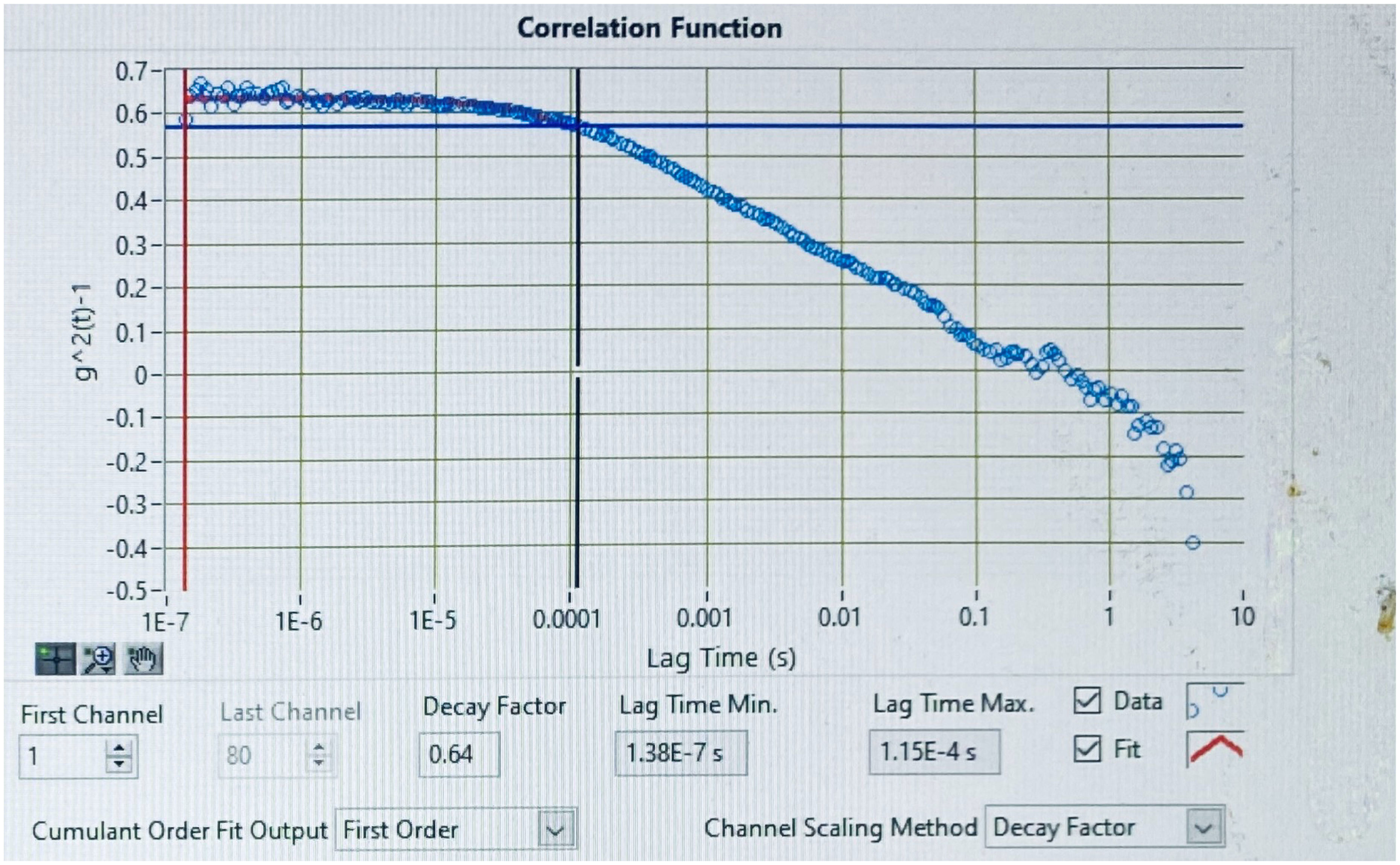
Patient diagnosed with Alzheimer's disease by DLS testing and confirmed by PET amyloid scan.
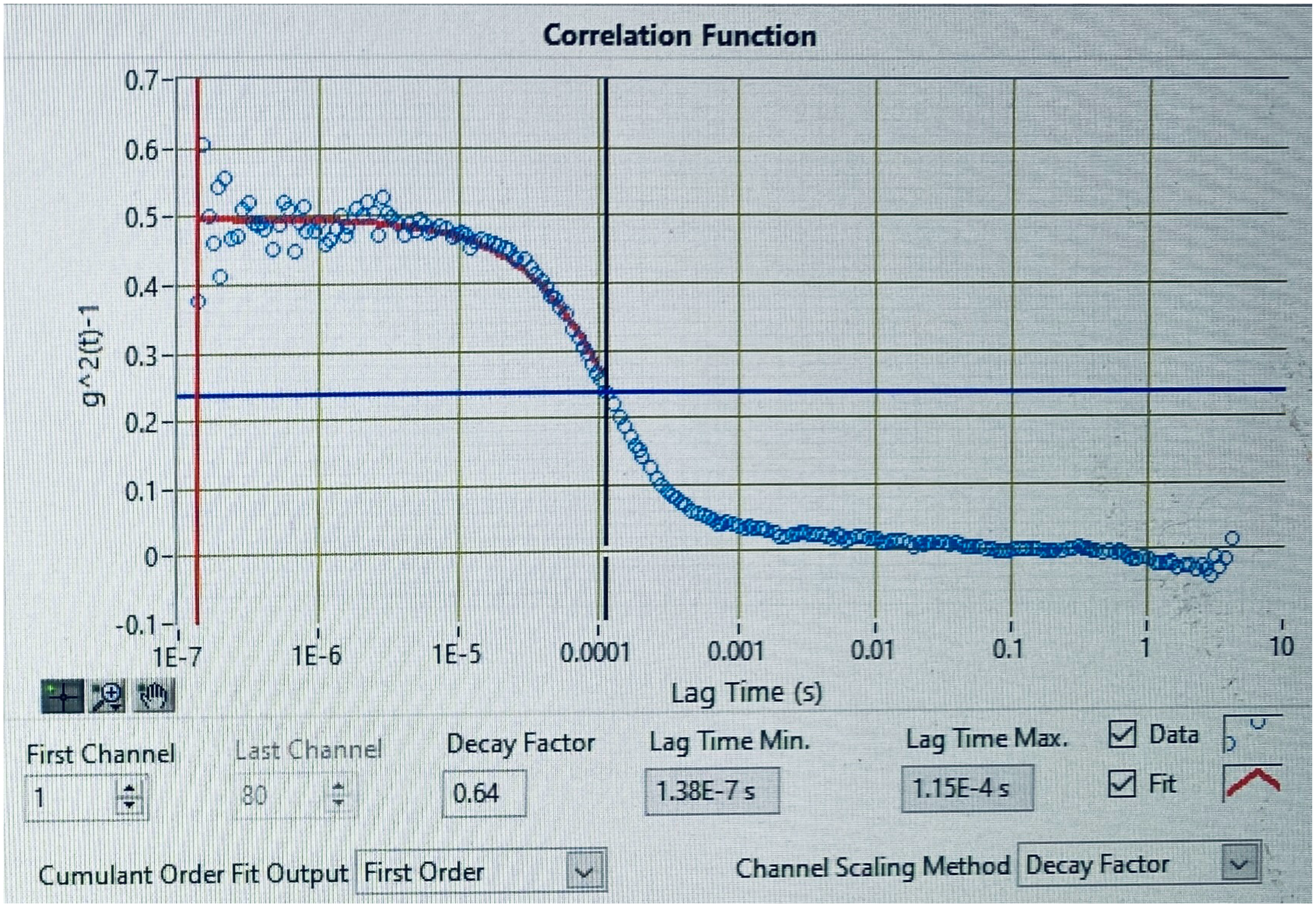
Patient without neurologic symptoms – Control group.
Discussion
The DLS test is easy to perform. One pupil is dilated with standard mydriatic agents, and the patient stares at a weak red light (much weaker than a direct ophthalmoscope), for 5 s. There are no disposable items involved, and the test costs only a few pennies to perform, making the technology potentially available to patients without health insurance and increasing the ability to screen patients
In the present paper, all patients had unremarkable retinal examinations. This was confirmed at the time of testing as the DLS detection beam is focused by the operator using a clinical retinal fundus camera.
Patients diagnosed with Alzheimer's disease undergoing DLS testing exhibit flattening of the initial line and their measurement begins above 0.6 g2(t)−1. The curve in non-Alzheimer's disease patients begins below 0.6 g2(t)−1 and demonstrates a quick descent.
The initial “straight line” observed in patients with Alzheimer's disease, indicates a slowing in the Brownian motion of the retinal proteins, as compared to the control group patient. This presumably represents a change in the velocity of the ganglion cells within the retinal nerve fiber layer. Postmortem analysis of the retina of Alzheimer's patients has demonstrated ganglion cell-inner plexiform retinal layer disfunction, which is eventually observed as retinal thinning by OCT testing.
Studies using OCT are affected by the health of the retina. Specifically, conditions such as diabetic retinopathy, age-related macular degeneration, and glaucoma affect OCT measurements. Further DLS testing will determine whether these conditions also effect the DLS results.
The development of a noninvasive, quantitative, and inexpensive test to make an “early” diagnose of Alzheimer's disease has the potential to lead to breakthroughs in drug development, the screening of large groups of patients, and allows a larger temporal treatment window before the onset of irreversible dementia.
Footnotes
Acknowledgments
The author thanks the patients and staff of 3 T Radiology who participated in this work.
Author contributions
Jeffrey Neill Weiss (Conceptualization; Data curation; Formal analysis; Investigation; Methodology; Project administration; Resources; Supervision; Validation; Visualization; Writing – original draft; Writing – review & editing).
Funding
The author received no financial support for the research, authorship, and/or publication of this article.
Declaration of conflicting interests
The author declared no potential conflicts of interest with respect to the research, authorship, and/or publication of this article.
Data availability
The data supporting the findings of this study are available from the author. The data is not publicly available due to privacy or ethical restrictions.