



Abstract
Background
Urinary formic acid (FA) has been reported to be a biomarker for Alzheimer's disease (AD). However, the association between FA and pathological changes in memory clinic patients is currently unclear.
Objective
This study aims to investigate associations between FA and pathological changes across different cognitive statuses in memory clinic patients.
Methods
A cohort of patients with mild cognitive impairment (MCI-Aβ- n = 37, MCI-Aβ+ n = 33), AD dementia (n = 39), and cognitively normal subjects (CN-Aβ- n = 98, CN-Aβ+ n = 50) were included. Comprehensive neuropsychological assessment, urinary FA, AD-related plasma biomarkers, MRI scans, [18F]-flurbetapir and [18F]-FDG PET scan data were collected from all participants.
Results
Urinary FA levels were higher in patients with MCI and AD than in CN subjects and higher in Aβ+ (CN- Aβ+, MCI-Aβ+, AD dementia) subjects than in Aβ-subjects (CN- Aβ-, MCI-Aβ-). Urinary FA was positively associated with cerebral Aβ deposition and negatively associated with glucose metabolism, both at the global level and in multiple regions of interest cortical regions in participants with different cognitive statuses. Additionally, urinary FA levels were positively correlated with the severity of white matter hyperintensities and hippocampal atrophy. Urinary FA combined with age, Mini-Mental State Examination, plasma p-tau181, and neurofilament light chain could be used to predict Aβ deposition in the brain.
Conclusions
Urinary FA is associated with brain pathological changes in memory clinic patients, including cerebral Aβ deposition, glucose metabolism, white matter hyperintensities, and hippocampal atrophy. It could be used as a biomarker for the early diagnosis of AD and predicting Aβ deposition.
Introduction
Alzheimer's disease (AD) is a progressive neurodegenerative disease typically characterized by memory loss and cognitive decline. Amyloid-β (Aβ) accumulation, tau deposition and neurodegeneration are the most representative pathological features of AD. As many clinical trials for the treatment of symptomatic AD have failed, 1 the focus on AD is shifting from reversing the clinical manifestations of advanced AD to early diagnosis and intervention in the prodromal stage. Detection of Aβ in the brain by amyloid positron emission tomography (PET) or measurement of Aβ42 in the cerebrospinal fluid (CSF) is considered to be the most effective method for early diagnosis of AD.2,3 However, both methods are difficult to implement for large-scale screening at the population level due to their invasive procedure or high cost. Therefore, there is a substantial need to develop biomarkers that are simple, effective, low-cost, non-invasive and associated with Aβ deposition.
Currently, attempts have been made to find these relatively specific peripheral biomarkers that might be more applicable for large-scale preliminary screening, early diagnosis, and predict Aβ deposition in the brain. Most research has been focused on peripheral blood markers. An increasing body of evidence suggests that plasma biomarkers are diagnostically meaningful and are associated with clinical progression in AD (Aβ42, Aβ42/40, total tau (t-tau), and p-tau181). 4 Another highly desirable source of AD biomarkers is urine, as it can be easily and non-invasively collected in relatively large quantities.5–7 In addition, it contains metabolites that reflect responses to pathophysiological conditions of injury and oxidative stress generated from other biological events at the system level.8,9 Urinary metabolite formaldehyde and formic acid (FA) were reported to be associated with AD.10–14 When exogenous formaldehyde enters the human body, it is metabolized into FA in the liver and erythrocytes, and then excreted in the urine and feces or through the respiratory system.15,16 Our previous preliminary studies showed that formaldehyde and FA in urine were higher in subjects with mild cognitive impairment (MCI) and AD compared to those with normal cognitive ability. Urinary FA is a potential biomarker for AD screening and early diagnosis.10,11
There are a series of pathologic changes in the pathogenesis of AD. According to the 2018 NIA-AA framework for AD, the definition of AD is shifting from clinical manifestations to biomarkers in living people. Biomarkers of AD are grouped into of Aβ deposition (A), pathologic tau (T), and neurodegeneration (N). Biomarkers of neuronal degeneration or injury are further measured by elevated CSF tau, decreased FDG uptake on PET, and atrophy on structural magnetic resonance imaging (MRI). 3 The amyloid cascade hypothesis of AD posits that cortical Aβ deposition is related to downstream neuronal dysfunction and cognitive impairment. 17 Glucose uptake in the brain decreases because of normal aging but this decline is accelerated in AD patients. 18 In addition, there are other changes in MRI associated with AD, including white matter hyperintensities (WMH) and regional atrophy of the medial temporal lobe (MTL). WMH, frequently seen in older adults, are usually considered participate in the vascular factor contributing to cognitive impairment and dementia. WMH are more prevalent and severe in AD patients compared to non-demented older adults19,20 and are associated with an increased risk of AD. 21 The MTL is an early affected site in AD-related neurodegeneration. 22 Regional atrophy of the MTL structures detected with MRI is also considered a reliable diagnostic marker for AD. 23 However, the association between FA and above pathological changes in memory clinic patients is currently unclear.
To further substantiate the reliability of urinary FA as a biomarker for the early diagnosis of AD, it is critical to accurately and thoroughly understand the relationship between urinary FA and brain pathological changes in the course of AD. In the present study, our aim was to investigate whether the urinary FA (1) differed among cognitively normal (CN), MCI, and AD dementia participants from memory clinic; (2) differentiated between Aβ positives and Aβ negatives in CN and cognitive impairment subjects; (3) associated with brain amyloid deposits and glucose metabolism; (4) associated with pathological changes of brain structure, including white matter lesions and hippocampus atrophy; (5) could be combined with blood markers to predict Aβ deposition in CN and cognitive impairment subjects.
Methods
Participants
A total of 257 participants were collected from Sixth People's Hospital, Shanghai, China through the outpatient memory clinic from January 1, 2022, to December 31, 2022, including 148 CN, 70 individuals with MCI, and 39 patients with AD dementia. The patients with MCI were in accordance with Jak/Bondi's criteria.24,25 The patients with AD were diagnosed according to the NIA-AA criteria with positive Aβ deposits.26,27 The inclusion criteria include: participants aged 50 to 75 years, who had completed at least 6 years of education, were fluent in Chinese, and had normal vision and hearing to complete cognitive tests. Participants were excluded if they abused alcohol or other substances. All participants underwent comprehensive clinical and neuropsychological evaluations, blood and urine sample collection, cranial MRI scan, [18F]-florbetapir PET, and [18F]-FDG PET scanning.
The study was approved by the Ethical Committee for Medical Research of the Shanghai Sixth People's Hospital Affiliated to Shanghai Jiao Tong University School of Medicine. Participants signed an informed consent form after being fully informed.
Neuropsychological assessment
Comprehensive clinical and neuropsychological valuations were assessed as previously described.25,28,29 Briefly, general cognitive performance was assessed by Mini-Mental State Examination (MMSE), Chinese version of Montreal Cognitive Assessment-Basic (MoCA-BC) and Chinese version of Addenbrooke's Cognitive Examination III (ACE-III-CV). In addition, six neuropsychological scores in four core tests were carried out to assess memory (Auditory Verbal Learning Test [AVLT] 30-min delayed free recall and AVLT recognition), language (Animal Verbal Fluency Test [AFT] and 30-item Boston Naming Test [BNT]) and attention/executive function (Shape Trail Test Part A and B [STT-A and STT-B]). All the neuropsychological assessments were carried out in Mandarin Chinese by trained raters.
MRI image acquisition
All participants underwent MRI scans with a 3.0 T MRI scanner (Prisma 3.0 T, Siemens, Erlangen, Germany) at the Shanghai Sixth People's Hospital in accordance with the manufacturer's approved guidelines. The severity of overall WMH was assessed according to the modified Fazekas WMH scale score. The scale is divided into four levels ranging from 0 to 3. Visual inspection of the volume of the MTL through the hippocampus (at the level of the anterior pontine tegmentum) yielded subjective MTA scores ranging from 0 (no atrophy) to 4 (severe atrophy). 23
PET image acquisition and processing
All PET scans were completed by PET/CT system (Biography 64 PET/CT, Siemens, Erlangen, Germany) at the PET center of Huashan Hospital Affiliated to Fudan University within one month after neuropsychological test. The methods of [18F]-florbetapir PET and [18F]-FDG PET scanning acquisition were described in our previous report.30,31 Using the Amyvid read protocol and the procedure applied in our previous study, the amyloid PET images were interpreted through visual inspection by three experienced raters by consensus. 30
The process for analysis by region of interest (ROI) and voxel-wise analysis was detailed in the previous study.31,32 The bilateral cerebellar crus and pons was used as a reference area to calculate voxel-wise SUVr of [18F]-florbetapir and [18F]-FDG respectively. The global cortex SUVr of [18F]-florbetapir was defined as the sum of the following 7 ROIs: the posterior cingulate, precuneus, lateral temporal, medial temporal, frontal and lateral parietal, occipital lobes. The global cortex SUVr of [18F]-FDG was defined as the sum of the following 8 ROIs: the hippocampus, frontal, lateral parietal, lateral temporal, medial temporal, occipital, posterior cingulate and precuneus lobes.
Measurement of urinary formic acid
Clean catch midstream urine samples were collected from all participants. The levels of FA in collected urine samples were determined by the Formate Assay Kit (ab111748, Abcam, Cambridge, UK), following the protocol from the manufacturer. Ten microliter of urine was used per well, and the absorbance at 450 nm was measured on a 96-well microplate reader (SpectraMax Paradigm Multi-Mode, Molecular Devices, San Jose, CA, USA). The concentration of FA was calculated according to the standard curve.
Plasma biomarker measurements
Plasma Aβ40, Aβ42, total tau (t-tau), phosphorylated tau181 (p-tau181), and neurofilament light chain (NfL) from the cohort were analyzed by the Single Molecular Array (Simoa) HD-1 Analyzer platform (Quanterix, Billerica, MA, USA) according to the manufacturer's instructions as previously described. 33
Statistical analysis
Continuous variables were described as mean ± SD and categorical variables were described as number (percentage). Comparison among CN, MCI, and AD dementia groups were performed using χ2 test for categorical variables and One-way analysis of variance (ANOVA) for continuous variables such as scores on neuropsychological assessments, age, and education years. One-way ANOVA were followed by Bonferroni's post-hoc comparisons tests. Group comparisons between Aβ negative (Aβ-) and Aβ positive (Aβ+) were performed using χ2 test for categorical variables and the student t-test for continuous variables. For inter-group comparisons, ANCOVA was used to adjust for confounding factors, such as APOE ε4 status for the comparison of FA among CN, MCI, and AD dementia groups, and age for the comparison of FA between Aβ- and Aβ+ groups. For correlation analysis between urinary FA and cognitive performance, association coefficients were calculated by partial correlation after adjusting for age, sex and education. For ROI-wise analysis, correlations were assessed using the Spearman rank test. Multiple linear regression models were used to assess the correlation between urinary FA and cranial MRI pathology. Age, sex, educational level, APOE genotype, and MTA scores/Fazekas scale scores were set as independent variables and urinary FA as the dependent variable. A multiple linear regression model was also performed to assess the predictors of Aβ deposition, and clinical characteristics, urinary FA, and plasma biomarkers were set as dependent variables. Akaike Information Criterion (AIC) and Bayesian Information Criterion (BIC) were used for model selection in regression analysis. All tests were 2-tailed, with a significance level of p < 0.05. All statistical analyses were conducted using Statistical Package for the Social Sciences 22.0 Software (SPSS 22.0).
Results
Characteristics of participants
Demographic and clinical data of the whole study population were summarized in Table 1. A total of 257 subjects were enrolled and all participants had finished urinary FA, amyloid PET and FDG PET examination (Table 1). There were no significant differences in age and sex among the groups. The educational level of AD dementia patients was significantly lower than that of CN subjects (9.9 ± 3.4 versus 12.2 ± 2.6, p < 0.001). The proportion of APOE ɛ4 carriers was significantly higher in AD dementia patients compared with CN and MCI subjects (51.3% versus 29.1%, 51.3% versus 32.9%, respectively; p = 0.032). The amyloid PET positive prevalence for CN and MCI groups was 33.8% and 47.1%, respectively. All the amyloid PET images were positive for subjects with AD dementia.
Demographics, neuropsychological tests, [18F]-florbetapir PET imaging and urinary FA levels for CN, MCI, and AD dementia groups.

CN: cognitively normal; MCI: mild cognitive impairment; AD: Alzheimer's disease; APOE: apolipoprotein; MMSE: Mini-Mental State Examination; MoCA-BC: Chinese version of Montreal Cognitive Assessment-Basic; ACE-III-CV: Chinese version of Addenbrooke's Cognitive Examination III; AVLT: Auditory Verbal Learning Test; AFT: Animal Verbal Fluency Test; BNT: Boston Naming Test; STT-A and B: Shape Trail Test Part A and B; Aβ: amyloid-β; FA: formic acid.
One-way ANOVA.
χ2 tests.
Changes of urinary FA in memory clinic patients and its association with cognitive abilities
In our cohort, urinary FA levels were inversely associated with cognitive performance, meaning that the worse the cognitive ability, the higher the level of FA in the urine (Table 1 and Figure 1A). Urinary FA levels were higher in MCI patients compared to the CN subjects (p < 0.05) and in AD dementia patients compared to MCI patients (p < 0.001). After adjustment for APOE ε4 status by ANCOVA, the difference in urinary FA level among the three groups still existed (p < 0.001). In addition, among all subjects, urinary FA levels were significantly higher in males compared with females (p < 0.05), at 0.211 ± 0.097 mM (n = 92) and 0.187 ± 0.083 mM (n = 165), respectively.
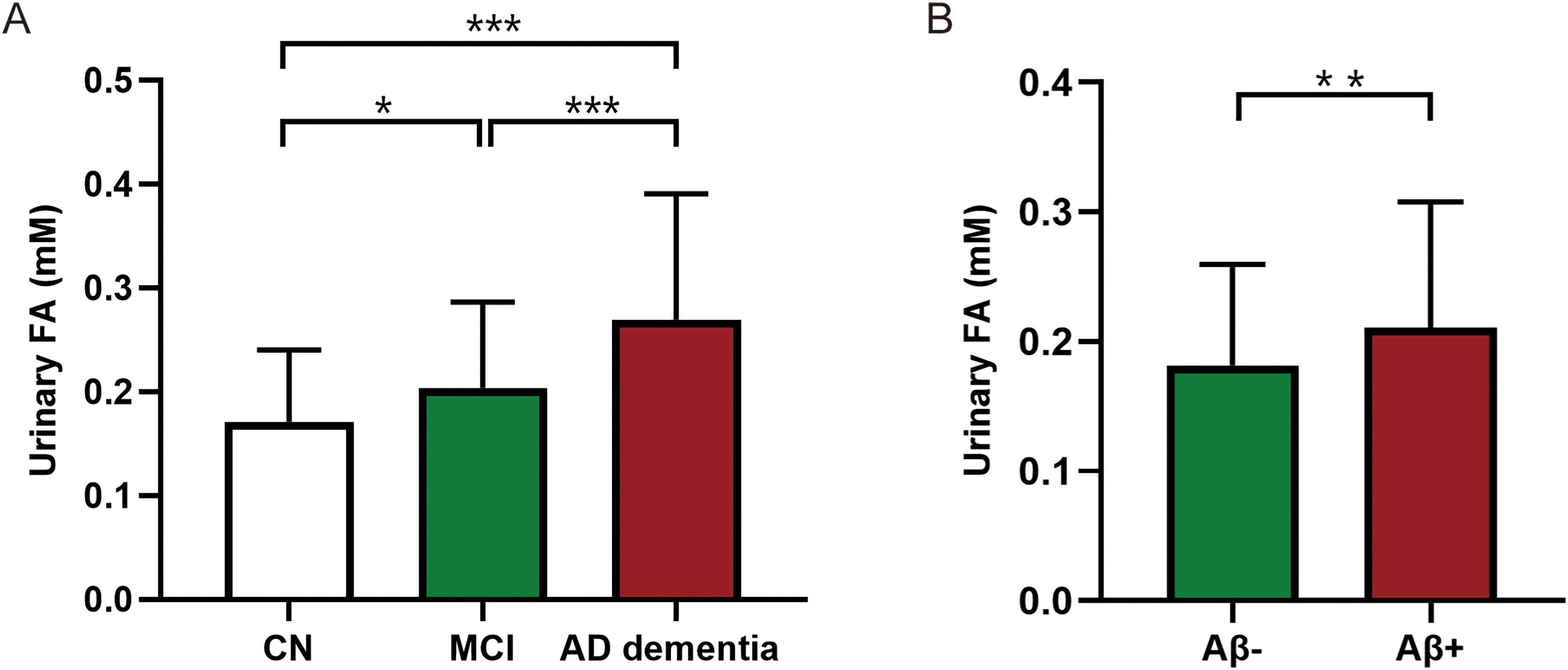
Changes of urinary FA across different cognitive statuses and its association with Aβ pathology. (A) Comparison of urinary FA levels among the CN, MCI, and AD dementia groups. (B) Comparison of urinary FA levels between the Aβ- and Aβ+ groups. *p < 0.05, **p < 0.01, ***p < 0.001. FA: formic acid; CN: cognitively normal; MCI: mild cognitive impairment; AD: Alzheimer's disease.
We further estimated the relationship between urinary FA levels and cognitive ability, including general cognitive scores and scores for memory, language and attention/executive functions. Because our aim was to study the process of cognitive decline, for all subsequent analyses we included CN, MCI, and AD dementia subjects. After adjustment for age, sex, and education by partial correlation analysis, urinary FA levels were negatively correlated with MMSE scores (R = −0.31, p < 0.001), MoCA-BC scores (R = −0.31, p < 0.001), and ACE-III-CV scores (R = −0.33, p < 0.001). Urinary FA levels were negatively correlated with AVLT-delayed recall (R = −0.24, p < 0.001), AVLT-recognition (R = −0.26, p < 0.001), AFT (R = −0.24, p < 0.001) and BNT (R = −0.18, p < 0.01), and were positively correlated with STT-A (R = 0.17, p < 0.05). Urinary FA did not correlate with STT-B. Thus, urinary FA levels showed a negative association with general cognition ability, memory and language functions, and attention/executive functions.
Association of urinary FA with cerebral Aβ pathology
As participants were heterogeneous given their Aβ deposits, we evaluated the association of urinary FA levels with Aβ deposits measured by Aβ PET. Cognitively normal subjects, patients with MCI and AD dementia were classified as Aβ positive or Aβ negative according to [18F]-florbetapir PET image results. Compared to Aβ- participants, Aβ+ participants were significantly older and performed significantly worse of general cognitive abilities (Table 2). In CN and cognitively impaired patients, urinary FA levels were significantly higher in Aβ+ subjects than in Aβ- subjects (p < 0.01, Figure 1B and Table 2). Due to the difference in age between Aβ+ and Aβ- groups, we further exclude the effect of age. In this model, urinary FA was used as the dependent variable, Aβ grouping as the independent variable, and age as the covariate. After adjustment for age, the difference in urinary FA levels between Aβ- and Aβ+ subjects persisted (p < 0.01, Supplemental Table 1).
Association of urinary FA with Aβ pathology.

Aβ-: amyloid-β negative; Aβ+: amyloid-β positive; APOE: apolipoprotein; MMSE: Mini-Mental State Examination; MoCA-BC: Chinese version of Montreal Cognitive Assessment-Basic; ACE-III-CV: Chinese version of Addenbrooke's Cognitive Examination III; FA: formic acid.
t test.
χ2 test.
We next estimate the relationship between urinary FA levels and Aβ deposition based on ROI-wise and voxel-wise analysis in all participants with different cognitive statuses. ROIs refer to the frontal, lateral parietal, lateral temporal, medial temporal, occipital, posterior cingulate, and precuneus lobe. Based on ROI-wise analysis, the results showed that urinary FA levels were positively correlated with Aβ deposition in global cortex (Figure 2A and Table 3), frontal, lateral parietal, lateral temporal, occipital, posterior cingulate, and precuneus lobe, and were not significantly related to Aβ deposition in the MTL (Table 3). Based on voxel-wise analysis, the results showed that Aβ deposition in a wide range of brain regions was positively correlated with FA (Figure 2C), which was consistent with the correlation with ROI.
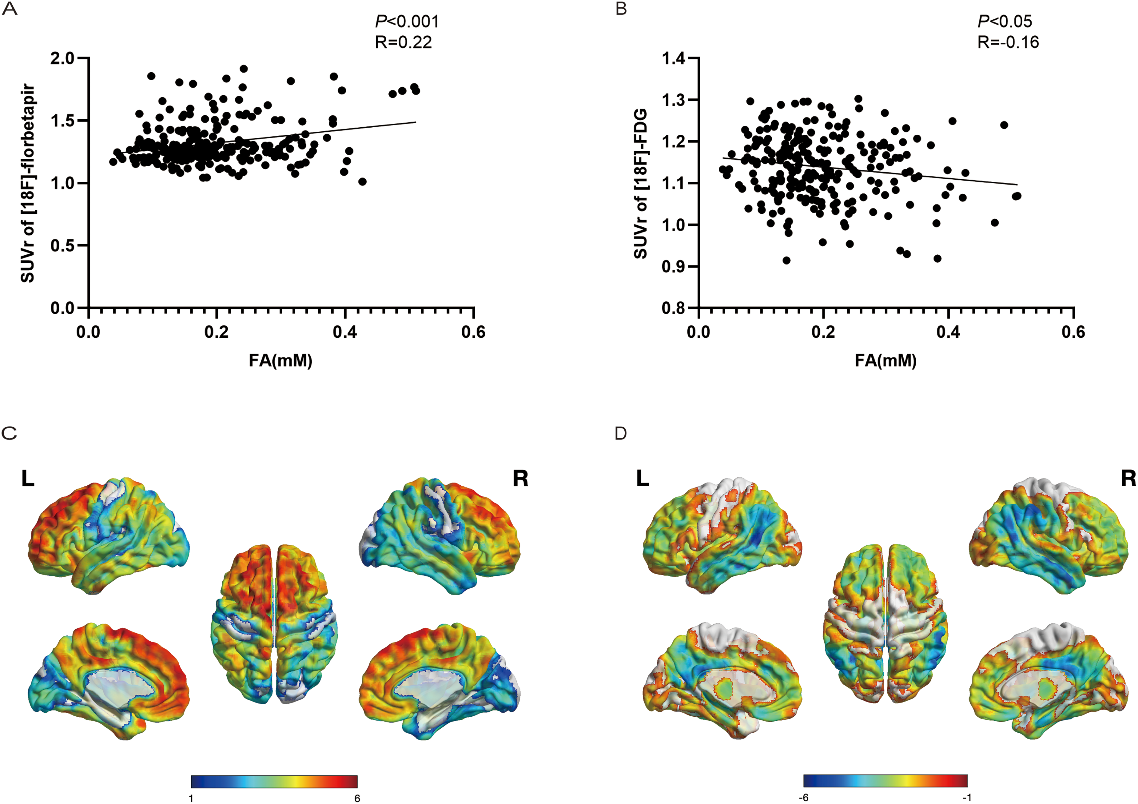
Association of urinary FA with Aβ deposition and glucose metabolism. (A) The Spearman correlations between urinary FA and global Aβ deposition. (B) The Spearman correlations between urinary FA and global glucose metabolism. (C) Association of urinary FA with Aβ deposition based on voxel-wise analysis. (D) Association of urinary FA with glucose metabolism based on voxel-wise analysis. The color bars indicate t-values, L: left; R: right.
Association of urinary FA with Aβ deposition and glucose metabolism based on ROI-wise analysis.

FA: formic acid; SUVr: standard uptake value ratios; ROIs: regions of interest.
Association of urinary FA with cerebral glucose consumption
In order to clarify whether there was a correlation between urinary FA levels and glucose metabolism in all participants with different cognitive statuses, we next used the correlation analysis to explore the relationship between urinary FA and glucose metabolism based on ROI-wise and voxel-wise analysis. Eight ROIs were as follows: the hippocampus, frontal, lateral parietal, lateral temporal, medial temporal, occipital, posterior cingulate and precuneus lobes. The results demonstrated that higher urinary FA was associated with lower global cerebral glucose consumption in memory clinic patients (Figure 2B and Table 3). Based on ROI-wise analysis, a negative correlation between urinary FA and glucose metabolism was observed in most cortical regions (hippocampus, lateral parietal, lateral temporal, medial temporal, posterior cingulate, frontal and precuneus lobe) except for the occipital lobe (Table 3). Based on voxel-wise analysis, the results showed that glucose metabolism in a wide range of brain regions was negatively correlated with FA (Figure 2D), which was consistent with the correlation with ROI.
Association of urinary FA with severity of hippocampal atrophy and white matter hyperintensities
We performed multiple linear regression analyses to analyze the relationship between urinary FA and the degree of hippocampal atrophy. The MTL atrophy is assessed visually with the medial temporal lobe atrophy (MTA) scale. 34 All subjects were included for subsequent analysis. In the multivariate analysis, we included age, sex, APOE genotype, and MTA score as independent variables and urinary FA as the dependent variable. After excluding the effects of age, sex, and APOE genotype, urinary FA levels were positively correlated with MTA scores (β = 0.270, p < 0.001, Supplemental Table 2).
Furthermore, we investigated the relationship between urinary FA levels and WMHs. The severity and distribution of WMHs was assessed by the visual rating of the Fazekas scale.35,36 In the multivariate analysis, we included age, sex, APOE genotype, and Fazekas scale score as independent variables and urinary FA as the dependent variable. After adjusting for basic characteristics (i.e., age, sex, and APOE ε4 status), there was a significant positive correlation between urinary FA levels and Fazekas scores (β = 0.245, p < 0.01, Supplemental Table 3).
The global Aβ deposition in the AD continuum can be predicted by urinary formic acid and other markers
As detection of Aβ in the brain by PET or CSF is complicated and expensive, periphery plasma biomarkers, including tau, NfL, and Aβ are increasingly being used to define and stage AD. 37 Urinary FA is a simple, low-cost and non-invasive potential biomarker for AD, and we wanted to explore whether urinary FA is also one of the predictors of Aβ. Therefore, we further explored whether urinary FA in combination with peripheral AD-related plasma markers and clinical features could be used to predict Aβ deposition in cognitively normal and cognitively impaired people. In the multiple linear regression analysis, global SUVr of [18F]-florbetapir PET was set as the dependent variable and the variables of demographics, general neuropsychological scores, urinary FA and peripheral blood markers (age, sex, APOE genotype, MMSE, MoCA-BC, ACE-III-CV, plasma Aβ40, Aβ42, Aβ42/40, t-tau, p-tau181, p-tau181/t-tau, p-tau181/Aβ42, and NfL) were included as independent variables. We used stepwise regression method to screen out the optimal variables that could predict Aβ deposition, and finally retained five significant variables: age, MMSE, plasma p-tau181, plasma NfL and urinary FA, named model 1. The AIC and BIC of model 1 are −246.8 and −222.6, respectively. We found that urinary FA combined with age, MMSE, p-tau181, and NfL could be used to predict global Aβ deposition in the brain (adjusted R2 = 0.31, p < 0.001). Associations between these variables and brain Aβ deposition were shown in Table 4. P-tau181 showed the strongest association with Aβ deposition (β = 0.346), and urinary FA showed the moderate association with Aβ deposition (β = 0.134).
The predictive model of urinary FA in combination with peripheral AD-related plasma markers and clinical features for global Aβ deposition in CN, MCI, and AD dementia patients a .

MMSE: Mini-Mental State Examination; p-tau181: phosphorylated tau181; NfL: neurofilament light chain; FA: formic acid; CN: cognitively normal; MCI: mild cognitive impairment; AD: Alzheimer's disease; Aβ: amyloid-β.
The variables were determined by using stepwise regression analysis. (Adjusted R2 = 0.313, p < 0.001)
Independent variables included in the analysis are as follows: age, sex, APOE genotype, MMSE score, MoCA-BC score, ACE-III-CV score, plasma Aβ40, Aβ42, t-tau, p-tau181, Aβ42/40, p-tau181/t-tau, p-tau181/Aβ42, p-tau181/Aβ42, NfL.
In order to validate the contribution of FA to the prediction model, we further conducted three multiple linear regression, named model 2, model 3, and model 4. All three models used global SUVr of [18F]-florbetapir PET as the dependent variable. Model 2 included both peripheral AD-related plasma markers and urinary FA as independent variables (plasma Aβ40, Aβ42, Aβ42/40, t-tau, p-tau181, p-tau181/t-tau, p-tau181/Aβ42, NfL, and urinary FA), and the AIC, BIC and adjusted R2 of model 2 were −221.2, −179.9, and 0.25, respectively. Model 3 only included peripheral AD-related plasma markers as independent variables (plasma Aβ40, Aβ42, Aβ42/40, t-tau, p-tau181, p-tau181/t-tau, p-tau181/Aβ42, and NfL), and the AIC, BIC and adjusted R2 of model 3 were −214.7, −176.9, and 0.23, respectively. Model 4 only included urinary FA as independent variable, and the AIC, BIC and adjusted R2 of model 4 were −186.8, −176.2, and 0.04, respectively. In summary, the performance of model1 was optimal and the results of models 2–4 confirmed that plasma markers combined with FA could increase the predictive value. FA alone had some predictive value but does not perform well enough, so it may be more appropriate to use it to increase the predictive value of clinical characteristics and plasma markers for Aβ deposition.
Discussion
AD is a neurodegenerative disorder with a long preclinical and prodromal phase, with pathophysiologic changes occurring years before clinical manifestations and potentially causing irreversible brain damage.38,39 Therefore, the availability of early and reliable diagnosis markers of the disease would allow its early detection and taking preventive measures to avoid neuronal loss. 40 In this study, we confirmed that urinary FA levels were inversely associated with cognitive performance, and altered early in cognitive impairment. It was associated with brain pathological changes in memory clinic patients, including cerebral Aβ deposition, glucose metabolism, WMHs and hippocampal atrophy. Urinary FA combined with age, MMSE, plasma p-tau181, and NfL could be used to predict Aβ deposition in the brain. Our study provides further pathological support for the use of urinary FA as a urine biomarker for the early screening of AD and prediction of Aβ deposition.
Urine contains cellular components, biochemical compounds and proteins that reflect the metabolic and pathophysiologic conditions of an individual. 41 It has become a highly desirable source of biomarkers for AD. Previous studies have identified a number of promising markers in urine. For example, Yilmaz's study identified 11 urinary metabolites that were significantly altered in MCI and AD patients. 42 Alzheimer-associated neuronal thread protein (AD7c-NTP) in urine has been demonstrated to be increased in MCI and AD, 43 and the presence of this protein in urine predicts Aβ plaques in patients with MCI. 44 Our previous studies have shown that formaldehyde and FA in urine have the potential to be novel biomarkers for early diagnosis of AD.10,11 Although there have been considerable advancements in the field, the lack of evidence linking the above-mentioned markers to pathologic changes in AD poses significant challenges to the use of such biomarkers in clinical practice.
The current study made four major findings. Firstly, the results showed that higher levels of urinary FA were associated with worse cognitive performance, including general cognition and three cognitive domains. In previous studies, the diagnostic criteria for AD dementia were based on clinical diagnosis, whereas in the present study, the diagnostic criteria for AD dementia were according to NIA-AA biological diagnostic criteria with positive Aβ deposition. Secondly, our study demonstrated higher urinary FA level was associated with higher brain Aβ deposition. Interestingly, this association was so widespread, encompassing not only the global cortex, but also many of the brain's ROIs, including the frontal, lateral parietal, lateral temporal, occipital, posterior cingulate, and precuneus lobe. In addition, our study showed that higher urinary FA level was associated with lower cerebral glucose consumption in the global cortex as well as most cortical ROIs, including the hippocampus, lateral parietal, lateral temporal, posterior cingulate, frontal, and precuneus lobes. To our knowledge, this was the first report about the relationship of urinary FA with cerebral Aβ deposition and glucose metabolism based on voxel-wise and ROI-wise analysis in normal and cognitively impaired populations, which provides further pathological support for urinary FA to be used as a biomarker for the early diagnosis of AD. Thirdly, we have revealed for the first time the association of urinary FA with WMH and hippocampal atrophy. Fourthly, we found that urinary FA combined with age, MMSE, plasma p-tau181, and NfL could be used to predict the cerebral global Aβ deposition in the process of AD. As detection of Aβ in the brain by PET or CSF is complicated and expensive, FA in urine could be used as a simple, low-cost and non-invasive biomarker to predict cerebral Aβ deposition for large-scale screening. In general, our study provided further evidence supporting the reliability of FA in urine as a biomarker for the early diagnosis of AD and screening of Aβ deposition in the brain. Since FA levels have been less studied in AD, this limits us to make further in-depth comparisons. Additional studies are needed to confirm or refute our findings.
There are some shortcomings in this study. Firstly, the limitations of this study are the relatively small sample size and the recruitment of subjects from only one center. Multicenter studies involving a larger number of subjects are needed to confirm the results of this study. Secondly, our study is limited by the small sample size, which does not allow us to characterize the dynamics of urinary FA across the AD continuum, i.e., Aβ+ NC, Aβ+ MCI and AD dementia. Thirdly, tau protein deposition is also a major neuropathologic biomarker of AD. The correlation between tau and FA was not explored in this study. Fourthly, some of the correlations found in this study were weak.
Also, we used stepwise regression, which may lead to deficiencies in variable inclusion and interpretation. Therefore, the results of the study need to be interpreted with caution and await further validation in cohort studies with larger sample sizes. In addition, longitudinal studies of the relative timing of changes in uric FA and the onset of neurodegeneration and structural changes in the brain would be helpful in understanding the causal direction of this relationship.
Conclusion
Urinary FA altered early in cognitively impaired patients from memory clinic. It was associated with cognitive abilities, cerebral Aβ deposition, glucose metabolism, WMHs, and hippocampal atrophy. Detection of the urinary FA is expected to be a feasible, noninvasive, and economical approach for monitoring the degree of cognitive deterioration in AD. These findings also provide pathological support for the use of urinary FA as a biomarker for the early screening of AD and prediction of Aβ deposition.
Supplemental Material
sj-docx-1-alz-10.1177_13872877241309117 - Supplemental material for Urinary formic acid is associated with cerebral amyloid deposition and glucose metabolism in memory clinic patients
Supplemental material, sj-docx-1-alz-10.1177_13872877241309117 for Urinary formic acid is associated with cerebral amyloid deposition and glucose metabolism in memory clinic patients by Ying Wang, Liangying Zhu, Kun He, Liang Cui, Fengfeng Pan, Yihui Guan, Rongqiao He, Fang Xie and Qihao Guo in Journal of Alzheimer's Disease
Footnotes
Acknowledgments
The authors would like to sincerely thank the patients, caregivers, and researchers who participated in the study.

Authors contributions
Ying Wang (Data curation; Formal analysis; Funding acquisition; Methodology; Writing – original draft); Liangying Zhu (Software; Writing – review & editing); Kun He (Methodology; Software); Liang Cui (Writing – review & editing); Fengfeng Pan (Writing – review & editing); Yihui Guan (Software; Supervision); Fang Xie (Conceptualization; Data curation; Methodology; Supervision); Rongqiao He (Conceptualization; Data curation; Methodology; Supervision); Qihao Guo (Conceptualization; Data curation; Funding acquisition; Methodology; Supervision).
Funding
The author(s) disclosed receipt of the following financial support for the research, authorship, and/or publication of this article: This work was supported by the National Natural Science Foundation of China (82171198), Shanghai Municipal Science Technology Major Project (2018SHZDZX01), and STI2030-Major Projects (2022ZD0213800).
Declaration of conflicting interests
The author(s) declared no potential conflicts of interest with respect to the research, authorship, and/or publication of this article.
Data availability
The data that support the findings of this study are available from the corresponding author upon reasonable request.
Supplemental material
Supplemental material for this article is available online.
References
Supplementary Material
Please find the following supplemental material available below.
For Open Access articles published under a Creative Commons License, all supplemental material carries the same license as the article it is associated with.
For non-Open Access articles published, all supplemental material carries a non-exclusive license, and permission requests for re-use of supplemental material or any part of supplemental material shall be sent directly to the copyright owner as specified in the copyright notice associated with the article.
