



Abstract
Background
Cathepsins, a family of lysosomal proteases, have been implicated in Alzheimer's disease (AD) pathogenesis through their involvement in amyloid-β protein precursor processing and neuroinflammation. However, the specific roles of different cathepsins in AD remain unclear.
Objective
This study aimed to investigate the genetic associations and potential causal relationships between cathepsins and AD, using Mendelian randomization (MR) to explore their roles as biomarkers and therapeutic targets.
Methods
A two-sample MR analysis was conducted using genome-wide association study data for AD and cathepsins. Genetic variants associated with cathepsin expression were used as instrumental variables. Forward MR assessed the causal effect of cathepsins on AD, while reverse MR explored the impact of AD on cathepsin levels. Colocalization analysis was performed to identify shared genetic variants between cathepsins and AD.
Results
Cathepsin H was significantly associated with an increased risk of AD (p = 0.0034, OR = 1.04), with consistent results across multiple MR methods. Colocalization analysis revealed a significant genetic overlap between Cathepsin L1 and AD (PP.H4 = 100%), suggesting a shared genetic basis.
Conclusions
Cathepsin H may be a potential risk factor for AD, while Cathepsin L1 shows promise as a therapeutic target and biomarker due to its genetic overlap with AD. Further research is needed to explore the mechanisms by which these cathepsins influence AD progression and to assess their therapeutic potential in diverse populations.
Keywords
Introduction
Alzheimer's disease (AD) stands as a leading cause of dementia, significantly affecting individuals and healthcare systems globally.1–3 It is characterized by the relentless decline in cognitive functions, prominently affecting memory, executive function, and language. 4 The pathological features of AD include the accumulation of amyloid-β (Aβ) plaques and tau protein tangles in the brain, which disrupt neuronal communication and lead to neuronal death.5,6 The onset of symptoms typically occurs in individuals over 65 years of age, gradually worsening from mild forgetfulness to severe cognitive impairments. 7 It not only affects the quality of life for patients but also places considerable strain on caregivers and healthcare systems. 8 As the global population ages, the prevalence of AD continues to rise, leading to increased social and economic burdens.9,10 Although a combination of genetic, lifestyle, and environmental factors are known to be involved, the causes of AD are not fully understood.
Cathepsins, a group of lysosomal proteases, play vital roles in protein degradation and turnover.11,12 Recent AD research highlights cathepsins for their crucial involvement in both amyloid formation and neuroinflammation processes. For instance, Cathepsin B and D are known to be involved in the proteolytic processing of the amyloid-β protein precursor (AβPP), a key process in the formation of Aβ plaques, a hallmark of AD.13,14 Observation of elevated Cathepsin B levels in AD-affected brains underscores its potential impact on disease progression. 15 Similarly, Cathepsin D may be a marker of nerve fiber degeneration. The expression level of Cathepsin D was found to be significantly increased in the frontal cortex of AD patients and correlated with the severity of neurofibrillary tangles. 16 Interestingly, recent studies have also suggested that Cathepsin D might play a neuroprotective role in certain conditions by promoting the clearance of Aβ, though its activity can shift to pro-inflammatory and neurodegenerative effects depending on the stage of the disease. Hans-Gert Bernstein found that cathB overexpression could improve learning and memory ability in AD mouse models, suggesting that it may have a role in improving neuronal function. 17 Recent studies have also shed light on the roles of other cathepsins. As a β-secretase, cathepsin S promotes the release of Aβ peptide, stimulates microglial activation, leads to microglial migration, and promotes neuroinflammation and disease progression. 18 Recent insights reveal the critical role of cathepsins in the development of AD, which might be potential therapeutic targets.19,20 Due to the direct involvement of Cathepsins B, D, S, and L1 in Aβ processing and neuroinflammation, the roles of these cathepsins in AD are well-documented.
Cathepsins Z, F, G, H, and O play crucial roles in processes of cellular stress responses, lysosomal function, and immune system modulation, which are important for the progression of AD.21,22 However, the contributions of these cathepsins remain less understood and warrant further investigation to elucidate their precise roles in AD pathogenesis. This balanced view of the neuroprotective and neurodegenerative roles of cathepsins underscores the importance of understanding the context-specific effects of these proteases in AD. Further studies exploring both their beneficial and harmful effects could help identify therapeutic windows for targeting cathepsins in AD treatment.
Mendelian randomization (MR) uses genetic variants as instrumental variables to infer causal relationships between risk factors and health outcomes, emulating the randomization process of clinical trials. 23 This approach reduces issues such as confounding and reverse causation common in observational studies. 24 MR leverages genetic data fixed at birth, which is unaffected by subsequent disease processes or environmental factors, providing clear insights into causal directions and strengths. Its application using public genetic databases allows for extensive hypothesis testing across diverse conditions and populations. 23 MR is invaluable where randomized controlled trials are impractical or unethical and complements findings from various research methods, enhancing the reliability of epidemiological inferences.23,25 And we hypothesize that specific cathepsin plays a causal role in AD pathology and can be used as a biomarker or therapeutic target.
To investigate the causal links between cathepsins and AD due to its unique capacity to infer causality from observational data, we employed MR analysis to explore the bidirectional relationships between nine cathepsins (Z, L1, B, F, G, H, O, S, D) and AD. In the forward analysis, we planned to assess the potential of these cathepsins as risk factors for AD. Conversely, the reverse analysis was used to explore the changes of these cathepsins in AD.
Methods
In our study, we utilized a two-sample MR approach to examine the causal links between nine specific cathepsins and AD within a given population. Ethical approval and patient consent were not prerequisites for our analysis, as the analysis was based on earlier research. Our findings and methodology were meticulously detailed in alignment with the Strengthening the Reporting of Observational Studies in Epidemiology (STROBE) guidelines to ensure comprehensive and transparent reporting. 26
Data sources
Genetic data utilized genetic data for AD was sourced from the Open GWAS database. The dataset, identified as “ ebi-a-GCST90027158”, was published in 2022. It is categorized as a binary trait and encompasses a European population comprising both males and females. The dataset involves data from 39,106 cases and 46,828 controls, with a total of 20,921,626 single nucleotide polymorphisms (SNPs). This extensive dataset allows for a comprehensive analysis of the genetic predisposition to AD The dataset is accessed athttps://gwas.mrcieu.ac.uk/datasets/ebi-a-GCST90027158/.
The data on Cathepsin Z, L1, B, F, G, H, O, S, and D and its subtypes used in our study were obtained from the Open GWAS database summary encompassing 46,281 individuals and involved more than 5 million SNPs.
Genetic instrument selection
As illustrated in Figure 1A, we conducted MR analysis based on three essential assumptions: first, the genetic variants used as instrumental variables should be robustly associated with cathepsin levels which is the exposure of interest; second, these variants should not be linked to confounders of the relationship between cathepsin levels and AD progression; third, the influence of these variants on AD risk should be mediated exclusively through their impact on cathepsin levels. 27 The reverse analysis for the effect of AD on Cathepsin is illustrated in Figure 1B.
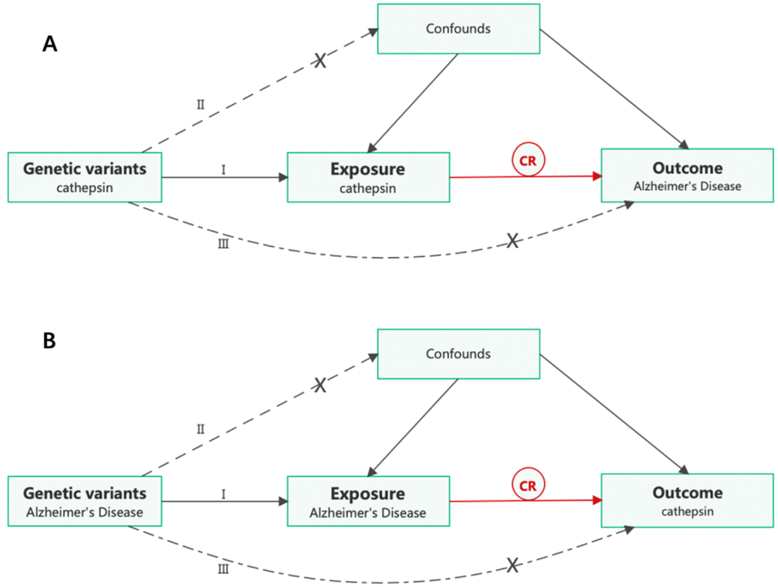
Diagram of Mendelian randomization (MR) study design. (A) The causal estimation of cathepsin on Alzheimer's disease (AD). (B) The causal estimation of cathepsin on AD. I) The genetic variants selected as instrumental variables (IVs) should be strongly associated with the risk factor of interest. II) The genetic variants used as IVs should not be associated with any confounders. III) The IVs should affect the risk of the outcome merely through the risk factor, not via any alternative pathways. CR, causal relationship.
Instrument selection
For the selection of instrumental variables (IVs), we identified genetic variants significantly associated with the expression of cathepsins from genome-wide association studies (GWAS). To ensure robustness, we used a genome-wide significance threshold of p < 5 × 10^-6 for cathepsin expression, consistent with MR studies focusing on expression traits, and p < 5 × 10^-8 for AD as the outcome when acting as an exposure. 28 Variants were pruned for linkage disequilibrium (LD) using a threshold of r2 < 0.001 and kb > 10,000, ensuring independence of the SNPs.27,29 In addition, we quantified the strength of the genetic instrument for all SNPs with an F-statistic calculated as (β2/se2) and we applied an F-statistics threshold of >10 for SNP selection to ensure robust instrument strength and reduce the risk of weak instrument bias. Power calculations indicated that the sample size and number of SNPs used would allow for sufficient power to detect the expected effect sizes of Cathepsin L1 on AD risk. We used MRPRESSO to screen the final IVs for the presence of abnormal SNPs. The statistical power was calculated according to Brion's method. 30
Mendelian randomization analysis
To explore the causal links between specific cathepsins and AD, MR analysis was conducted using the “TwoSampleMR” package in R, and genetic variants were taken as IVs.
Our primary method was the inverse variance weighted (IVW) approach, which assumes that all genetic variants are valid instruments. IVW provides an efficient estimate under the assumption that there is no horizontal pleiotropy. While MR is a powerful tool for causal inference, it is limited by the assumption of no horizontal pleiotropy, which can bias the results. We used MR-Egger and MR-PRESSO as sensitivity analyses to assess the presence of pleiotropy, ensuring robustness in our causal estimates. We also applied the Weighted Median and Weighted Mode estimators, which are more robust in cases where some genetic variants may not be valid instruments.28,31 MR-Egger regression tests for the presence of pleiotropy through the intercept, where a non-zero intercept suggests horizontal pleiotropy. 28 Weighted Median is reliable if at least 50% of the SNPs are valid IVs, and the Weighted Mode method assumes that the largest consistent SNP cluster provides valid estimates. These multiple methods collectively offer robustness, with p-values below 0.05 considered statistically significant. 32 Pleiotropy and Heterogeneity: We addressed horizontal pleiotropy by utilizing MR-Egger regression and testing the Egger intercept for deviation from zero. We also conducted heterogeneity tests (Cochran's Q) to assess variability in SNP effects across all MR models. 33 Lastly, the significance thresholds for forward and reverse MR analyses were aligned with existing MR guidelines, ensuring robust and reliable causal inference. To identify shared genetic variants between cathepsins and AD, we conducted a colocalization analysis. This analysis helps determine whether the genetic variants associated with cathepsin levels are also involved in AD, suggesting a potential shared biological mechanism. We utilized the coloc R package to perform the colocalization analysis, applying the Bayesian framework to calculate posterior probabilities (PP) for different hypotheses: H0: No association with either trait; H1: Association with cathepsin expression only; H2: Association with AD only; H3: Association with both traits but due to independent variants; H4: A shared genetic variant influences both traits. A high posterior probability for H4 (PP.H4 > 80%) indicates strong evidence for colocalization, suggesting that cathepsin expression and AD share a common genetic basis. We used publicly available GWAS summary statistics for AD and cathepsins, applying a region-based approach (±100 kb around significant single nucleotide polymorphisms, SNPs) to test for colocalization.
Results
The results of forward analysis
As shown in Figure 2, the results showed that Cathepsin F had an average effect size of 0.002229 with a mean p-value of 0.614575 (IVW: p = 0.1703, OR = 1.03, 95%CI = 0.99–1.07), indicating a non-significant association with AD. Similar results were obtained in Cathepsins B, G, L1, O, S, Z, and Cathepsin D with p-values all above the statistical significance (p > 0.05).
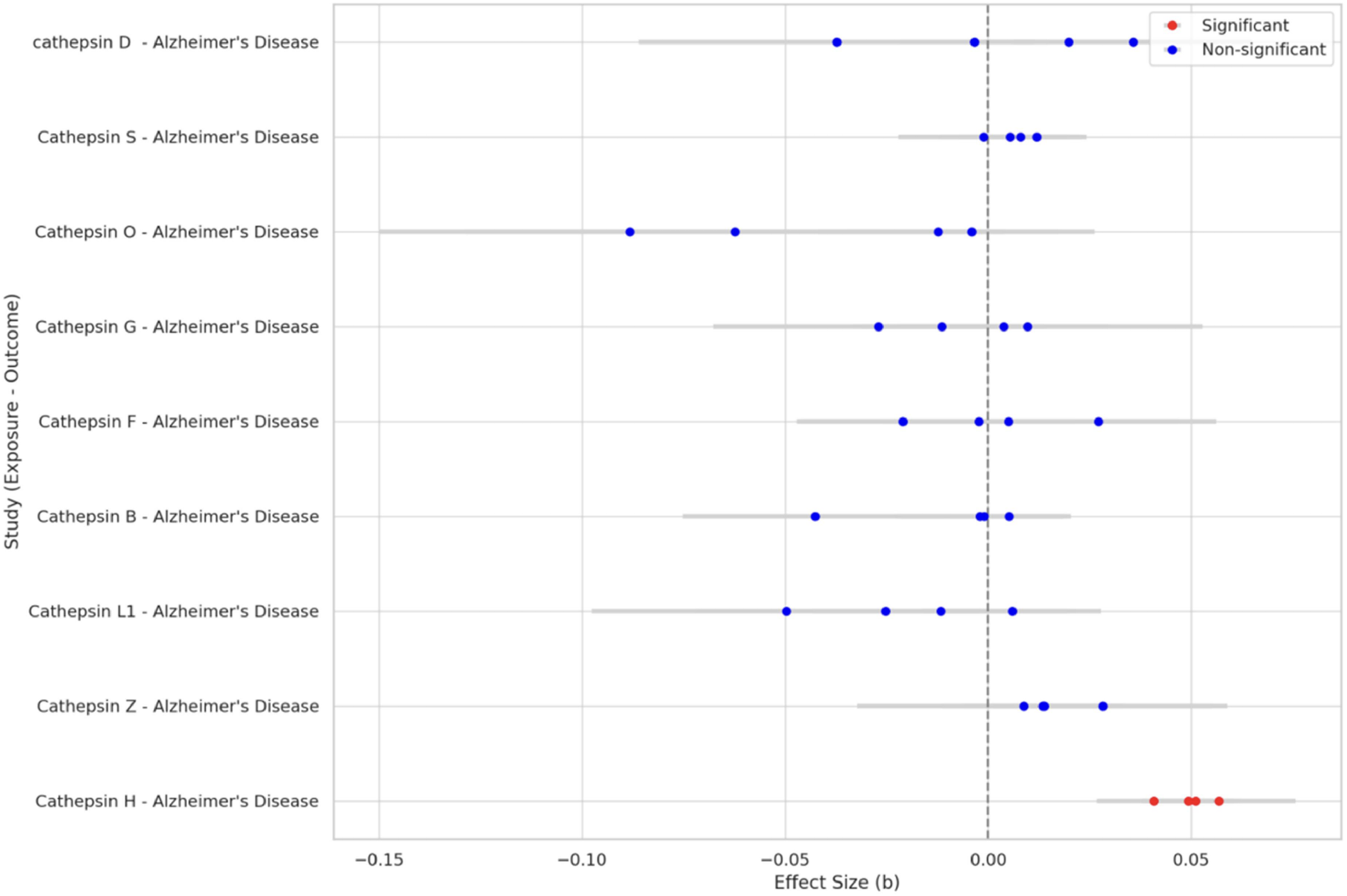
This forest plot displays individual MR results for various cathepsins, using red points to indicate statistically significant associations (p < 0.05) and blue for non-significant ones, with each point's position reflecting the effect size and the line its standard error.
Meanwhile, the results demonstrated that Cathepsin H was significantly associated with AD, with a mean effect size of 0.049467 and a p-value of 0.004851 (IVW: p = 0.003441, OR = 1.04, 95%CI = 1.01–1.07). Consistent associations were further corroborated by the MR-Egger (p = 0.01473, OR = 1.06, 95%CI = 0.93–1.10) and Weighted median approaches (p = 1.61*10−5, OR = 1.05, 95%CI = 1.03–1.07).
Since several MR methods including MR Egger, Weighted Median, IVW, and Weighted Mode were used to explore the association between Cathepsin H and AD, we conducted a multiple testing correction to control the false discovery rate (FDR), utilizing the Benjamini-Hochberg (BH) procedure. After adjusting for multiple comparisons using the BH method, the p-values for MR Egger, Weighted Median, IVW and Weighted Mode were corrected to 0.01473, 6.44 × 10⁻⁵, 0.004588, and 0.002432, respectively.
Furthermore, the results of both MR-Egger intercept and MR-PRESSO global tests showed no evidence of directional pleiotropy for any of these causal associations, as detailed in Table 1. Notably, when the IVW method was applied, no evidence of causal associations between other types of cathepsins and AD was obtained. The data for more methods are shown in Supplemental Table 1.
Causal association of cathepsins on AD estimated by univariable MR analysis.
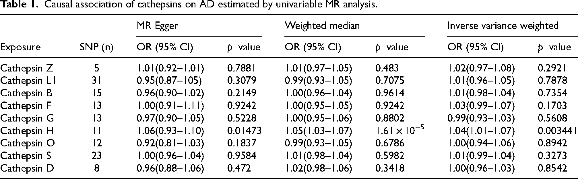
The Odds Ratio (OR) of MR analysis was 1.06 (95% CI: 0.93–1.10) for MR Egger, 1.05 (95% CI: 1.03–1.07) for Weighted median, 1.04 (95% CI: 1.01–1.07) for IVW, and 1.05 (95% CI: 1.03–1.08) for Weighted mode. These findings suggested a modest b association between increased levels of Cathepsin H and the risk of developing AD, with most confidence intervals not crossing the null value, particularly noteworthy in the Weighted median and Inverse variance weighted analyses.
The results of reverse MR analyses
As detailed in Table 2, Cathepsin H showed a significant association with AD when analyzed using MR-Egger (p = 0.006881, OR = 0.81, 95%CI = 0.70–0.94) and Weighted median approaches (p = 0.008153, OR = 0.84, 95%CI = 0.74–0.96), indicating a protective effect against AD. However, when reversing the directionality—with Cathepsin H as the outcome and AD as the exposure—the significance was not observed in the IVW analysis (p = 0.1633, OR = 0.94, 95%CI = 0.86–1.03), suggesting that the causal relationship might not be reciprocal or that Cathepsin H's levels were not influenced by AD in a statistically significant manner.
Causal association of cathepsins(outcome) on AD estimated by univariable MR analysis.

The results of reverse MR analysis demonstrated that AD elevated the levels of cathepsin D (IVW: p = 0.03791, OR = 0.88,95% CI = 0.78–0.99, and weighted median: p = 0.009109, OR = 0.79, 95% CI = 0.66–0.94) and cathepsin L1(IVW: p = 0.009143, OR = 0.94,95% CI = 0.89–0.98, and weighted median: p = 0.02905, OR = 0.93, 95% CI = 0.87–0.99). Moreover, the p-values of the MR-Egger intercept and MR-PRESSO global test showing no signs of directional pleiotropy (0.826 and 0.804, respectively) (Supplemental Table 2). No evidence supported a causal association between any other various types of cathepsins (Except cathepsin D and L1) and AD. The data for more methods are shown in the Supplemental Figures. The negative effect size suggested that lower levels of Cathepsin L1andD might be associated with an increased risk of AD according to Figure 3.

Forest plots of univariate Mendelian randomization analyses of AD and cathepsin risk. We performed an IVW, MR Egger, Weighted median, and Weighted mode analysis to assess the causal relationship between AD and cathepsins. (Statistically significant results are highlighted in red, with error lines representing 95% CI.).
We explored the causal impact of Cathepsin H levels on the risk of AD, employing a multi-faceted statistical approach to ensure the robustness and reliability of our findings. Initially, the forest plot analysis illuminated a statistically significant correlation between several genetic variants associated with Cathepsin H levels and the susceptibility to AD, suggesting a potential causal relationship. Then, we conducted a leave-one-out sensitivity analysis, which to confirmed the stability of the aforementioned findings. Additionally, the results of scatter plot analysis also suggested that changes in Cathepsin H levels may significantly affect AD (Figure 4).
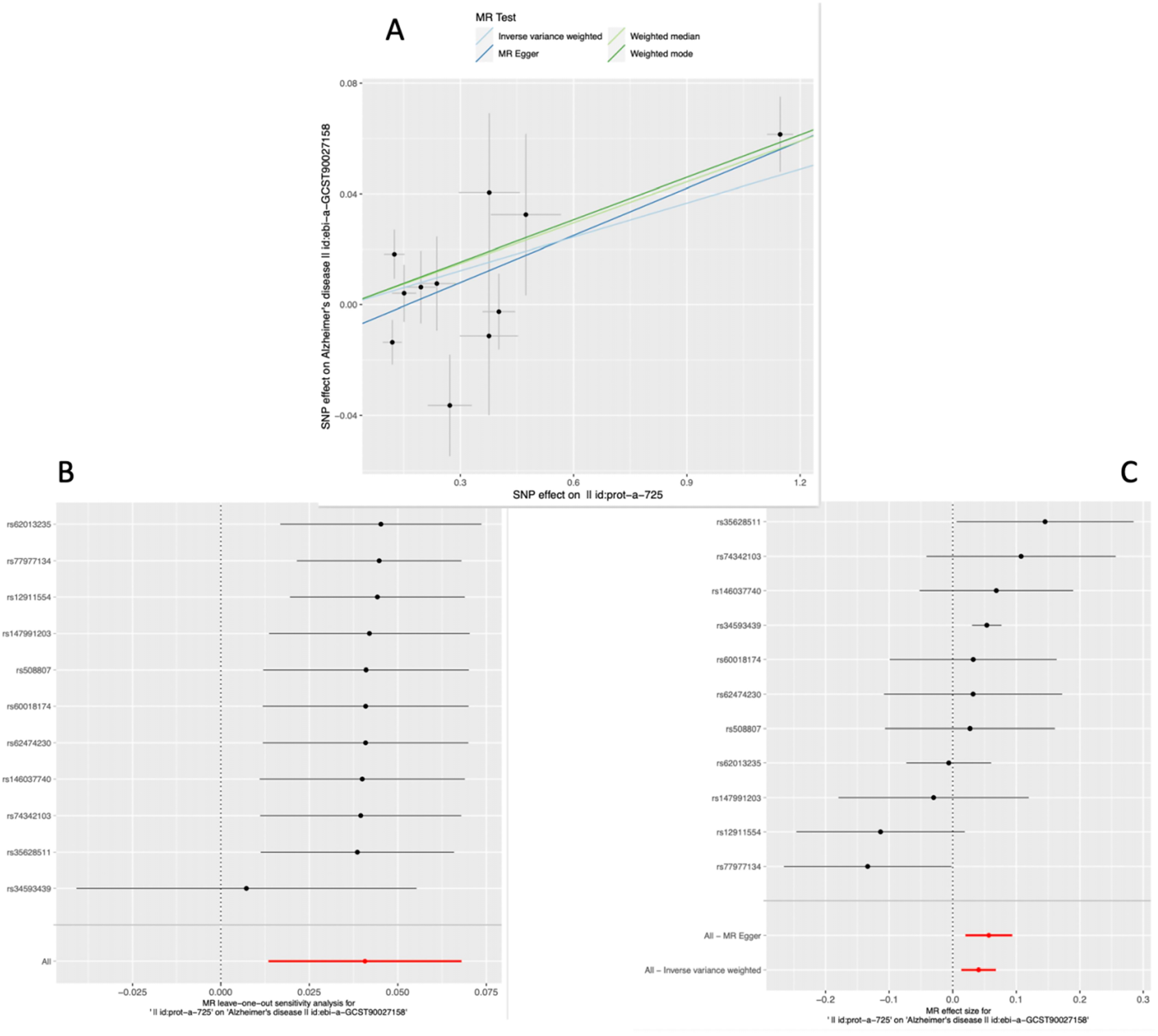
(A) Forest plot: shows the effects of different genetic variations on AD risk, suggesting a potential causal link between Cathepsin H levels and AD. The combined estimate indicates the overall effect size, with confidence intervals providing the uncertainty around this estimate. (B) Leave-One-Out Analysis: Demonstrates the robustness of the study's findings by showing that no single genetic variant disproportionately influences the result. This suggests that the observed association between Cathepsin H levels and AD risk is not due to outlier SNPs. (C) Scatter Plot: Illustrates a consistent relationship across multiple Mendelian randomization methods, supporting the hypothesis that higher or lower levels of Cathepsin H might influence the risk of developing AD.
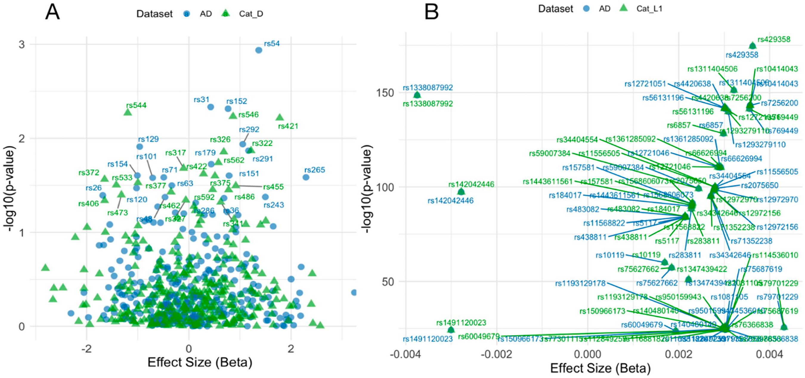
(A) Colocalization Scatter Plot (AD versus Cat_L1). The scatter plot shows the effect sizes (Beta) against -log10(p-values) for shared genetic variants between AD and Cathepsin L1. Significant SNPs are highlighted. (B) Colocalization Scatter Plot (AD versus Cat_D). The scatter plot shows the effect sizes (Beta) against -log10(p-values) for shared genetic variants between AD and Cathepsin D. Significant SNPs are highlighted.
Based on the previous forest plot results, inverse MR Analysis found that there may be a relationship between AD as exposure and cathepsin D and L1 as outcomes. We performed correlation analyses (including forest plots, funnel plots, leave-one-out plots, and scatter plots) to reveal significant genetic associations between AD as exposure and Cathepsin D and L1 levels as outcome.
In summary, the forward MR analysis focusing on the impact of cathepsins on AD, Cathepsin H was significantly associated with an increased risk of developing AD, suggesting its potential role as a risk factor in AD pathogenesis. This was supported by findings across various statistical methods, including MR-Egger and Weighted median approaches, which consistently indicated a modest association between elevated levels of Cathepsin H and AD risk. Conversely, the reverse MR analysis explored the effect of AD on the levels of cathepsins, revealing that AD could significantly influence the levels of Cathepsin D and L1, suggesting that the progression or presence of AD might impact these cathepsin levels. Notably, the results showed a significant association between AD and reduced levels of Cathepsin D and L1, indicating a potential protective effect against AD for these cathepsins.
Colocalization analysis
Colocalization analysis was performed to identify shared genetic variants between AD and key cathepsins (H, D, and L1). This analysis helps in understanding whether the genetic variants associated with cathepsin levels are also linked to AD, indicating a potential causal relationship and shared biological pathways between these traits.
The colocalization results revealed that Cathepsin H and Cathepsin D showed no significant genetic overlap with AD, with posterior probabilities (PP.H4) for shared causal variants being extremely low (6.23e-13% and 1.24e-16%, respectively). In contrast, Cathepsin L1 demonstrated a strong colocalization with AD, with a PP.H4 of 100%, indicating a significant shared genetic basis between Cathepsin L1 levels and AD risk (Figure 5).
Discussion
Our MR analysis revealed a significant association between elevated levels of Cathepsin H and an increased risk of developing AD. There are no studies that have specifically focused on cathepsin H and AD, and we speculate that the potential role of Cathepsin H in AD may be elucidated by its involvement in broader neurodegenerative processes such as neuroinflammation and oxidative stress, but this needs to be verified by more experiments. For example, Hook et al. studies have highlighted cathepsins’ involvement in hippocampal damage response and in other neurodegenerative diseases like Parkinson's disease, which share pathological features with AD, such as protein aggregation and lysosomal dysfunction.15,34 We emerging genetic studies using MR highlight the nuanced influences of genetic factors in complex diseases like AD, supporting the relevance of investigating less understood genes like Cathepsin H. This underscores the importance of further experimental research to explore how Cathepsin H could modulate AD pathogenesis, potentially leading to novel therapeutic targets.
In our reverse MR analysis, we observed that AD may influence the levels of Cathepsin D, suggesting a potential feedback mechanism where the presence of AD could enhance Cathepsin D expression or activity. This finding presents a novel aspect of Cathepsin D's role in AD, contrasting with the traditional view where it is primarily seen as influencing the disease's onset and progression.35,36 Cathepsin D directly participates in the cleavage of the AβPP, influencing the production of Aβ peptides. This activity is crucial because it affects the aggregation propensity and toxicity of Aβ.36,37 The proteolytic action of Cathepsin D on AβPP can generate either Aβ fragments that are prone to aggregation, contributing to plaque formation, or shorter peptides that are less amyloidogenic.35,38 Studies suggest that alterations in Cathepsin D expression or activity could leading to increased neuronal vulnerability and death. For example, overactivation of Cathepsin D might lead to excessive breakdown of cellular components, including vital proteins and organelles, exacerbating neurodegenerative processes. 38 There is also evidence that excessive or dysregulated activity of Cathepsin D might contribute to neuronal death and AD progression. 15 Our results add complexity to this picture by indicating that AD pathology itself may alter Cathepsin D levels, which could either exacerbate or mitigate disease progression depending on the context and stage of the disease.
Similarly, our reverse MR analysis suggests that AD impacts the levels of Cathepsin L1, underscoring a potentially adaptive or compensatory role of this enzyme in response to the disease. This observation hints at a complex interplay where AD might not only be influenced by Cathepsin L1 but may also modulate its expression as part of the disease's pathology. Schechter et al. found that Cathepsin L1's role in the normal lysosomal degradation pathways, with less emphasis on its involvement in neurodegenerative diseases like AD.39,40 For example, Hook et al. revealed that elevated Cathepsin H activity correlated with increased Aβ deposition. 15 Cathepsin L1 has been implicated in inflammatory processes within the central nervous system. It can modulate the activity of cytokines and other inflammatory mediators, which are critical in the neuroinflammatory processes observed in AD. Aberrant Cathepsin L activity could exacerbate or fail to properly regulate inflammation, thereby contributing to neuronal damage and the progression of AD. 40 Cathepsin L contributes to AD by degrading nuclear lamina components such as lamin B1, leading to nuclear instability, apoptosis, and exacerbating neurodegenerative processes in AD. 41 However, our findings suggest that changes in Cathepsin L1 levels could be a response to the neuronal damage incurred during AD progression, potentially serving as a protective mechanism to mitigate further damage.
These findings are particularly significant as they suggest that therapeutic interventions targeting Cathepsin D and L1 might need to consider the disease state and progression to be effective. In AD brains, pro-cathepsin L (pCPL) was found to be highly expressed in fully activated microglia, suggesting that its activity is linked to microglial activation and immune dysfunction in AD, 42 but this strategy may need adjustment as the disease progresses and begins to influence Cathepsin D and L1 levels differently. This dual role of the Cathepsin as both a potential contributor and a consequence of AD pathology emphasizes the need for a dynamic therapeutic approach, tailored to individual disease profiles and stages.
While our study did not find significant causal relationships for Cathepsins B, F, G, O, S, and Z with AD, this highlights the selective nature of protease involvement in AD and suggests that not all cathepsins play a central role in its pathogenesis. Cathepsin B, for instance, has been implicated in both the formation and degradation of Aβ plaques. Research suggests that it might contribute to the amyloidogenic pathway by cleaving the AβPP in a way that favors the production of Aβ, a key pathological marker of AD.43,44 Similarly, Cathepsin S has been identified for its role in neuroinflammation, a critical aspect of AD pathology. It is believed to facilitate the breakdown of the blood-brain barrier, leading to increased migration of pro-inflammatory cells into the brain, thus exacerbating neuroinflammatory responses and potentially accelerating neuronal damage. 14 In addition, Cathepsin G, though less studied in the context of AD, has been associated with other neurodegenerative processes and could play a role in modulating immune responses within the central nervous system. 45
Our findings echo these observations, highlighting the critical nature of these cathepsins in influencing neuronal vulnerability and AD progression, as well as underscoring the complexity of protease activity's balance in the disease mechanism.15,36,37 The bidirectional MR analysis employed in our study elucidates the intricate relationship between AD and cathepsins, suggesting that the disease's development might not only result from genetic and environmental factors but also from alterations in the expression of proteases implicated in AD's pathology. Future studies should continue to explore these cathepsins’ potential roles, considering the diverse functions of cathepsins in the brain and their implications for AD.
In order to explore the AD and protease Cathepsin H, D and more detailed genetic link between L1, we made colocalization analyses. The colocalization analysis revealed a significant genetic overlap between Cathepsin L1 and AD, suggesting a shared genetic basis and a potential causal relationship. This significant overlap indicates that Cathepsin L1 may play a crucial role in AD pathology and could serve as a promising biomarker and therapeutic target. On the other hand, Cathepsin H and Cathepsin D showed no significant genetic overlap with AD, indicating that these proteases might operate through distinct genetic pathways unrelated to AD. These findings highlight the specificity of Cathepsin L1 in AD, providing a strong rationale for focusing on this protease in future research. Further investigations are necessary to elucidate the mechanisms by which Cathepsin L1 influences AD progression and to validate its potential as a biomarker and therapeutic target.
The potential for targeting Cathepsin L1 therapeutically is supported by its involvement in Aβ degradation and neuroinflammation, two critical pathways in AD progression. This finding opens avenues for stage-specific intervention strategies, where cathepsins could be modulated at early or specific stages of AD to prevent the formation of amyloid plaques or reduce neuroinflammation, ultimately slowing disease progression. For example, inhibitors or enhancers of Cathepsin L1 could be developed to modulate its activity depending on the disease stage, as its early involvement in amyloid degradation might be beneficial, while prolonged activity could exacerbate neuroinflammation.
However, there are some limitations that should be acknowledged. First, the assumptions inherent in MR, such as no unmeasured confounding and the reliability of genetic instruments as proxies for protease levels, may affect the validity of our causal inferences. Second, our analysis is limited to the data from populations predominantly of European descent, which may not be generalizable to other ethnic groups. Thus, future studies should aim to replicate these findings in diverse populations, which could reveal population-specific genetic variants that affect cathepsin function and AD risk. This approach will not only validate our findings but also help in tailoring therapeutic strategies to specific populations. Third, given the complex regulation of protease activity and its context-dependent roles in AD pathology, our findings should be interpreted with caution and verified in diverse biological models. In addition, limitations include potential pleiotropy and the assumption of instrument validity, which may affect the causal interpretations. 46 Another limitation is the reliance on publicly available summary statistics from GWAS. The use of summary-level data may limit the granularity of our analysis, particularly regarding potential confounders that could not be directly adjusted for, such as population structure or gene-environment interactions.
Further investigation into how Cathepsin L1 interacts with tau protein could provide novel insights into its broader role in AD pathogenesis. The potential therapeutic implications of targeting Cathepsin L1 are promising. Given the significant colocalization between Cathepsin L1 and AD observed in our analysis, experimental studies are warranted to validate these findings and explore the feasibility of targeting Cathepsin L1 in therapeutic interventions.
Conclusion
Our study reveals crucial genetic links between AD and proteases Cathepsin H, D, and L1, providing insights into their potential roles in AD pathology. The colocalization analysis highlighted a significant genetic overlap between Cathepsin L1 and AD, suggesting a shared genetic basis and potential causal relationship. In contrast, Cathepsin H and Cathepsin D showed no significant genetic overlap with AD, indicating distinct genetic pathways. These findings suggest that Cathepsin L1 may serve as a promising biomarker for AD and a potential therapeutic target.
The results from the MR analysis further support the genetic link between Cathepsin L1 and AD, strengthening the evidence for its role in disease progression. However, these findings are just the beginning of understanding the complex interplay between these proteases and AD pathology. Further research is needed to elucidate the precise mechanisms through which these cathepsins influence disease progression, assess their potential as biomarkers for early diagnosis, and explore therapeutic interventions targeting these enzymes. Future studies should explore the potential for population-specific genetic variations to influence cathepsin activity and AD risk, especially given that this study focused primarily on populations of European ancestry. Expanding research to include diverse populations will help uncover broader, more generalizable insights into cathepsin function in AD.
Moreover, clinical trials investigating inhibitors or enhancers of cathepsin activity, particularly for Cathepsin L1, could provide further translational relevance to our findings. Considering the role of cathepsins in neuroinflammation and Aβ degradation, these proteases may be targeted at specific stages of AD to prevent or slow disease progression. However, off-target effects must be carefully monitored, given the involvement of cathepsins in other physiological processes. Future research should focus on experimental validations of the Cathepsin L1-AD relationship using cell and animal models. Additionally, the potential therapeutic utility of targeting Cathepsin L1 should be explored in clinical trials, particularly in its role in amyloid processing and neuroinflammation modulation.
Supplemental Material
sj-docx-2-alz-10.1177_13872877251314058 - Supplemental material for Genetic colocalization of cathepsins H, D, and L1 with Alzheimer's disease: Implications for biomarker and therapeutic target discovery
Supplemental material, sj-docx-2-alz-10.1177_13872877251314058 for Genetic colocalization of cathepsins H, D, and L1 with Alzheimer's disease: Implications for biomarker and therapeutic target discovery by Yu-Ting Dong, Xiao Luo, Li-Li Zhang, Yi-Meng Gong, Dan Wang, Dong-Ling Zhong, Yu-Xi Li, Xiao-Min Ma, Rong-Jiang Jin and Juan Li in Journal of Alzheimer's Disease
Supplemental Material
sj-xlsx-3-alz-10.1177_13872877251314058 - Supplemental material for Genetic colocalization of cathepsins H, D, and L1 with Alzheimer's disease: Implications for biomarker and therapeutic target discovery
Supplemental material, sj-xlsx-3-alz-10.1177_13872877251314058 for Genetic colocalization of cathepsins H, D, and L1 with Alzheimer's disease: Implications for biomarker and therapeutic target discovery by Yu-Ting Dong, Xiao Luo, Li-Li Zhang, Yi-Meng Gong, Dan Wang, Dong-Ling Zhong, Yu-Xi Li, Xiao-Min Ma, Rong-Jiang Jin and Juan Li in Journal of Alzheimer's Disease
Supplemental Material
sj-xlsx-4-alz-10.1177_13872877251314058 - Supplemental material for Genetic colocalization of cathepsins H, D, and L1 with Alzheimer's disease: Implications for biomarker and therapeutic target discovery
Supplemental material, sj-xlsx-4-alz-10.1177_13872877251314058 for Genetic colocalization of cathepsins H, D, and L1 with Alzheimer's disease: Implications for biomarker and therapeutic target discovery by Yu-Ting Dong, Xiao Luo, Li-Li Zhang, Yi-Meng Gong, Dan Wang, Dong-Ling Zhong, Yu-Xi Li, Xiao-Min Ma, Rong-Jiang Jin and Juan Li in Journal of Alzheimer's Disease
Footnotes
Acknowledgments
Our data are taken from the publicly available GWAS database. We thank the genetics consortiums for making the GWAS catalog data publicly available.

Author contributions
Yu-Ting Dong (Conceptualization; Data curation; Funding acquisition; Project administration; Resources; Software); Xiao Luo (Resources; Software; Validation; Visualization; Writing – original draft); Li-Li Zhang (Investigation; Methodology; Project administration; Validation; Writing – review & editing); Yi-Meng Gong (Investigation; Methodology; Project administration; Resources; Software); Dan Wang (Funding acquisition; Investigation; Supervision; Validation); Dong-Ling Zhong (Conceptualization; Formal analysis; Funding acquisition; Visualization; Writing – original draft); Xi-Yu Li (Investigation; Methodology; Resources; Validation; Visualization); Xiao-Min Ma (Investigation; Methodology; Project administration); Rong-Jiang Jin (Funding acquisition; Methodology; Project administration; Resources); Juan Li (Project administration; Resources; Software; Validation; Visualization; Writing – original draft).
Funding
The authors disclosed receipt of the following financial support for the research, authorship, and/or publication of this article: We acknowledge Tianjin science and technology plan projects (grant number 18PTLCSYOOO60) and Yunnan province innovation team of prevention and treatment for brain diseases with acupuncture and Tuina (grant number 202405AS350007) for the funding of this research.
Declaration of conflicting interests
The authors declared no potential conflicts of interest with respect to the research, authorship, and/or publication of this article.
Supplemental material
Supplemental material for this article is available online.
References
Supplementary Material
Please find the following supplemental material available below.
For Open Access articles published under a Creative Commons License, all supplemental material carries the same license as the article it is associated with.
For non-Open Access articles published, all supplemental material carries a non-exclusive license, and permission requests for re-use of supplemental material or any part of supplemental material shall be sent directly to the copyright owner as specified in the copyright notice associated with the article.
