



Abstract
Background
Synaptic transmission dysfunction is associated with a range of neurological disorders, including Alzheimer's disease (AD). However, the role of γ-aminobutyric acid (GABA)-mediated synaptic inhibition in AD has not been fully explored.
Objective
We studied basal, GABA-activated slow spontaneous synaptic currents (sIPSCs) in dentate gyrus (DG) granule cells in the dorsal hippocampus of an AD mouse model (tg-APPSwe) and investigated insulin's modulatory effects.
Methods
GABA-activated slow sIPSCs were recorded in the DG granule cells by whole-cell patch-clamp recordings in dorsal hippocampal brain slices from 5–6 (adult) and 10–12 (aged) months old wild-type (WT) and AD mice, in the presence or absence of insulin (1 nM).
Results
The median 10–90% rise time of slow sIPSCs significantly decreased with age (10–12 months vs. 5–6 months) only in AD mice. The median amplitude of the slow sIPSCs was decreased in adult and aged AD mice as compared to WT mice whereas the slow sIPSCs frequency was only reduced in the aged WT mice. The median 63% decay time and total current density of the slow IPSCs was significantly decreased in the aged AD mice as compared to both WT mice and to the adult AD mice. Insulin application exerted no effect on slow sIPSCs properties in any of the animal groups.
Conclusions
The characteristics of the slow sIPSCs recorded in DG granule cells of dorsal hippocampus from WT and AD mice are altered by age- and disease-state, whereas insulin has negligible effects.
Introduction
Gamma-aminobutyric acid (GABA) is the predominant inhibitory neurotransmitter in the brain. It binds and activates two different types of receptors- the ionotropic GABAA and the G-protein coupled GABAB receptors generating GABA-mediated neuronal inhibition. The phasic GABAA receptors inhibition can be separated into fast and slow GABAA receptor mediated synaptic currents.1,2 The kinetics of these currents are not only different, but they are also evoked by distinct presynaptic GABAergic interneurons.1,3,4 The slow, spontaneous inhibitory postsynaptic currents (sIPSCs) in the principal neurons of the hippocampus are evoked in response to release from the neurogliaform interneurons and Ivy cells that release GABA by volume transmission.1,3 Several roles for neurogliaform interneurons and Ivy cells have been proposed including influencing neuronal network synchrony and oscillatory activity.1,3
It is well-established that the dorsal hippocampus (posterior hippocampus in primates) is involved in spatial learning and memory, 5 and a gradual decline in cognitive function is one the hallmarks of Alzheimer's disease (AD). In a preclinical AD mouse model (APPNL-G-F), abnormal neuronal hyperactivity in brain regions including dorsal hippocampus may contribute to impaired memory retrieval. 6 The dentate gyrus (DG) plays a crucial role in regulating cortical inputs to the hippocampus, preventing overexcitation of the hippocampal Cornu Ammonis (CA) regions. This regulation is achieved through the relatively low excitability of DG granule cells, which is attributed to both their intrinsic properties and the local GABAergic microcircuits within the dentate gyrus. 7 While fast and tonic GABA-activated sIPSCs in DG granule cells have been well-characterized,7,8 slow sIPSCs mediated by the volume transmission have received less attention.
We and others have previously shown that the fast sIPSCs in hippocampal neurons are shaped by many factors including age, metabolic hormones like insulin and the amyloid-β protein (Aβ),4,8–10 but less is known how these conditions affect the slow IPSCs. Here we used an AD transgenic model (Tg-APPSwe mice) to explore if the slow sIPSCs were affected by age, insulin or disease. The tg-APPSwe mouse model carries a mutant version of the amyloid precursor protein (APP) that contains the Swedish mutation. 11 This genetic alteration leads to elevated levels of the Aβ in these mice. In the APPSwe mice, the accumulation of Aβ within neurons is observed starting at 5–6 months of age. 12 By around 12 months of age, these mice also develop extracellular Aβ plaques, as well as increased microgliosis and astrogliosis in the hippocampus.11,12 That insulin regulates peripheral glucose homeostasis is well established, but insulin is also increasingly recognized as a factor required for healthy brain function.13,14 In the DG granule cells region within the dorsal hippocampus, the effects of insulin on the fast sIPSCs are influenced by both the age and the stage of disease progression in wild-type (WT) and APPSwe mice. 8 Here, we extended these studies with additional analyses of the same dataset as used by Hammoud et al. 8 and examined the slow sIPSCs in adult (5–6 months) and aged (10–12 months) WT and APPSwe mice. Our results show that the slow sIPSCs are reduced in the aged APPSwe mice but are not significantly modulated by insulin.
Methods
Animals
All experiments were conducted in accordance with the local ethical guidelines and protocols approved by the Uppsala animal ethical committee, Swedish law and regulations based on the Directive 2010/63/EU, C129/14, C112914/15. The present study utilized adult (5–6 months old) and aged (10–12 months old) male and female C57BL/6J mice, as well as tg-APPSwe mice (bred on a C57BL/6J background). The tg-APPSwe mice were bred at Uppsala University by crossing male heterozygous tg-APPSwe with female C57BL/6J. 8 Age-matched male and female C57BL/6J mice were used as WT controls. The animals were maintained on a 12-h light/12-h dark cycle with ad libitum access to water and food. The number of animals used in experiments was minimized.
To record GABA-activated currents from dorsal hippocampal DG granule cells, the following number of hippocampal slices were use: 36 slices from 6 WT at 5–6 months of age; 41 slices from 7 tg-APPSwe at 5–6 months of age; 47 slices from 8 WT mice at 10–12 months of age; and 50 slices from 13 tg-APPSwe mice at 10–12 months of age. From each slice, activity of a single neuron was recorded.
Genotyping
The genotyping technique was performed as described previously in details. 8 Tg-APPSwe mice overexpress transgene with human APP (isoform 695) bearing the Swedish mutation (KM670/671NL) under the murine Thy1 promoter. 12 Mouse tail tip samples were subjected to the “HotSHOT” rapid genomic DNA isolation method to enable detection of the gene of interest. 15 A PCR master mix was made with JumpStartTM Taq DNA Polymerase (Catalog Number D6558, Sigma-Aldrich) and primer pair (APP –TYI-1-GAATCCAAGTCGGAACTCTT; APP-SQ6rw-TGTCAGGAACGAGAAGGGCA). The PCR reaction was carried out under the following parameters: an initial heating at 94°C for 2 min, followed by 30 cycles of 94°C for 15 s, 63°C for 15 s, and 72°C for 30 s, and a final incubation at 72°C for 10 min. Electrophoresis was then performed on a 1% agarose gel and the expected PCR product size was 400 bp.
Hippocampal brain slice
The hippocampal slices were produced in accordance with the methods previously reported with slight modifications.16,17 At the day of experiment, the animal was euthanized by cervical dislocation and decapitated. The brain was then rapidly removed and placed in an ice-cold NMDG-based solution containing (in mM): 93 NMDG, 2.5 KCl, 1.2 NaH2PO4, 30 NaHCO3, 20 HEPES, 25 D-glucose, 10 MgSO4, 0.5 CaCl2, 5 sodium ascorbate, 2 thiourea, 3 sodium pyruvate, 12 N-acetyl-L-cysteine, which was oxygenated with 95% O2 and 5% CO2. The pH of this solution was adjusted to 7.3–7.4 using HCl and the osmolarity was maintained between 300–305 mOsm. Dorsal hippocampal slices were defined in the coronal plane according to Paxinos and Franklin. 18 Coronal hippocampal slices, 350 µm thick, were then cut using a vibrating microtome Leica VT1200 S (Leica Microsystems AB, Germany). The first 3–4 consecutive hippocampal slices form the dorsal pole were collected8,17,19 and submerged in the NMDG-based solution for 10 min at 32°C. Afterward, the slices were transferred to a HEPES-holding solution (20–22°C) for at least 1 h prior to use. The HEPES-holding solution contained (in mM): 92 NaCl, 2.5 KCl, 1.2 NaH2PO4, 30 NaHCO3, 20 HEPES, 25 D-glucose, 2 MgSO4, 2 CaCl2, 5 sodium ascorbate, 2 thiourea, 3 sodium pyruvate, 12 N-acetyl-L-cysteine, pH 7.3–7.4 adjusted with NaOH when oxygenated with carbogen; osmolarity 305–308 mOsm.
Electrophysiology
Whole-cell voltage-clamp recordings were performed on DG granule cells located primarily in the infrapyramidal blade of the dorsal hippocampus. The recordings were conducted at holding potential of −60 mV using Multipatch 700B amplifier controlled by pClamp 10.5 software (Molecular Devices, USA). 20 All recordings were carried out at room temperature (20–22°C) under continuous perfusion (1.5–2 ml/min) of ACSF containing (in mM): 119 NaCl, 2.5 KCl, 1.3 MgSO4, 1 NaH2PO4, 26.2 NaHCO3, 2.5 CaCl2, 11 D-glucose and 3 kynurenic acid (pH 7.3–7.4 equilibrated with 95% O2 and 5% CO2; osmolarity 300–303 mOsm adjusted with sucrose). The borosilicate glass patch pipettes had a resistance of 3.5–4.1 MΩ when filled with an intracellular solution containing (in mM): 140 CsCl, 8 NaCl, 2 EGTA, 0.2 MgCl2, 10 HEPES, 2 MgATP, 0.3 Na3GTP, 5 QX314Br, pH 7.2 adjusted with CsOH, osmolarity 285–290 mOsm. Inhibitory postsynaptic currents (sIPSCs) were recorded from the granule cells for at least 5 min after a 10-min baseline stabilization period, in the continuous presence of 3 mM kynurenic acid in the ACSF solution to block excitatory synaptic transmission. Insulin (1 nM) was either acutely applied to the slices for at least 10 min or the slices were preincubated with insulin (1 nM) at room temperature for at least 30 min prior to recording. Only neurons with a stable baseline throughout the entire recording were included into the analysis. Circulation insulin levels range between pM and low nM range,21–23 and insulin enters the brain from the blood involves a saturable transport system. 24 Therefore, we used 1 nM insulin in the study to avoid unspecific activation of other receptors, e.g., insulin-like growth factor-1 receptor (IGF1R). 25
Data analysis
The synaptic currents were analyzed using the Mini Analysis software (Synaptosoft), with the detection threshold set at a level 5 times greater than the baseline noise and checked visually. 17 Spontaneous IPSCs having a rise time (10–90%) >5 ms were considered as slow sIPSCs. Frequency, median amplitude (pA), median rise time (10–90%) (ms), median decay time (63%) (ms) and median charge transfer (pC) for single sIPSC were analyzed by the Mini Analysis software automatically. To estimate total slow synaptic current (sIPSC T ) density for each cell, we normalized total slow synaptic current (frequency x charge transfer; pA) to cell membrane capacitance value (pF), obtained after getting into the whole-cell mode. Ordinary two-way ANOVA with Fisher's LSD multiple comparison test was used to determine statistical significance (GraphPad Prism 10 Software, USA). Any p-value lower than 0.05 was interpreted as statistical significance.
Reagents
The chemicals for electrophysiological experiments were obtained from Sigma-Aldrich (Germany), except for insulin (human, recombinant (yeast), Cat. No. 11376497001, Roche Diagnostics GmbH, Mannheim, Germany).
Results
Fast and slow sIPSCs recorded in DG granule cells of the dorsal hippocampus
In our recordings from dorsal hippocampal DG granule cells, we identified two distinct sub-populations of GABA-activated sIPSCs (Figure 1(a)).The currents were clearly differentiated and grouped into fast and slow sIPSCs based on their kinetic properties (Figure 1(a)).The fast and slow kinetics can be distinguished by; the fast sIPSCs having rise time (10–90%) ≤5 ms and decay time (63%) <20 ms (black circles) and the slow sIPSCs having rise time (10–90%) >5 ms and decay time (63%) ≥20 ms (green circles) (Figure 1(b)). To characterize the properties of the slow sIPSCs and examine whether they are altered with age and disease progression, we studied the sIPSCs in the dorsal hippocampal DG granule cells of APPSwe mice before (5–6 months) and during (10–12 months) Aβ plaque accumulation as well as in age-matched WT mice.4,11,12 The majority of the sIPSCs recorded, in both 5–6 months and 10–12 months age groups in WT and APPSwe mice, were fast synaptic currents, ranging from about 84% to 87% (Figure 1(c)). Nevertheless, a significant number (13–16%) of slow events were also recorded (Figure 1(c)). In this study we focus on the slow sIPSCs, as we have previously described the characteristics of the fast sIPSCs in these mice. 8 The median rise time (10–90%) of slow sIPSCs was markedly decreased at 10–12 months as compared to 5–6 months only in APPSwe mice (Figure 1(d)). The median decay time (63%) of the slow IPSCs was significantly reduced in APPSwe mice aged 10–12 months as compared to both WT mice and younger APPSwe mice aged 5–6 months (Figure 1(e)). Additionally, the median decay time (63%) of slow sIPSCs decreased with age (10–12 months versus 5–6 months) in WT mice (Figure 1(e)). The kinetic characteristics of the fast and slow sIPSCs are in accordance with previous reports.2,4

Fast and slow spontaneous IPSCs ratio and their kinetic parameters in DG granule cells of dorsal hippocampus in 5–6 or 10–12 months tg-APPSwe mice and wild-type (WT) mice. (a) Example of current recording from DG granule cells from 10–12 months old WT mice showing representative fast and slow IPSCs. (b) A graph showing the median 63% decay time plotted against the median 10–90% rise time of sIPSCs estimated for each individual DG granule cell recorded from 10–12 months old WT mice. Based on the kinetics parameters two sub-populations of sIPSCs are defined: fast with rise time ≤5 ms and decay time <20 ms (black circles) and slow with rise time >5 ms and decay time ≥20 ms (green circles). (c) A bar graph showing the percentage of the fast (gray) and slow (green) sIPSCs in DG granule cells of dorsal hippocampus in WT and tg-APPSwe mice at different age recorded in ACSF. (d, e) Kinetic parameters of slow sIPSCs. Summary plots for the median 10–90% rise time (d) and the median 63% decay time (e) of slow sIPSCs recorded from dorsal DG granule cells in hippocampal brain slices from WT and tg-APPSwe mice, age groups: 5–6 months old and 10–12 months old. Data are presented as scatter dot plot for individual values and box and whiskers plot with median value plotted as a line and the mean values shown as ‘+’. Outliers are defined by the Tukey method and marked as filled dot plot (black circles). Statistical analyses are performed by excluding outliers and only statistically significant differences are marked on the graph. Ordinary two-way ANOVA with Fisher's LSD multiple comparison test was applied. Vhold = −60 mV.
Slow sIPSCT density is reduced in aged APPSwe mice
We further characterized properties of slow sIPSCs, including frequency, median amplitude, median charge transfer and total current density (sIPSC T density) recorded from dorsal DG granule cells in the WT and APPSwe mice aged 5–6 and 10–12 months. Examples of current traces of the slow sIPSCs are presented in Figure 2(a) and (b) for mice aged 5–6 and 10–12 months, respectively. When examined at 5–6 months of age, DG granule cells in APPSwe mice exhibited a marked reduction in the median amplitude of the slow sIPSCs compared to WT mice (Figure 2(d)). However, no notable differences were observed in the frequency, or total current density between the two groups (Figure 2(c) and (e)). In contrast, when assessed at 10–12 months of age, the median amplitude and total current density were considerably reduced in the APPSwe mice compared to the WT mice (Figure 2(d) and (e)), whereas the frequency remained similar (Figure 2(c)). Interestingly, the total current density of the slow IPSCs decreased with age (10–12 versus 5–6 months) in WT and APPSwe mice (Figure 2(e)). The frequency of the slow IPSCs was decreased with age only in WT mice (Figure 2(c)). These results indicate an age- and Aβ plaque accumulation-dependent reduction of slow IPSCs in APPSwe mice.
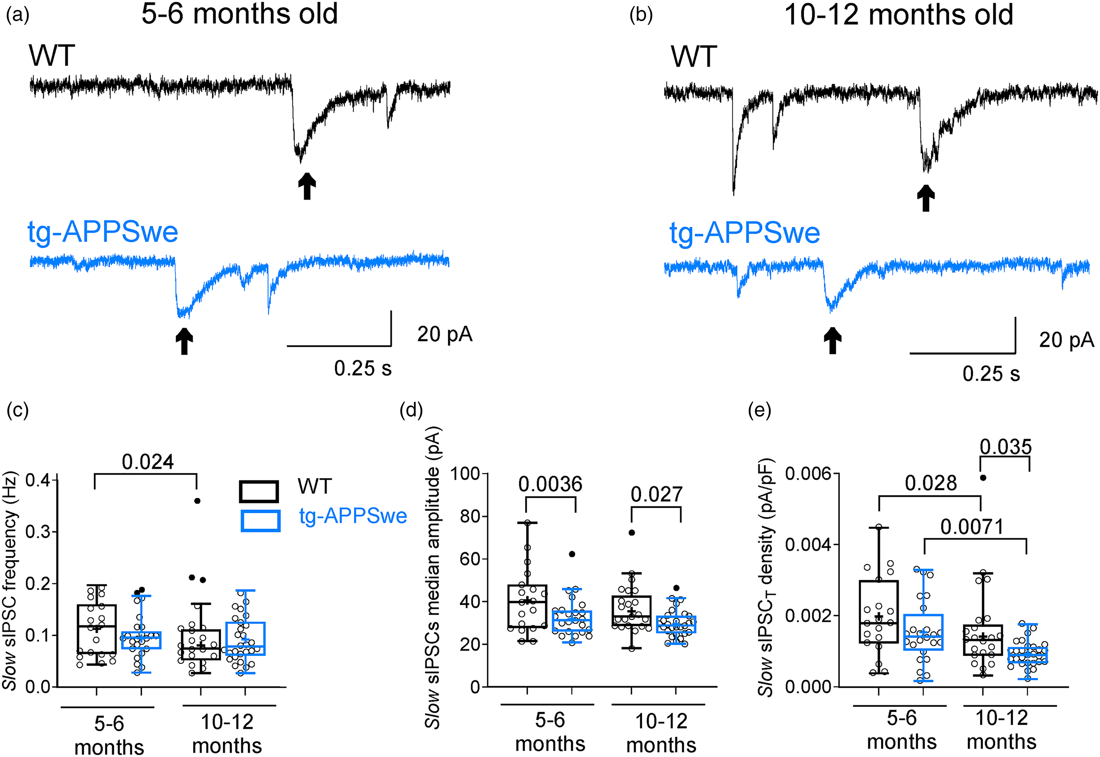
Characteristics of the slow sIPSCs in DG granule cells of the dorsal hippocampus in 5–6 and 10–12 months old tg-APPSwe mice and their WT mice. Representative voltage-clamp current traces of spontaneous IPSCs in dorsal DG granule cells from 5–6 (a) and 10–12 months old (b) WT (black segment, upper panel) and tg-APPSwe (blue segment, lower panel) mice under control conditions. Arrows indicate slow sIPSCs recorded from DG granule cells. Summary for the mean frequency (c), the median amplitude (d), and the total current density (e) of the slow sIPSCs in DG granule cells of WT and tg-APPSwe mice recorded from dorsal hippocampal slices under control conditions. Data are presented as scatter dot plot for individual cells and box and whiskers plot with median value plotted as a line and mean values shown as ‘+’. Outliers are defined by the Tukey method and marked as dot plot (filled black circles). Statistical analyses are performed by excluding outliers. Ordinary two-way ANOVA with Fisher's LSD multiple comparison test was applied. All experiments were performed with parallel controls from the same animal/age group. Vhold = −60 mV.
Effect of insulin on slow sIPSCs in WT and APPSwe mice
We have previously shown that insulin may modulate GABA-mediated fast sIPSCs and tonic currents in APPSwe mice aged 10–12 months. 8 Therefore, we examined whether insulin affected the slow sIPSCs properties recorded in granule cells of DG from different animal groups. Insulin did not alter the percentage of fast and slow sIPSCs (Figure 3(a)) or significantly affect the median rise time (10–90%) (Figure 3(b)), median decay time (63%) (Figure 3(c)) or total current density of the slow sIPSCs (Figure 3(d)). Additionally, insulin had no notable effect on frequency, median amplitude, or charge transfer (Table 1) in the WT and APPSwe mice aged 5–6 and 10–12 months.
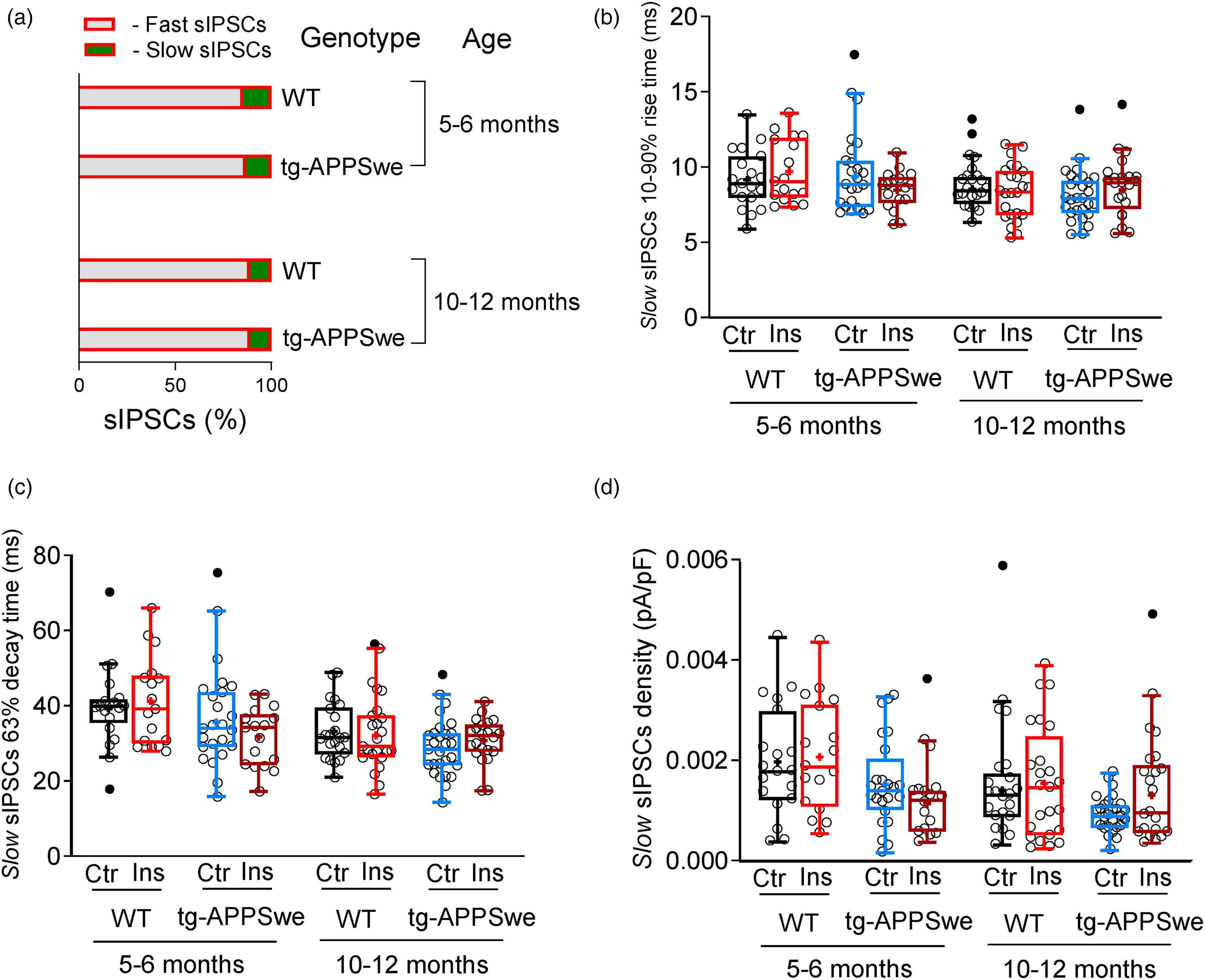
Insulin has negligible effect on the slow sIPSC T density in DG granule cells of the dorsal hippocampus in WT and tg-APPSwe mice. (a) A bar graph showing the percentage of the fast (gray) and slow (green) sIPSCs in DG granule cells of dorsal hippocampal slices treated with insulin (1 nM) in WT and tg-APPSwe mice at different age. (b, c) Kinetic parameters of slow sIPSCs. Summary plots for the median 10–90% rise time (b) and the median 63% decay time (c) of slow sIPSCs recorded from dorsal DG granule cells in hippocampal brain slices under insulin (1 nM) application from WT and tg-APPSwe mice, age groups: 5–6 months old and 10–12 months old. (d) Summary statistics for the total current (sIPSC T ) density of slow sIPSCs in dorsal DG granule cells of WT and tg-APPSwe mice 5–6 and 10–12 months old recorded from hippocampal slices after pre-incubation with 1 nM insulin (Ins, red). All experiments were performed with parallel controls from the same animal/age group. Data are presented as scatter dot plot for individual cells and box and whiskers plot with median values plotted as a line and mean values shown as ‘+’. Outliers are defined by the Tukey method and marked as dot plot (filled black circles). Statistical analyses are performed by excluding outliers. Ordinary two-way ANOVA with Fisher's LSD multiple comparison test was applied to examine the insulin effect. Vhold = −60 mV.
Insulin does not have effect on GABA-mediated slow sIPSC parameters in the dorsal DG granule cells.

Ordinary two-way ANOVA with Fisher's LSD multiple comparison test was used to compare insulin-treated cells with control cells in each animal group. The number of recorded cells ranges between 17 and 28 in each group. No statistical significance (significant level at 0.05) was detected in all comparisons. Data are presented as mean ± SEM.
Discussion
Here we studied in DG granule cells the GABA-activated slow sIPSCs in WT and tg-APPSwe mice. The results clearly show that properties of the slow sIPSCs have already started to change in APPSwe mice aged 5–6 months and are markedly manifested in the older, 10–12 months old APPSwe mice. The findings align with decreased synaptic strength or loss of synapses in the aged APPSwe mice in comparison with WT mice.26,27 However, the slow sIPSCs are not noticeably affected by insulin, which contrasts with the modulation of the fast sIPSCs and tonic currents that was clearly manifested in the APPSwe mice aged 10–12 months. 8
The slow sIPSCs are elicited by neurogliaform interneurons, which are a subtype of slow-spiking GABAergic interneurons that signal through a slow form of volume transmission that often lacks distinct postsynaptic anatomical specialisations.1,3,4 Selective change in the sIPSCs emerged in the APPSwe mice aged 5–6 months where the slow sIPSCs but not the fast sIPSCs were altered as compared to WT mice. 8 The fast sIPSCs changed later and were altered in the APPSwe mice aged 10–12 months. 8 That both the fast and the slow sIPSCs amplitudes and total current densities were reduced whereas the frequency was unaltered, may indicate a decline in the number of surface GABAA receptors expressed in the DG granule cells in the APPSwe mice aged 10–12 months. However, the tonic extrasynaptic current was increased from the age of 5–6 months in the APPSwe mice. 8 Together these findings suggest the reduction in the sIPSC T densities in the APPSwe mice aged 10–12 months is selective and may be related to smaller synapses or fewer GABAA receptors located at the postsynaptic sites reached by the neurogliaform interneurons-GABA signaling. In addition, GABA spillover from parvalbumin-expressing interneurons can also evoke slow sIPSCs in newborn DG neurons. 4 A deficit of hippocampal DG neurogenesis in APPSwe mice28,29 potentially contributes to the reduction of slow sIPSCs.
The observed net increase in neuronal excitatory-to-inhibitory ratio in mouse AD models is believed to be linked to the association of Aβ with synapses, which in turn leads to impaired synaptic function and disrupted network activity.9,30,31 Loss of inhibitory synapse has also been reported based on postmortem brain samples from AD patients. 26 Our observations of decreased slow sIPSC T density in the 10–12 months APPSwe mice in comparison with WT mice, further supports the finding of attenuated inhibitory synaptic tone in APPSwe mice.
The influence of insulin signaling in neurons and its impact on GABA-activated currents has been studied in neurons in rat and mouse hippocampus,8,17,32–34 rat prefrontal cortex,35,36 insular cortex 37 and mouse cerebellar granule cells. 38 In general, insulin strengthens the GABA signaling but the level is dependent on age, cell-type, GABAA receptor composition and distribution and tissue location. We have previously shown that insulin increases the fast sIPSCs current density but reduces tonic currents in hippocampal DG granule cells of APPSwe mice aged 10–12 months. 8 At the time when plaques have formed, insulin appears to support normal inhibitory synaptic transmission in the APPSwe mice. Interestingly, the effect of insulin on the slow sIPSC T density was negligible in the APPSwe mice.
In a transgenic mouse model of AD harboring the Swedish mutation (tg-APPSwe), a decrease in the GABA-evoked slow synaptic current density develops in the DG granule cells of dorsal hippocampus at 10–12 months of age. Insulin has no effects on the slow sIPSCs in contrast to the fast sIPSCs and tonic extrasynaptic currents in these neurons. This difference suggests that insulin selectively regulates the different types of GABA-activated currents.
Footnotes
Acknowledgments
We would like to thank Prof. Marco Capogna for first introducing us to the slow IPSCs.

Author contributions
Olga Netsyk (Conceptualization; Data curation; Formal analysis; Visualization; Writing – original draft; Writing – review & editing); Sergiy V Korol (Formal analysis; Visualization; Writing – review & editing); Jin-Ping Li (Funding acquisition; Resources; Writing – review & editing); Bryndis Birnir (Conceptualization; Formal analysis; Funding acquisition; Resources; Visualization; Writing – original draft; Writing – review & editing); Zhe Jin (Conceptualization; Formal analysis; Funding acquisition; Visualization; Writing – original draft; Writing – review & editing).
Funding
The authors disclosed receipt of the following financial support for the research, authorship, and/or publication of this article: The study was funded by Swedish Research Council grants 2018–02952 and 2015-02417 to BB, Excellence of Diabetes Research in Sweden (EXODIAB) to BB and ZJ and 2018-02503 to JPL, Gun och Bertil Stohnes Stiftelse (2024) and O.E. och Edla Johanssons vetenskapliga Stiftelse (2024) to ZJ.
Declaration of conflicting interests
The authors declared no potential conflicts of interest with respect to the research, authorship, and/or publication of this article.
Data accessibility
The dataset that supports the findings of this study are available from the corresponding author upon reasonable request.