



Abstract
Background
Research suggests that modifying risk factors may prevent or delay up to 40% of dementia cases, including Alzheimer's disease (AD). Thus, understanding the potential of healthful dietary patterns, like the Mediterranean diet (MD), in AD prevention is crucial. While supplementation of individual Mediterranean foods has demonstrated efficacy in reducing AD biomarkers and cognitive impairment in rodents, the effects of a comprehensive MD warrant further investigation. Additionally, while rodent studies often use a “Western diet” as a model for the typical American diet (TAD), these diets generally exceed the macronutrient densities of typical American consumption, particularly in fats and carbohydrates.
Objective
To better reflect human diets, we developed two diets for mice that more closely mirrored the macronutrient composition of the traditional MD or the TAD, each with matched macronutrient profiles (50% kcal from carbohydrates, 35% kcal from fat, 15% kcal from protein), and distinct food sources from Mediterranean regions or the U.S., respectively.
Methods
Male C57BL/6J mice were randomly assigned to one diet (MD or TAD) at weaning (21 days of age), which they consumed for six months.
Results
Compared to the TAD, MD animals had lower body weight, abdominal and hepatic fat, serum TNF-α, and central Aβ1–42, while also exhibiting enhanced exploratory behavior, reduced anxiety-like behavior, and preserved spatial memory. The MD also protected against LPS-induced central inflammation and BDNF loss.
Conclusions
These findings suggest that a comprehensive MD provides protection against metabolic and AD-related markers in wildtype mice, despite matched caloric availability to the TAD.
Keywords
Introduction
Historically, Alzheimer's disease (AD) prevalence in the United States has consistently outstripped that of countries bordering the Mediterranean Sea in Southern Europe. For example, in 2018, approximately 10% of Americans over the age of 65 had AD, 1 whereas their Southern European counterparts had a comparatively lower incidence, at 6.88%. 2 Research on human subjects has identified several risk factors linked to an increased risk of AD, such as sedentary lifestyle, 3 obesity,4–6 and poor diet.7–10 Based on these findings, the observed lower rate of AD prevalence in Southern Europe may be linked to the traditional Mediterranean diet (MD). For example, strict adherence to the MD has been associated with reduced weight gain and obesity,11–14 along with diminished pro-inflammatory mediators,15–17 amyloid-beta (Aβ),9,18 brain atrophy,10,19–21 cognitive decline,22–28 and risk of AD and related dementias28,29 in middle-aged and elderly adults. This largely plant-based diet offers longevity benefits, due to key dietary factors such as high fiber content, low glycemic load, high monounsaturated fatty acid (MUFA) intake, and abundant plant polyphenols.30–32
In contrast, the typical American diet (TAD), a largely animal-based diet, is suffused with high levels of saturated fatty acids (SFA) and simple carbohydrates, while simultaneously devoid of many essential nutrients. 33 For example, recent evidence highlights a significant nutrient deficiency among Americans: merely 10% meet daily fruit and vegetable recommendations, 34 and only 7.4% achieve the recommended fiber goal. 35 Both obesity36–38 and AD38–40 prevalence are rapidly increasing in the U.S. Numerous studies using a “Western diet” that incorporate exaggerated levels of SFA, and about 42–60% kilocalories (kcal) from total fat,41–48 provoke obesity-related biomarkers,41,42 inflammation,44,45 Aβ and beta-secretase 1 (BACE1),44,46,49 along with spatial memory impairment42,50–54 in C57BL/6J mice. However, the effects of a TAD reflecting the average macronutrient profile of Americans have not been thoroughly studied in rodent research. 55 Given the extensive existing studies on “Western diets,” further research is needed to understand the TAD's impact on metabolic markers and AD susceptibility. Additionally, while prior animal studies have demonstrated that individual MD components, like olive oil, prevent or mitigate AD pathologies, such as hippocampal and cortical Aβ1–42 in wildtype mice 56 and transgenic models of AD,57,58 few have aimed to replicate human Mediterranean dietary patterns in the context of AD research.
To address these crucial gaps in understanding diet-related AD risks, we designed two experimental diets to mimic either the MD or TAD, by providing the average macronutrient densities and diverse food sources typical of Mediterranean regions, including Greece and Italy,26,30,32 or the United States.59,60 Additionally, we designed the diets to reflect the typical percentage of kilocalories from different fat sources, including SFAs, MUFAs, and polyunsaturated fatty acids (PUFAs).32,61,62 Further, we mimicked the typical omega-6 to omega-3 fatty acid ratio in the MD and the TAD,61,63,64 while also accounting for typical insoluble and soluble fiber content, as the MD is fiber-rich compared to the TAD. 61 Finally, we matched the macronutrient composition of both diets to control for energy availability. Through this distinctive approach that better reflects reality for human populations, we investigated the effects of early-life implementation and long-term consumption of the MD or TAD on AD-related markers and comorbidities in wildtype mice. In Experiment 1, we hypothesized that the MD would protect mice against (1) excessive weight gain, (2) increased abdominal and hepatic fat, (3) peripheral and central inflammation, (4) Aβ1–42 in the cortex and hippocampus, and (5) behavioral and cognitive deficits, compared to the TAD.
In Experiment 2, an additional group of mice was utilized to examine the effects of lipopolysaccharide (LPS) treatment on the central gene expression of AD biomarkers. As wildtype mice do not typically produce high levels of pro-inflammatory cytokines without an inflammatory stimulus, we utilized LPS, a toll-like receptor 4 (TLR4) agonist, to induce an innate immune response, as established by prior research.65–70 Our laboratory has repeatedly demonstrated that peripheral LPS injection induces mRNA expression of pro-inflammatory cytokines and reduces expression of brain-derived neurotrophic factor (BDNF) in the dorsal hippocampus of C57BL/6J mice. 67 Prior research has also established that LPS treatment rapidly induces a more robust inflammatory response in the dorsal hippocampus compared to the ventral hippocampus.71,72 Therefore, we hypothesized that the MD would protect mice against elevated levels of LPS-induced gene expression of the pro-inflammatory cytokines TNF-α, IL-1β, and IL-6, in addition to the amyloid-β protein precursor (AβPP) cleavage enzyme beta-secretase 1 (BACE1). Additionally, we hypothesized that the MD would protect against IL-10 and BDNF loss following LPS administration, compared to the TAD.
General methods for experiments 1–2
Subjects
The current study utilized male wildtype C57BL/6J mice bred at Texas Christian University's (TCU) vivarium (from stock animals initially obtained from The Jackson Laboratory, Bar Harbor, ME). All procedures adhered to the Guide for the Care and Use of Laboratory Animals (National Research Council, 2011), and were approved by the TCU Institutional Animal Care and Use Committee (IACUC Protocol #2021-14). Mice were housed in standard polycarbonate cages with pelleted, paper-chip bedding and compressed cotton square nestlets (LabSupply, Fort Worth, TX) in groups of three to four, under a 12-h light/dark schedule.
Experimental design and timeline
At postnatal day (PND) 21, mice were weaned and randomly assigned to one of two experimental diets: a Mediterranean diet (MD) or typical American diet (TAD) (ns = 23) for six months, with access to food and water ad libitum. At six months of age, mice underwent behavioral testing, and at six months and 21 days of age, tissue and serum were collected.
Experimental diet formulations
The MD and the TAD were created in pelleted form, and purchased from Research Diets, Incorporated (New Brunswick, NJ). See Table 1 for experimental diet formulations and Supplemental Material for a complete list of diet ingredients.
Experimental diet formulations.

Diets were purchased from Research Diets (New Brunswick, NJ).
Methods for experiment 1
Measures of food consumption and body weight
Food consumption was measured weekly by weighing and recording the total amount of food in each cage's hopper every Friday morning. On Monday mornings, the remaining food in each hopper was weighed and recorded, and average consumption was calculated. Additionally, body weight was measured and recorded once per week.
Open field testing
At six months of age, mice underwent open field testing in enclosed chambers (27 × 27 cm) equipped with infrared beams to measure horizontal and vertical movement (Med Associates Incorporated, St Albans, VT). The open field chamber lights were maintained at 5.5 lux. Prior to testing, two zones were defined: the center zone (304.85 cm2) and the outer zone (423.39 cm2). Time spent in these zones was utilized to assess anxiety-like behavior, with more time in the center zone indicating reduced anxiety-like behavior. 73 Mice were placed in the chamber for 10 min, and vertical counts (rearing behavior), ambulatory distance (cm), time in the center zone (seconds), and average speed of ambulatory episodes (cm per second) were measured.
Elevated zero maze
Mice were tested in the elevated zero maze to further measure anxiety-like behavior, as validated by previous research. The elevated zero maze (platform 50 cm tall, circular diameter 60 cm) consisted of four alternating quadrants: two open and two enclosed by walls. The testing room lights were maintained at 45–48 lux. At the beginning of the test, each mouse was placed in an open quadrant, facing a closed quadrant, and given five minutes of uninterrupted time to move. Time spent in each quadrant was used to measure anxiety-like behavior, with more time in the open quadrants indicating reduced anxiety-like behavior. 74 A video camera was mounted on the ceiling to record the total time spent in the open quadrants versus the enclosed quadrants using EthoVision XT software (Noldus Information Technology, Leesburg, VA).
Object-location memory task
The object-location memory task (OLM) was utilized to assess spatial learning and memory. Arena settings were created with EthoVision XT software (Noldus Information Technology) and recorded with a ceiling-mounted video camera. OLM was comprised of habituation (2 sessions, 4 h apart, no objects), training (2 sessions, 4 h apart, two identical objects), and testing, involving the relocation of one object to the opposite corner (new spatial location). The testing room lights were maintained at 45–48 lux. During each session, mice had ten minutes to freely explore. See Figure 1 for OLM timeline. Exploration time was recorded when the mouse's nose entered the predetermined zone (three cm radius surrounding the object). Post-testing, the percentage of time spent exploring the object in the new location was calculated utilizing a previously-used formula. 42 Object location exploration was also assessed during training to ensure unbiased location preference.

Object-Location memory task. During each session, mice were placed in the OLM arena and provided ten minutes of uninterrupted exploration. The arena walls provided two distinct visual cues (one wall had a blue triangle, and one wall had three green stripes). During the training session, experimenters recorded the percentage of time that mice spent exploring two identical objects. During testing, one object was moved to the opposite corner of the chamber, and the percentage of time exploring the object in the new location was measured. This image was created using BioRender.
Tissue and serum collection
After six months consuming the prescribed diets, mice were fasted for six hours prior to euthanasia, and blood was immediately collected. Following blood collection, liver, spleen, and white adipose tissue were removed, weighed, and stored in neutral buffered formalin. Additionally, both hemispheres of the frontal cortex and hippocampus were collected and lysed in a protease inhibitor-containing cocktail (PRO-PREP, Bulldog Bio, Portsmouth, NH).
Peripheral cytokine analysis
A VPLEX Custom Mouse Cytokine Pro-Inflammatory Panel 1 multiplexing kit and QuickPlex SQ 120 instrument (Meso Scale Diagnostics, Rockville, MD) were utilized to measure the following analytes in serum: mouse TNF-α, IL-1β, IL-6, IL-10, and IFN-γ. Sample replicates with an intra-assay coefficient of variation of over 25% were excluded from the analysis, as advised by MSD technical support. See Supplemental Material for complete assay methodology.
Liver histology
Livers were collected from each mouse and divided for hematoxylin and eosin (H&E) and Oil Red O staining. Paraffin embedded sections of liver were collected on Millenia Command Adhesion Slides (StatLab, McKinney, TX) and stained with H&E. 75 Frozen sections of livers were also collected on Millenia Command Adhesion Slides (StatLab) and stained with Oil Red O, per manufacturer's instructions (StatLab). All images were collected on a Nikon Eclipse 90i microscope (Nikon Instruments, Melville, NY) and analyzed using NIS-Elements AR 4.60.00 software (Nikon Instruments).
Aβ1–42 ELISA
Protein assays (DC Protein Assay; Bio-Rad Laboratories, Hercules, CA) were conducted to calculate the total amount of protein in each sample, and protein concentrations were standardized. A mouse Aβ1–42 ELISA (Invitrogen, ThermoFisher Scientific, Waltham, MA) was conducted, per manufacturer's instructions, and read on a plate reader (BMG LabTech FLUOstar Omega, Cary, NC). Sample replicates with an intra-assay coefficient of variation over 25% were excluded from the analysis.
Statistical analyses
All data were analyzed with Statistical Package for Social Sciences (SPSS; Version 29.0, IBM, Armonk, NY). Normality was assessed prior to running statistical analyses, and transformations were performed to correct for violations of homogeneity of variance. Two-tailed independent samples t-tests and mixed-design analyses of variance (ANOVAs) were conducted to analyze behavioral and biological dependent variables. All outliers were detected utilizing SPSS's interquartile range, and for all analyses, an alpha level of p ≤ 0.05 was considered significant.
Methods for experiment 2
Subjects and experimental design
Following 6 months of diet administration (MD or TAD), male C57BL/6J mice were randomly assigned to one of two treatment conditions: one intraperitoneal (i.p.) injection of LPS (E. coli serotype O26:B6; Sigma-Aldrich, St Louis, MO) at a dose of 250 μg/kg of body weight, or an equivalent volume of sterile saline four hours prior to tissue collection. Injections were performed between 0800 h and 1100 h. This LPS model has previously been utilized in our laboratory. 67 Four hours after injections, both hemispheres of the dorsal hippocampus were collected under RNase-free conditions utilizing RNaseZap™ (Invitrogen, ThermoFisher Scientific, Wilmington, DE), and immediately placed in RNAlater™ Stabilization Solution (Invitrogen, ThermoFisher Scientific, Wilmington, DE). Tissue was stored at −80°C prior to RNA isolation.
Quantitative reverse-transcription polymerase chain reaction (qRTPCR)
Central gene expression for TNF-α, IL-1β, IL-10, IL-6, BDNF, BACE-1, and β-actin (internal control gene) was measured in the dorsal hippocampus, utilizing qRTPCR. First, RNA was isolated from hippocampal tissue utilizing a Maxwell LEV simplyRNA Purification kit (Promega Corporation, Madison, WI), and measured for quantity and purity with a NanoDrop™ OneC Microvolume Spectrophotometer (ThermoFisher Scientific, Wilmington, DE). Subsequently, reverse transcription was performed utilizing Script Reverse Transcriptase Supermix (Bio-Rad, Hercules, CA) and a 7500 Real-Time PCR Thermal Cycling System (Applied Biosystems, Foster City, CA). Expression levels of mRNA for the target genes were then measured using iTaq Universal Probes Supermix (Bio-Rad) and PrimePCR Probe Assay probes (Bio-Rad, Hercules, CA). Each gene sample was run in triplicate, using a control sample from mice on the MD that had been treated with saline. The relative normalized expression of cDNA from the fluorescence data was normalized to β-actin, relative to the control sample, utilizing the CFX Connect Real-Time PCR Detection System (Bio-Rad, Hercules, CA).
Statistical analyses
Analyses of variance (ANOVAs) were conducted utilizing Prism (GraphPad Software, Inc., San Diego, CA), to examine the effects of diet condition (MD or TAD) and treatment (LPS or saline) on gene expression. An alpha level of p ≤ 0.05 was considered significant. If a sample had a quantitation cycle (Cq) standard deviation of 0.25 or higher, the outlier of the triplicates was removed from the dataset. If the Cq standard deviation was still more than 0.25 following this correction, the sample was excluded from data analysis.
Experiment 1 results
Average food and kilocalorie consumption
Two mixed-design ANOVAs revealed a significant main effect of diet duration (number of months on diet) on daily food consumption (grams), F(5,60) = 9.988, p ≤ 0.001,
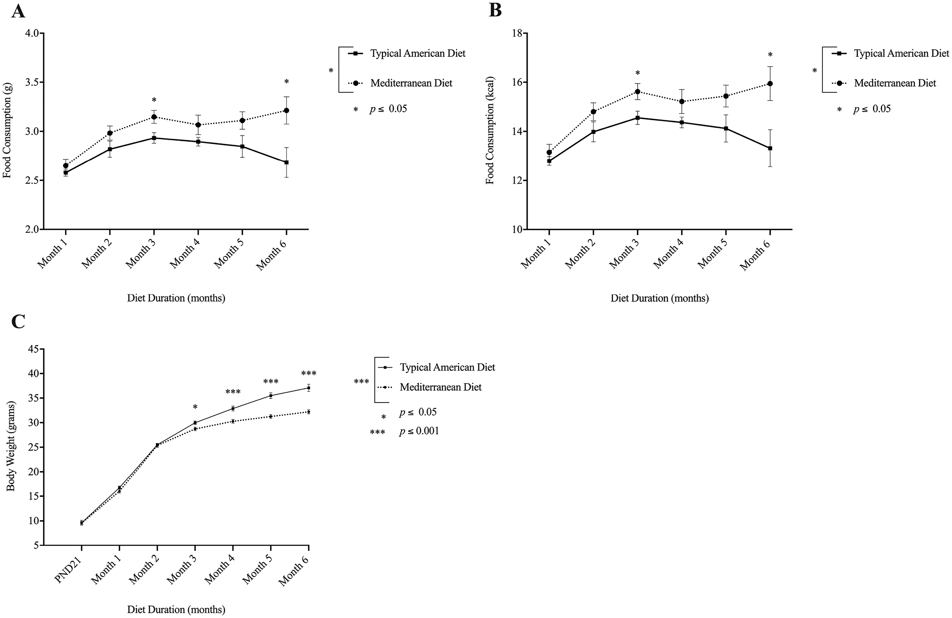
Mice on the MD consumed more daily kilocalories, but had lower body weight than mice on the TAD. (A) A mixed-design ANOVA revealed a significant main effect of diet condition (MD vs. TAD) on food consumption, such that mice on the MD consumed more food than those on the TAD, p = 0.028. (B) Mice assigned to the MD consumed more kilocalories per day than those on the TAD, p = 0.028. (C) There was a significant main effect of diet condition on body weight, p ≤ 0.001, in which males on the MD weighed less than those on the TAD. Bars represent ± SEM. Significant differences (p ≤ 0.05) are designated by *.
Body weight
A mixed-design ANOVA revealed a significant main effect of diet duration (number of months on diet) on body weight (grams) over the course of six months of diet administration, F(6, 240) = 2013.004, p ≤ 0.001,
Organ and white adipose tissue weight
An independent samples t-test revealed a significant difference in white adipose tissue weight, t(27) = −9.194, p ≤ 0.001, and liver weight, t(27) = −4.355, p ≤ 0.001, in which mice on the TAD had heavier white adipose tissue and livers than those on the MD. The magnitude of the difference in the means of white adipose tissue weights (mean difference = -1.397, 95% CI [−1.085, −1.709]) was large (η2 = 0.758), and the magnitude of the difference in the means of liver weights (mean difference = -0.523, 95% CI [−0.277, −0.769]) was large (η2 = 0.413). However, there was no difference in spleen weight, t(33) = -1.583, p = 0.123. See Supplemental Material.
Liver health following diet administration
We imaged four, randomly-selected liver sections from three mice in each diet condition (MD or TAD). An independent samples t-test revealed a significant difference in the percentage of pixelated area that was unstained following H&E staining, in which mice on the MD had less percentage of pixelated area unstained compared to those on the TAD, t(21) = 39.173, p ≤ 0.001. The data violated homogeneity of variance, and a reflect and square root transformation was performed to correct this. The magnitude of the difference in the means of unstained area in the liver (mean difference = -22.486, 95% CI [−23.605, -21.368]) was large (η2 = 0.986). See Figure 3.

The MD prevented hepatic fat compared to the TAD. (A) H & E-stained liver from a mouse on the MD. (B) H & E-stained liver from a mouse on the TAD. (C) An independent samples t-test revealed a significant difference in the percentage of pixelated area that was unstained following H & E staining, p ≤ 0.001. (D) Oil-Red O-stained liver from a mouse on the MD. (E) Oil-Red O-stained liver from a mouse on the TAD; lipids are stained red. Bars represent ± SEM. Significant differences (p ≤ 0.05) are designated by *.
Peripheral cytokine production in serum
An independent samples t-test revealed that mice on the TAD produced significantly more TNF-α (pg/mL) compared to those on the MD, t(32) = -3.790, p ≤ 0.001. One sample was removed from the analysis due to an intra-assay coefficient of variation (CV) value over 25%, as recommended by MSD's technical team. The magnitude of the difference in the means (mean difference = -2.12, 95% CI [−3.253, −0.979]) was large (η2 = 0.31). Additionally, the difference in IL-1β (pg/mL) production between diet conditions was not significant, t(23) = -1.809, p = 0.084. Six samples were removed from the analysis due to CV values over 25%. Additionally, three outliers were removed from the analysis because they fell outside of the interquartile range determined by SPSS. The magnitude of the difference in the means (mean difference = -0.07967, 95% CI [0.0115, −0.171) was moderate (η2 = 0.125). Further, there was no significant difference in IL-10 (pg/mL) production between diet conditions, t(29) = -0.818, p = 0.420. Three outliers were removed from the analysis because they fell outside of the interquartile range determined by SPSS. Finally, an independent samples t-test failed to reveal a significant difference in IFN-γ (pg/mL) production between diet conditions, t(30) = 0.040, p = 0.968. Two samples were removed from the analysis due to CV values over 25%. See Figure 4. Although we also aimed to measure IL-6, numerous samples fell below the lowest detectable limit of the assay's standard curve, thus precluding analysis.
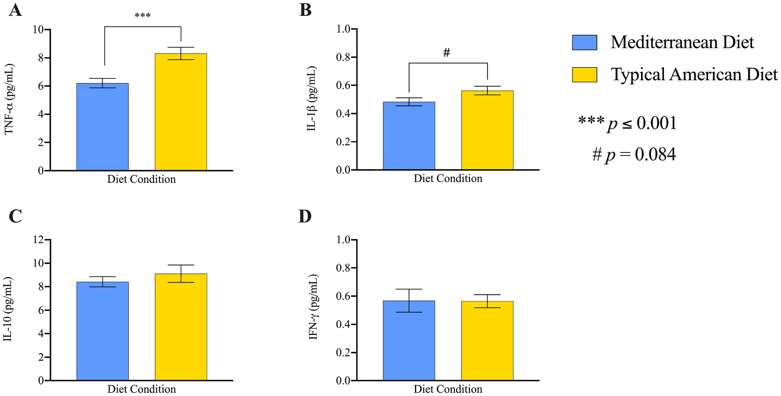
The MD protected against serum TNF-α, but not IL-1β, IL-10, or IFN- γ compared to the TAD. Independent samples t-tests were performed to compare the quantity of peripheral cytokines produced in serum following six months of diet. (A) Mice on the MD produced significantly less serum TNF-α compared to mice on the TAD, p ≤ 0.001. (B–D) There was no significant difference in IL-1β, IL-10, or IFN- γ production between diet conditions, ps > 0.05. (A–D) Bars represent mean ± SEM. Significant differences (p ≤ 0.05) are designated by *.
Aβ1–42 production in the cortex and hippocampus
An independent samples t-test revealed a significant difference in Aβ1–42 (pg/mg) in the cortex, t(22) = -3.067, p = 0.006, in which mice on the MD had less Aβ1–42 in the cortex, in comparison to those on the TAD. The data violated homogeneity of variance, and a log transformation was performed to correct this. The magnitude of the difference in the means (mean difference = -9.63, 95% CI [−3.10, −16.16,]) was very large (η2 = 0.299). Approximately 30% of variance was explained by diet. Two outliers were outside of the interquartile range determined by SPSS and removed from the analysis.
An independent samples t-test also revealed a significant difference in Aβ1–42 (pg/mg) in the hippocampus, such that mice on the MD had less hippocampal Aβ1–42 compared to those on the TAD, t(29) = -2.103, p = 0.044. The magnitude of the difference in the means (mean difference = -4.522, 95% CI [−0.124, −8.920]) was moderate (η2 = 0.132). Three outliers were removed from the analysis. Additionally, two samples were removed from the analysis due to intra-assay CV values over 25%. See Figure 5.
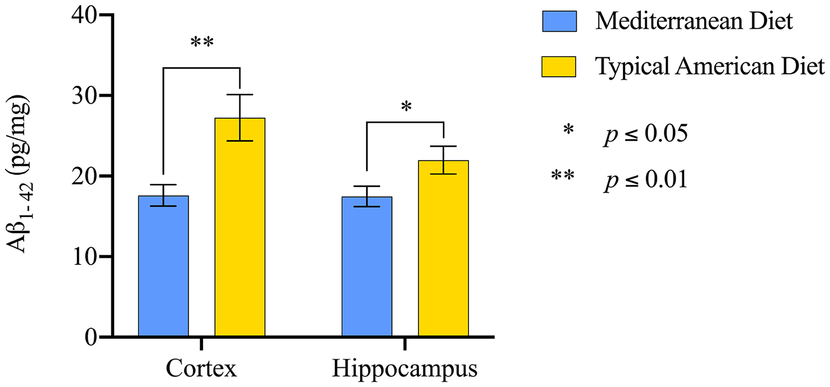
The MD protected against increased Aβ1–42 in the cortex and hippocampus compared to the TAD. Mice on the TAD had significantly more Aβ1– 42 (pg/mg) in the cortex, p ≤ 0.01, and hippocampus, p ≤ 0.05, compared to mice on the MD. Bars represent mean ± SEM. Significant differences (p ≤ 0.05) are designated by *.
Open field behavior
Vertical counts
An independent samples t-test revealed a significant difference in vertical counts t(27) = 2.767, p ≤ 0.01, such that mice on the MD exhibited significantly more vertical counts than mice on the TAD. The magnitude of the difference in the means during the total testing time (mean difference = 14.111, 95% CI [24.573, 3.648]) was very large (η2 = 0.221). Three outliers were detected utilizing SPSS and were removed from the analysis. See Figure 6.
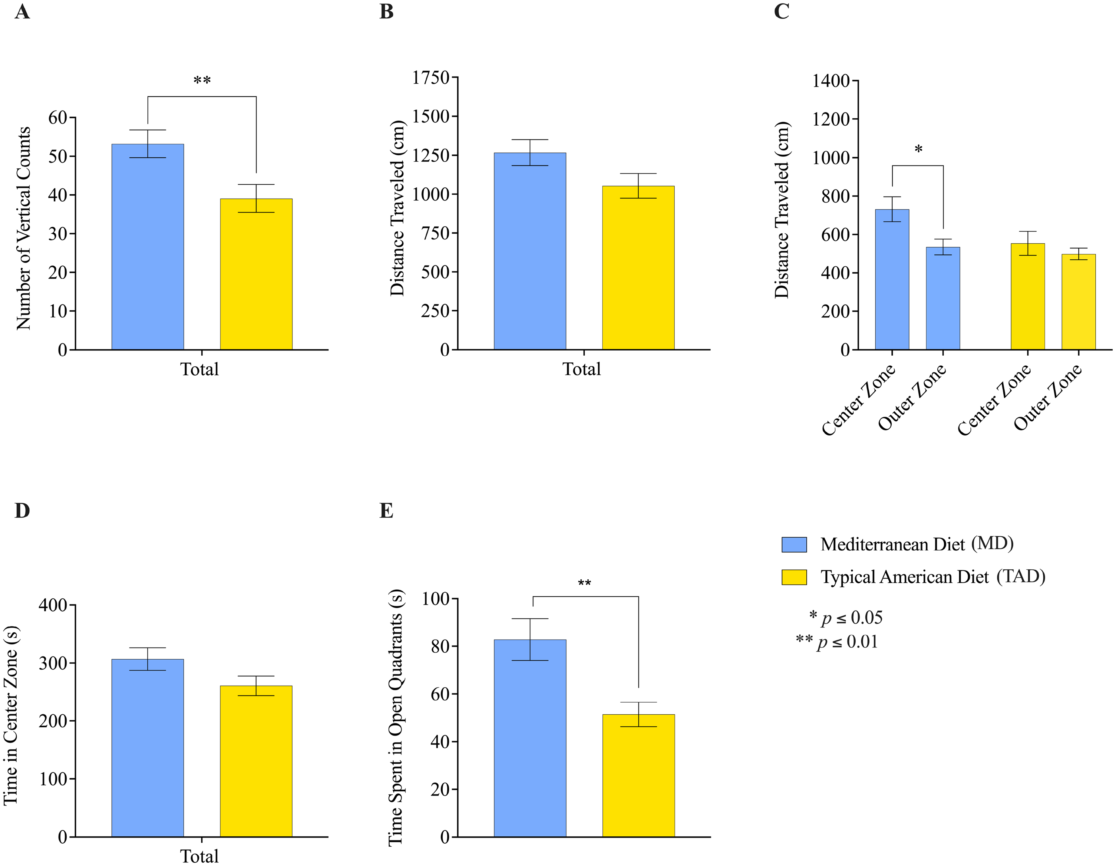
Locomotor activity, exploratory and anxiety-like behavior. (A–D) Multiple independent samples t-tests were performed to examine exploratory behavior and locomotor activity during the open field test. (A) Mice on the MD exhibited more vertical counts compared to mice on the TAD, p ≤ 0.01. (B) There was no significant difference in the total ambulatory distance traveled (cm/s) between the two diet conditions, p = 0.072. (C) A paired-samples t-test revealed that mice on the MD traveled significantly more (cm) in the center zone compared to the outer zone of the open field, p ≤ 0.05. (D) There was no significant difference in time (s) spent in the center zone of the open field between the two diet conditions, p ≥ 0.05. (E) Mice on the MD spent significantly more time in the open quadrants of the elevated zero plus maze in comparison to mice on the TAD, p ≤ 0.01. Bars represent mean ± SEM. Significant differences (p ≤ 0.05) are designated by *.
Distance traveled
An independent samples t-test did not reveal a significant difference in the total ambulatory distance traveled t(30) = 1.867, p = 0.072. The magnitude of the difference in the means during the total testing time (mean difference = 213.878, 95% CI [447.887, − 20.130]) was moderate (η2 = 0.104). However, when analyzing the total ambulatory distance traveled in the center zone, the difference in the distance traveled approached significance, t(30) = 1.961, p = 0.059. There was no difference in the total ambulatory distance traveled in the outer zone, t(30) = 0.702, p = 0.488. Additionally, a paired-samples t-test revealed that mice on the MD traveled a significantly farther distance in the center zone in comparison to the outer zone, t(16) = -2.777, p = 0.013, whereas there were no differences in the amount of distance traveled between the center and outer zone in mice on the TAD, t(14) = -0.954, p = 0.356. See Figure 6.
Time spent in the center zone of open field
An independent samples t-test did not reveal a significant difference in the amount of time spent in the center zone during testing, t(27) = 1.748, p = 0.092. See Figure 6.
Average speed
Elevated zero maze behavior
An independent samples t-test revealed that mice on the MD spent significantly more time in the open quadrants of the elevated zero maze in comparison to those on the TAD, t(31) = 3.121, p = 0.004. The data violated homogeneity of variance, and a log transformation was conducted to correct this. The magnitude of the difference in the means (mean difference = 0.214, 95% CI [0.354, 0.743]) was large (η2 = 0.239). One outlier was detected by SPSS and excluded from the analysis. See Figure 6.
Spatial learning and memory in OLM
Post-testing, the percentage of time spent exploring the object in the new location was calculated using the formula: new object location time / [new object location time + old object location time] x 100, as adopted from Heyward and colleagues. 42 As expected, an independent samples t-test revealed that there was no difference in the percentage of time spent exploring the new object location during training, t(18) = -0.787, p = 0.441. However, an independent samples t-test revealed that mice on the MD spent significantly more time exploring the object moved to the new location, compared to mice on the TAD, during testing, t(18) = 2.259, p = 0.037. The magnitude of the difference in the means (mean difference = 21.919, 95% CI [42.305, 1.532]) was large (η2 = 0.221). See Figure 7.
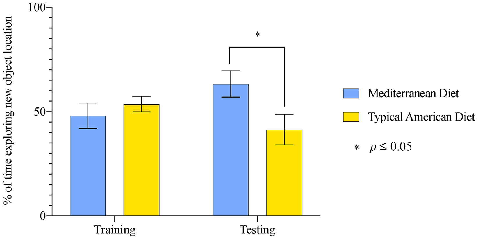
The MD preserved spatial learning and memory in the OLM test in comparison to the TAD. Mice on the MD spent significantly more time exploring the object moved to the new location, compared to mice on the TAD, during testing, p = 0.037. Bars represent ± SEM. Significant differences (p ≤ 0.05) are designated by *
Experiment 2 results
Gene Expression of BDNF and BACE1 in the Dorsal Hippocampus
A two-way ANOVA revealed a significant main effect of diet condition (MD or TAD) on hippocampal BDNF mRNA, F(1, 38) = 9.263, p = 0.004, in which mice on the MD had increased BDNF gene expression compared to those on the TAD. This effect was largely driven by LPS-treated mice, and post-hoc analyses revealed that mice on the MD had increased BDNF gene expression compared to those on the TAD following LPS treatment, p = 0.006. There was no significant main effect of injection (LPS or saline), F(1, 38) = 0.069, p = 0.794, on BDNF gene expression. In contrast to our hypothesis, a two-way ANOVA did not reveal a significant main effect of injection, F(1,39) = 3.485, p = 0.070, or diet condition, F(1,39) = 2.606, p = 0.115, on BACE1 gene expression. See Figure 8.
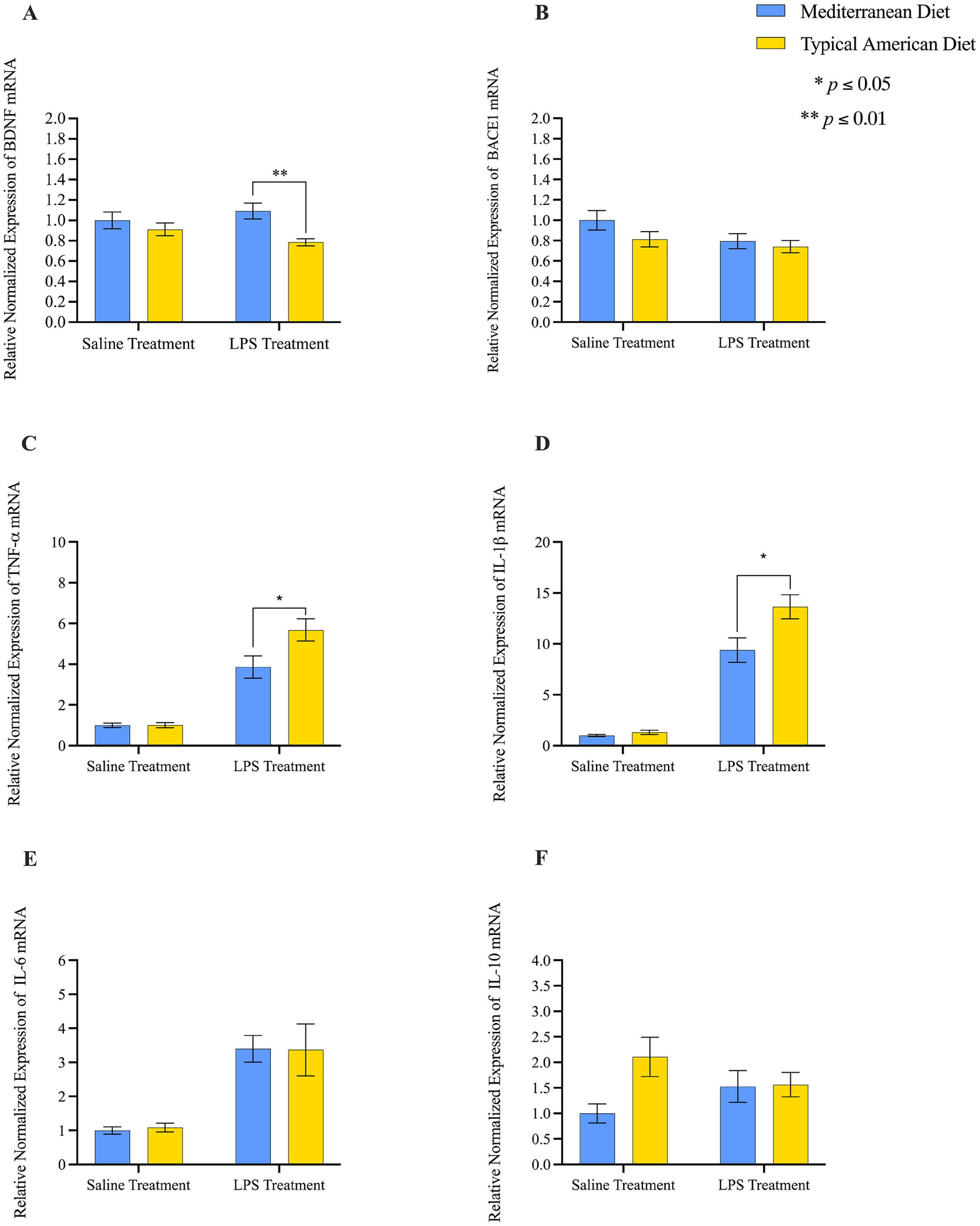
The MD protected against both elevated LPS-induced TNF-α and IL-1β gene expression, and LPS-induced BDNF loss compared to the TAD. (A) Mice on the TAD had significantly decreased gene expression of BDNF compared to male mice on the MD, p ≤ 0.05. (B) There was no significant main effect of diet on relative normalized gene expression of BACE1, p > 0.05. (C) Mice on the TAD had increased TNF-α gene expression compared to mice on the MD, p ≤ 0.05. (D) Mice on the TAD had increased IL-1β gene expression compared to MD fed counterparts, p ≤ 0.05. (E) There was no significant main effect of diet on relative normalized gene expression of IL-6, p > 0.05. (F) There was no significant main effect of diet on relative normalized gene expression of IL-10, p > 0.05. (A–F) Bars represent ± SEM. Significant differences (p ≤ 0.05) are designated by *.
Gene Expression of TNF-a, IL-1β, IL-6, and IL-10 in the Dorsal Hippocampus
A two-way ANOVA revealed a significant main effect of injection on TNF-α gene expression, F(1,38) = 73.55, p ≤ 0.001, such that LPS-injected mice had increased TNF-α gene expression, compared to saline-treated mice. Additionally, there was a significant main effect of diet condition, F(1,38) = 4.320, p = 0.045, in which mice on the TAD had increased TNF-α gene expression compared to MD-fed counterparts. This was further supported by a significant interaction between injection (LPS or saline) X diet condition (MD or TAD), F(1,38) = 4.242, p = 0.046, in which the combined effects of the TAD and LPS treatment significantly elevated TNF-α gene expression compared to the TAD and saline treatment. Additionally, post-hoc analyses revealed that the TAD and LPS treatment significantly increased TNF-α gene expression compared to the MD and LPS treatment, p = 0.019.
A two-way ANOVA also revealed a significant main effect of injection on IL-1β gene expression, F(1,35) = 113.4, p ≤ 0.001, such that LPS-treated mice had increased IL-1β gene expression compared to saline-treated mice. Additionally, there was a significant main effect of diet condition, F(1,35) = 5.484, p = 0.025, in which mice on the TAD had increased IL-1β gene expression compared to MD fed counterparts. Finally, the interaction between injection (LPS or saline) X diet condition (MD or saline) was significant, F(1,35) = 4.106, p = 0.050. Post-hoc analyses revealed that the TAD and LPS treatment significantly increased IL-1β gene expression compared to the MD and LPS treatment, p = 0.011.
Additionally, a two-way ANOVA revealed a significant main effect of injection on IL-6 gene expression, F(1,37) = 21.22, p ≤ 0.001, in which LPS-treated mice had increased IL-6 gene expression compared to vehicle-treated mice. However, contrary to our hypothesis, there was no significant main effect of diet condition on IL-6 gene expression, F(1,37) = 0.003, p = 0.958. Finally, a two-way ANOVA did not reveal a significant main effect of injection, F(1,22) = 0.001, p = 0.980, or diet condition, F(1,22) = 3.624, p = 0.070, on IL-10 gene expression. See Figure 8.
Discussion
To fill key gaps in the current understanding of diet-related AD risks, as well as explore potential, nutritional AD prevention strategies in animal research, we designed two macronutrient-matched diets (MD and TAD) reflecting comprehensive dietary patterns and average macronutrient intake in Mediterranean regions and the U.S. Although there is evidence that the MD is associated with a reduction in AD-related markers in human subjects research, the current study aimed to further explore the underlying biological mechanisms responsible for the potential, protective effects of the MD in mice. The current findings align closely with those observed in human subjects.
For example, research utilizing magnetic resonance imaging (MRI) has shown that close adherence to the MD is associated with protection from AD pathologies, including Aβ,9,18 brain atrophy,10,19–21 and cognitive decline22,23,25,26 in middle-aged and elderly adults. Specifically, high adherence to the MD has been associated with reduced ventricular enlargement and atrophy in the hippocampus and temporal cortex of middle-aged adults,10,19 while low adherence has been shown to increase atrophy and induce cortical thinning. 10 In further support of the current research, a recent study reported that long-term consumption of the MD was associated with reduced global AD pathology and Aβ in postmortem brains of elderly adults. 76
In further support of the current findings, research in elderly adults has shown that excessive consumption of refined grains and sugars is associated with increased Aβ production in the cortex and lower test scores on cognitive tests in cognitively healthy, elderly adults. 77 Further, human subjects research has demonstrated that a MD (with olive oil as the main fat source) has been associated with reduced peripheral pro-inflammatory mediators,16,17 while high quantities of SFA78–80 and red meat 81 have been associated with increased pro-inflammatory mediators in adults. For example, a Western-style diet, rich in SFA, was associated with increased pro-inflammatory gene expression in the adipose tissue of adults at risk of developing metabolic syndrome. 80
Although the previously discussed research has provided evidence that there is a connection between diet and AD susceptibility, there is limited research on the underlying biological and behavioral mechanisms of a long-term, comprehensive MD, and how it potentially diminishes AD susceptibly in rodents. Further, potential health consequences stemming from long-term adherence to the TAD remain inadequately explored in rodent studies. The overarching term “Western diet” lacks a precise definition, as prior studies have utilized numerous variations of the Western diet, including “high-fat”, “high-sugar”, or cafeteria diets, with exaggerated macronutrient densities that are not representative of human diets.82,83 Therefore, the current experiments addressed these limitations by exploring the impact of early-life implementation and long-term adherence to the more realistic MD or TAD on AD vulnerability in adulthood.
Prior to testing, we conducted a comprehensive literature review to ensure that the experimental diets accurately modeled dietary patterns observed in both Mediterranean and U.S. populations. Prior research indicates that the MD provides approximately 35–40% kcal from fat.30,32,84 Specifically, the MD provides about 19% kcal from monounsaturated fatty acids (MUFAs) and 5% kcal from polyunsaturated fatty acids (PUFAs), with a typical omega-6 to omega-3 ratio of 1:1 or 2:1.61,64,85 Additionally, the MD generally provides 15–20% kcal from protein, with its primary macronutrient source being complex carbohydrates, constituting approximately 40–48% of daily kcal intake.61,85 In contrast, the average American adult consumes about 34% kcal from fat, and the majority of macronutrients are provided by refined carbohydrates and sugars.33,59
We specifically designed this experiment to compare the MD and TAD without an additional group consuming a low-fat, “standard” diet. All diets were designed with the same percentage of kcals from macronutrients to account for macronutrient availability; thus, we did not add a low-fat, “standard,” diet. The “standard” diet typically provided to rodents in laboratory settings does not mimic a human diet, as they are grain-based and low in fat. 86 Moreover, standard laboratory diets for rodents are inconsistent in both macronutrient composition and typically only provide about 10% kcal from fat. 86 Numerous “standard” grain-based diets are available for rodents, and typically provide a wide variety of macronutrient sources. However, the improper use of “standard” diets in rodent research has recently become an important topic of discussion within the scientific community. A recently published article highlighted potential problems with widely-used rodent diets: “they [standard diets for rodents] are generally considered to maintain a healthy phenotype in the animal (though it can be argued ‘compared to what?’).” 86
The formulas for standard chow diets tend to fluctuate, and the ingredients are not strictly regulated. 86 For example, a previous experiment tested over thirteen different laboratory diets globally, and found contaminants such as glyphosate and heavy metals. 87 Thus, this large variation of macronutrient density ranges, ingredients, pesticides, and heavy metals in “standard” diets has considerable variability that could affect scientific data. In contrast, purified ingredient diets use highly refined ingredients, such as casein, fewer chemicals, and more consistent product testing. 86 Therefore, the current study compared two macronutrient-matched, purified diets to control for variability and prevent confounding variables, aiming to clearly demonstrate that the two experimental diets (MD and TAD) were driving differences in the physiological and behavioral dependent variables. Likewise, prior experiments examining the effects of diet on rodent physiology and behavior have also only utilized two experimental diets without the use of a “standard” rodent diet.45,88
Through this approach, we found that the MD protected mice from adverse alterations in: (1) body weight, (2) abdominal and hepatic fat, (3) serum TNF-α, (4) Aβ1–42 in both the cortex and hippocampus, (5) exploratory behavior, (6) anxiety-like behavior, and (7) spatial memory, compared to the TAD. Although our hypotheses were largely supported, we only saw a moderate, but not significant, increase in serum IL-1β in mice on the TAD, and did not find any differences in serum IL-10, or IFN-γ between diet conditions. Interestingly, although the diets were macronutrient-matched, mice on the MD consumed more food per day compared to mice on the TAD, indicating a potential appetitive preference. However, despite consuming more kilocalories, mice assigned to the MD had reduced body weight, white adipose fat, and hepatic fat compared to mice on the TAD. A previous study investigating the effects of the “Western” diet found that male C57BL/6J mice consumed less food when placed on a “Western” diet, in comparison to those fed a low-fat chow diet, most likely due to higher kilocalorie content in the “Western” diet. 89 However, contrary to most prior research, the two diets utilized in the current study were macronutrient-matched, and thus provided the same amount of energy availability.
Although prior research has established that high-fat “Western” diets (providing about 40–60% kcal from fat) induce excess weight gain compared to low-fat diets,42,55 the current study is one of the first to illustrate that the TAD induces weight gain in male C57BL/6J mice.
We infer that fatty acid ratios in the MD protected against increased body weight. The TAD was formulated to mimic the fatty acid ratios consumed by the typical American, and thus provided more SFA compared to the MUFA-rich MD utilized in the study. Although there is limited research on the effects of a comprehensive MD in rodents, some studies have found that olive oil supplementation protects rodents against excess body weight compared to SFA rich diets.57,90 When examining the effects of Mediterranean dietary factors on body weight, there are conflicting results in the scientific literature, as other studies have not found significant effects of MD factors on body weight in wildtype mice and transgenic mouse models of AD.91–93
However, despite these contradictory findings, we hypothesize that fatty acids in the MD contributed to protection against weight gain. For example, olive oil has been associated with reduced postprandial glucose and insulin, and increased glucagon-like peptide-1 (GLP-1) in insulin resistant patients. 94 GLP-1 plays an important role in the regulation of blood sugar and appetite, and GLP-1 agonists are commonly utilized to improve blood sugar and insulin levels, as well as reduce body weight, in diabetic patients. 95 Interestingly, the MD, rich in MUFAS, has been shown to increase basal GLP-1 in patients with diabetes mellitus type II, a risk factor and comorbidity of AD. 95
Mediterranean fatty acids have also been shown to decrease adipocyte formation in mice that were previously fed a “Western diet.” 96 For example, omega-3 is an important fatty acid in the MD that has been shown to increase GLP-1 in obese mice. 97 The ratio of fatty acids, such as the two PUFA subtypes, omega-6 and omega-3, has been associated with adipocyte formation and obesity.98,99 Prior research has found that the ratio of omega-6:omega-3 affects the production of prostaglandins, which are derived from omega-6-AA (arachidonic acid), and some prostaglandins have been shown to impact the ability for white fat cells to brown. 99 Brown fat is important for energy storage and usage, which contributes to a healthy body weight. 99 Thus, high levels of dietary omega-6 could potentially increase prostaglandins and prevent white fat cells from being converted into brown fat. 99 The recommended ratio of omega-6 to omega-3 is 1:1 or 2:1, which is typical of the MD. 64 In contrast, the average American typically consumes an omega-6 to omega-3 ratio of 15:1.33,64,98 This evidence provides support for the current findings, demonstrating that fatty acid ratios in the MD formula potentially contributed to the prevention of weight gain compared to the TAD.
We further explored the therapeutic capacity of the MD against the inflammatory trigger LPS, as prior studies have established that peripheral LPS administration provokes neuroinflammation that is associated with cognitive impairment.65,67–69 The results revealed that the MD was more effective at preventing an increase in LPS-induced gene expression of TNF-α and IL-1β, and loss of BDNF expression, in the dorsal hippocampus compared to the TAD. However, our hypotheses were only partially supported, as the MD was not effective at reducing LPS-induced BACE1 or IL-6 expression, and did not affect IL-10 mRNA, compared to the TAD.
To our knowledge, this is the first study to directly compare the long-term effects of a macronutrient-matched MD and TAD on obesity-related variables, AD biomarkers, behavior, and cognition in wildtype mice, modeling sporadic AD. Although the TAD has not been thoroughly explored, a few researchers have aimed to create a diet that models the average American like the current study. For example, despite their use of the term “Western diet,” two separate research teams employed diets that attempted to mirror the average American dietary pattern, similar to our TAD. For example, Johnson, Shively and colleagues recently utilized two macronutrient-matched diets for nonhuman primates that closely resemble those used in our experiments.100–104 Experimenters found that long-term consumption of their “Western diet” induced excess body fat, insulin resistance, hepatosteatosis, 103 anxiety,100,101 and the expression of pro-inflammatory genes, 101 compared to the MD in nonhuman primates.
Further, scientists focused on AD, 55 cancer, 83 and obesity 105 research have sought to develop diets for rodents that better reflect the average American diet. Like the present study, Graham and colleagues (2016) formulated an animal fat and protein-based diet for mice that mirrors the typical diet of Western societies. However, they compared this diet to a lower-fat, plant-based diet that was neither modeled after an MD, nor macronutrient-matched. 55 They also addressed an additional research gap by placing mice on the experimental diets at weaning, thereby enabling the observation of long-term effects of diet administration into adulthood, in contrast to the prevalent focus on short-term dietary interventions in prior studies. 55 The results of the current study support and extend these findings, demonstrating that long-term consumption of a TAD increases body weight and fat compared to a plant-based diet in C57BL/6J mice. Like the previously described experiment, 55 the TAD in the current study provided approximately 5–25% fewer kilocalories from fat compared to commonly used “Western diets” in research with C57BL/6J mice,41,42,44,45,47,48 yet it still increased body weight and adiposity compared to the MD. As obesity arguably represents the top modifiable risk factor for AD, 4 these findings in non-human primates 103 and C57BL/6J mice 55 highlight the risk of long-term TAD consumption on body weight, whereas the MD could be utilized as a potential strategy for weight gain prevention.
Additionally, our results demonstrate that the TAD, as opposed to the more exaggerated “Western diets”88,106,107 employed in the majority of scientific literature, is sufficient to increase fat deposition in the liver. Notably, metabolic dysfunction-associated steatotic liver disease (MASLD) has been associated with increased risk of AD 108 and cognitive decline in human subjects. 109 Although the underlying biological interplay between the liver and brain during AD development is still being explored, a recent study reported that partial blockage of blood to the liver hinders clearance of peripheral Aβ1–40 and Aβ1–42 in the body, and thus exacerbates central Aβ1–40 and Aβ1–42 in APP/PS1 mice. 110 Interestingly, recent studies have reported that the MD is associated with a reduced risk of MASLD, 111 and improved liver health in patients with MASLD. 112 Likewise, animal research has demonstrated that the MD protects against hepatosteatosis in non-human primates, 103 whereas plant-based diets have been shown to inhibit MASLD compared to a “Western diet” in C57BL/6J mice. 88 Collectively, our results, in conjunction with prior evidence, indicate that the MD reduces markers of MASLD, which is potentially linked to the MD-associated Aβ reduction.
Accordingly, we measured Aβ1–42, a hydrophobic amino acid consisting of 42 residues, due to its higher susceptibility to forming insoluble Aβ plaques compared to Aβ1–40. 113 Previous studies utilizing C57BL/6J mice have also utilized similar methodology, and only measured Aβ1–42.49,56,114 The current data revealed that the MD prevented elevated levels of Aβ1–42 in the cortex and hippocampus compared to the TAD. This finding aligns with previous studies demonstrating that high-fat “Western diets” induce Aβ1–42 in C57BL/6J mice, 44 and exacerbate amyloid plaque pathology in transgenic AD models.43,115–118
Similarly, a moderate “Western diet” exacerbated Aβ1–42 plaque burden in the hippocampus of APP/PS1 mice; however, the experimenters only measured Aβ1–42 in hippocampal lysates in APP/PS1 mice, and not in their wildtype counterparts. 55 Thus, we addressed this research gap in the current study, and demonstrated that the TAD induces Aβ1–42 production in wildtype mice without genetic predisposition to AD. In further support, prior studies have revealed the amyloid-reducing capacity of individual MD components, like olive oil, in transgenic models of AD57,58 and wildtype mice. 56 In addition, both fish oil and DHA supplementation have been shown to mitigate hippocampal and cortical Aβ production in transgenic mice.116,119–122 As the current study utilized a whole MD approach, it is one of the first to demonstrate that a comprehensive MD effectively protects against Aβ1–42 production in wildtype mice.
To further examine the relationship between peripheral inflammation and Aβ1–42, we measured cytokine production in serum, and cytokine gene expression in the dorsal hippocampus. The current findings underscored the anti-inflammatory effects of the MD against elevated peripheral TNF-α, in addition to LPS-induced TNF-α and IL-1β gene expression in the dorsal hippocampus. Prior studies have shown that long-term consumption of a high-fat diet exacerbates gene expression of central pro-inflammatory cytokines in wildtype mice45,46 and transgenic models of AD. 118 In contrast, research on MD components have shown that hydroxytyrosol effectively reduces peripheral pro-inflammatory mediators in C57BL/6J mice, 123 and olive oil supplementation attenuates LPS-induced inflammation in BALB/c mice,124,125 and C57BL/6J mice. 126
Although our hypotheses were largely supported, our hypotheses that the TAD would increase gene expression of BACE1 and IL-6, and decrease gene expression of IL-10 in the dorsal hippocampus were not supported. In contrast to the current results, prior evidence has shown that a “Western diet,” providing an exaggerated 60% kilocalories from fat, increases BACE1 in male C57BL/6J mice. 44 However, our TAD only provided 35% kilocalories from fat, making direct comparison between these studies challenging. Our null findings align with a previous study, in which a “Western diet”, providing 40% kilocalories from fat, did not affect pro-inflammatory cytokine and chemokine production compared to a low-fat diet in C57BL/6J mice. 45 However, when the experimenters compared an extremely high-fat (60% kcal from total fat) and high-SFA diet to a low-fat diet, they observed significant differences in TNF-α, IL-6, MCP-1, and BDNF in the cortex, 45 suggesting that higher SFA content engenders greater inflammation in the brain. Notably, more evidence has revealed that SFA provokes inflammation because it functions like a TLR4 agonist. 127 Indeed, treatment with a TLR4 inhibitor has been shown to mitigate inflammation and other harmful effects induced by a high-SFA diet in C57BL/6J mice. 44 While our diet mimicked the average SFA content in the American diet, rather than exaggerated amounts of SFA in commonly utilized high-fat “Western diets”, these results demonstrate that the TAD is also a pro-inflammatory diet. 45 While the inflammation induced by our TAD may be lower than that caused by commonly used “Western diets”, we demonstrate that the average American diet is sufficient to engender obesity and AD-related deleterious effects.
“Western diets” have also been shown to reduce proteins related to long-term potentiation and neuronal plasticity, such as synaptophysin, PSD95, and BDNF,45,46,128,129 whereas plant polyphenols and probiotics, commonly found in the MD, have been shown to protect against high-fat diet-induced BDNF loss46,129 in the cortex and hippocampus of mice. The current results align with prior findings, and demonstrate that a comprehensive MD effectively sustains BDNF gene expression after LPS treatment, compared to the TAD. Given the reduced serum BDNF levels and increased pro-inflammatory cytokines in AD patients,130,131 our results collectively suggest that the MD may prove protective against BDNF loss in an inflammatory state.
The results of behavioral testing revealed that mice on the TAD exhibited reduced exploratory behavior and locomotor activity compared to mice on the MD. This aligns with findings demonstrating that olive oil or fish oil supplementation increases locomotor activity in rats 90 and mice. Moreover, the MD brought about protective effects against spatial memory impairment in the OLM test. These results are consistent with prior studies highlighting the negative impact of “Western diets” on spatial learning and memory, specifically in the OLM task, 42 novel object recognition task, 48 T-maze,45,50 and Morris water maze,48,51 in mice. In contrast, Mediterranean dietary factors, like plant polyphenols, fatty acids, or butyrate, have been shown to improve learning and memory in transgenic models of AD93,132 and wildtype mice. 88 We also found that higher levels of cortical Aβ1–42 were moderately associated with spatial memory impairment, suggesting that the induction of Aβ1–42 by the TAD disrupts spatial learning and memory compared to the MD. These findings underscore our hypothesis that a comprehensive MD helps prevent the emergence of AD biomarkers associated with cognitive impairment, and thus, should be further explored as an AD prevention strategy, along with the mechanisms involved in its efficacy.
In addition to the current study, our laboratory is presently exploring the effects of the MD and TAD in female C57BL/6J mice; however, our preliminary results indicate that the effects are biologically and behaviorally sexually dimorphic. Similarly, recent studies have also reported that female C57BL/6J mice are less susceptible to the harmful effects of the “Western diet” compared to their male counterparts. For example, female C57BL/6J mice reportedly do not develop increased adiposity, obesity, glucose intolerance, insulin resistance, or behavioral and cognitive alterations following a “Western diet” compared to their male counterparts. 133 We intend to continue the investigation of the MD or TAD in female C57BL/6J mice, as well as potential protective mechanisms, such as estrogen and progesterone, against AD-related markers.
There were limitations in our current experiments that should be addressed in future research. First, we aimed to design two novel rodent diets that mimicked human diets in pellet form. Whole fruits, vegetables, and other components of the MD were not included in our diet formulation, although we tried to represent them as best as possible. For example, although we did not include whole fruits and vegetables, we included complex carbohydrates from brown rice, and dietary fiber from inulin, psyllium, and cellulose, to mimic nutrients that would be attained from fruits and vegetables. In the future, it would be important to incorporate fruits and vegetables in a comprehensive MD, like a formulation designed for non-human primates.100–104 An additional limitation is that we did not examine the effects of lifelong adherence to the MD or TAD in aged mice, or the ability of MD consumption later in life to counteract the effects of the early life TAD consumption. Therefore, we aim to address these limitations in our future experiments.
With estimates projecting a nearly two-fold increase in AD cases in the U.S. over the coming decades, the urgency of preventive health strategies is crucial. Better efforts are needed to develop rodent diets that are more translatable to humans, as this would enhance experimental designs in biomedical research and improve our understanding of potential, nutritional strategies for AD prevention. In this context, the current set of experiments stands out as one of the first investigations to establish that the TAD, designed to mimic average human dietary patterns in the U.S., nonetheless provokes AD-related pathology, even in the absence of human transgenes or exaggerated “Western diets” most often employed in the scientific literature. Collectively, these results generally support our hypothesis that long-term TAD consumption is linked to heightened sporadic AD susceptibility, whereas the MD protects against it.
Supplemental Material
sj-docx-1-alz-10.1177_13872877251319467 - Supplemental material for A Mediterranean-style diet protects against cognitive and behavioral deficits, adiposity, and Alzheimer's disease-related markers, compared to a macronutrient-matched typical American diet in C57BL/6J mice
Supplemental material, sj-docx-1-alz-10.1177_13872877251319467 for A Mediterranean-style diet protects against cognitive and behavioral deficits, adiposity, and Alzheimer's disease-related markers, compared to a macronutrient-matched typical American diet in C57BL/6J mice by Paige N Braden-Kuhle, Vivienne A Lacy, Kelly N Brice, Morgan E Bertrand, Hatice Buse Uras, Catherine Shoffner, Bridgette E Fischer, Ashish Rana, Jada L Willis, Gary W Boehm and Michael J Chumley in Journal of Alzheimer's Disease
Footnotes
Acknowledgments
Thank you to all the incredible undergraduate research assistants in the Neurobiology of Aging Laboratory at TCU.
ORCID iDs

Author contributions
Paige Nicole Braden-Kuhle (Conceptualization; Data curation; Formal analysis; Investigation; Methodology; Project administration; Writing – original draft; Writing – review & editing); Vivienne A Lacy (Conceptualization; Data curation; Investigation; Methodology; Project administration; Writing – original draft; Writing – review & editing); Kelly N Brice (Conceptualization; Data curation; Formal analysis; Investigation; Methodology; Visualization; Writing – original draft; Writing – review & editing); Morgan E Bertrand (Data curation; Investigation; Methodology; Writing – review & editing); Hatice Buse Uras (Investigation; Methodology); Catherine Shoffner (Investigation; Methodology); Bridgette E Fischer (Investigation; Methodology); Ashish Rana (Methodology); Jada L Willis (Conceptualization; Formal analysis; Funding acquisition; Supervision; Writing – review & editing); Gary W Boehm (Conceptualization; Data curation; Formal analysis; Funding acquisition; Investigation; Methodology; Supervision; Visualization; Writing – original draft; Writing – review & editing); Michael J Chumley (Conceptualization; Data curation; Formal analysis; Funding acquisition; Investigation; Methodology; Supervision; Visualization; Writing – original draft; Writing – review & editing).
Funding
The authors disclosed receipt of the following financial support for the research, authorship, and/or publication of this article: TCU Invests in Scholarship Grant, The transgenerational effects of diet on gene expression, microbiome, and prevention of Alzheimer's disease, 06/2022– 06/2023, $20,000, PIs: Dr Michael Chumley (TCU), Dr Gary Boehm (TCU), Dr Jada Willis (TCU), Dr Matthew Hale (TCU), and Dr Michael Allen (UNTHSC). Graduate and Undergraduate Science and Engineering Research Center Grants, Texas Christian University (2021–2023).
Declaration of conflicting interests
The authors declared no potential conflicts of interest with respect to the research, authorship, and/or publication of this article.
Supplemental material
Supplemental material for this article is available online.
References
Supplementary Material
Please find the following supplemental material available below.
For Open Access articles published under a Creative Commons License, all supplemental material carries the same license as the article it is associated with.
For non-Open Access articles published, all supplemental material carries a non-exclusive license, and permission requests for re-use of supplemental material or any part of supplemental material shall be sent directly to the copyright owner as specified in the copyright notice associated with the article.
