



Abstract
Background
Alzheimer's disease (AD) and age-related macular degeneration (AMD) place considerable health burden on affected individuals and significant economic burden on society.
Objective
This study aims to explore the shared cellular and molecular mechanisms underlying the pathogenesis of AD and AMD.
Methods
The investigation in this study is conducted via single-cell and bulk tissue transcriptomic analysis. Transcriptomic datasets of AD and AMD were obtained from the GEO database. The shared differentially expressed genes (DEGs) in control and AD- and AMD-affected samples were identified. Functional enrichment analysis for DEGs was subsequently performed. Then, the protein-protein interaction (PPI) network of these DEGs was established via the STRING database and hub genes of this network were identified by Cytoscape software. Single-cell transcriptomic analysis was performed using Seurat R package to explore their expression in different cell types.
Results
Differential analysis identified 127 shared DEGs of the two diseases, including 71 upregulated and 56 downregulated genes. Upregulated DEGs were enriched in inflammation, gliogenesis, cell apoptosis, and response to bacterial and viral infection and downregulated DEGs were enriched in mitochondrial function and energy production. PPI network and Cytoscape determined 10 hub genes, of which the NFKBIA gene was associated with the severity of both AD and AMD. Moreover, single-cell transcriptomic analysis showed that NFKBIA was highly expressed in microglia from disease-affected tissues.
Conclusions
The findings indicated that microglia with high NFKBIA expression were important contributors to the progression of both AD and AMD. Microglia-derived NFKBIA might serve as a potential therapeutic target for AD and AMD.
Keywords
Introduction
Along with the aging of population, the incidence of neurodegenerative diseases (NDDs) has increased globally and is currently a major public health concern in many countries. NDD is a spectrum of disorders involving functional and structural alterations of neurons, ultimately leading to neurological dysfunction. 1 Among them, Alzheimer's disease (AD) and age-related macular degeneration (AMD) place considerable health burden on affected individuals.
AD is the most common human NDD that causes progressive cognitive behavior decline, which is associated with the deposition of toxic proteins such as amyloid-β (Aβ) in memory-related brain areas. 2 In AD, extracellular Aβ deposition elicits the activation of brain immune cells, triggers oxidative stress, and may ultimately lead to neurodegeneration and neuronal loss.3,4 AD pathogenesis involves a complex set of pathogenic processes, such as Aβ-tau interactions, cytokine release from glial cells, and glial lipid accumulation. 5 AMD is a leading cause of severe visual impairment (VI) or irreversible blindness in the elders.6–8 Worldwide, the number of individuals affected with AMD keeps increasing.6,9 AMD subtypes consist of early and advanced AMD, the latter includes geographic atrophy (GA) and neovascular/wet AMD (nAMD/wAMD).10–12 For nAMD, anti-vascular endothelial growth factor (VEGF)11,12 via intravitreal injection have been used to slow the progression of the disease, despite that the response to anti-VEGF treatment varies. 10 For GA, effective treatment is still not available. Notably, the association between eye disorders and the incidence of dementia has been reported. 13 Especially, Feng et al. reported a significant association between AMD and AD. 13
Despite the different clinical manifestations of these two NDDs, mounting evidence suggests that they share common pathways in their pathogenesis, such as mitochondrial dysfunction, dysregulation of proteome homeostasis, inflammation, and oxidative stress.14,15 However, detailed information regarding this topic is still lacking and requires to be further elucidated. Therefore, this study aims to explore the shared cellular and molecular mechanisms underlying the pathogenesis of AD and AMD. As a result, NFKBIA was identified as a shared disease progression-related gene and NFKBIA + microglia were suggested to be important contributors to the progression of both AD and AMD. Nuclear factor kB (NF-kB) pathway plays an important role in the immune response and cell survival, via regulating the expression of proinflammatory genes, antiapoptotic proteins, and angiogenesis regulators. NFKBIA is a member of the NF-kB inhibitor family, with the biological power to inhibit NF-kB signaling. 16 In addition, the association between NFKBIA and inflammation has been noticed. 17 However, the potential role of NFKBIA in both AMD and AD has not been discussed. The findings will help us further understanding the etiology and pathophysiology of these two NDDs.
Methods
Data acquisition
The study design and flowchart of this study was shown in Figure 1. GSE1297 dataset provides the transcriptomic data of human hippocampus (n = 31) from control group and AD patients. GSE29801 dataset contains the transcriptomic data at bulk tissue level of human maculas (n = 118) from control group and AMD patients. 18 Transcriptomic data of AD-affected microglia (GSE158234 dataset) and AMD-affected maculas (GSE137537 dataset) at single-cell level were also obtained from GEO database. RNA sequencing data used in this study was publicly available and approved by the ethics committees of original institutions, following the tenets of the Declaration of Helsinki.
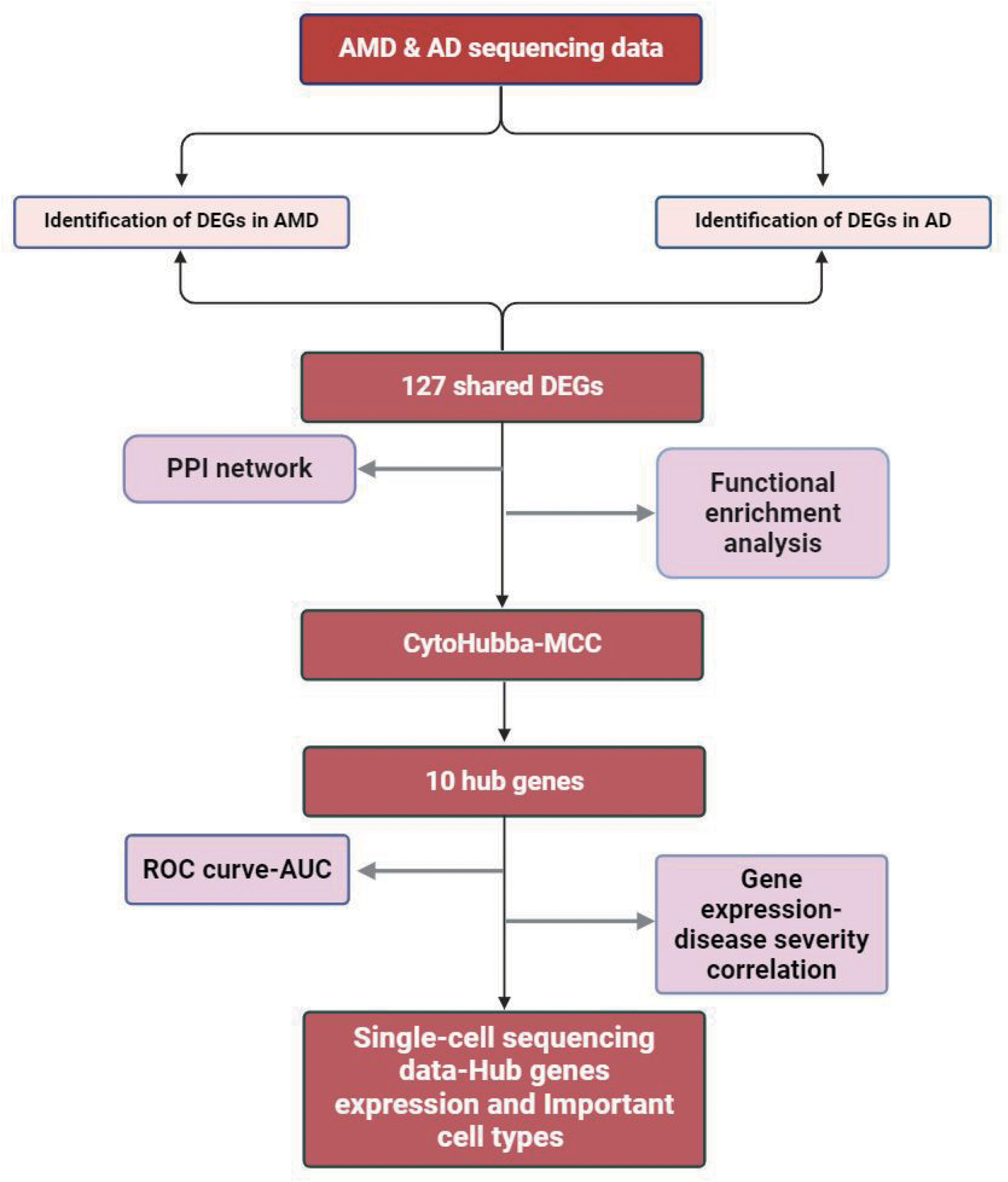
Study design and flowchart of this study. AMD: age-related macular degeneration; AD: Alzheimer's disease; DEGs: differentially expressed genes; PPI: protein-protein interaction; MCC: maximal clique centrality; ROC: receiver operating characteristic; AUC: area under curve.
Differentially expressed genes (DEGs) analysis
The DEGs between control and disease-affected samples were identified using the criteria: adjusted p < 0.05 and |log (FC)| > 1. Functional enrichment analysis for DEGs was conducted via the Metascape database (https://metascape.org/gp/index.html).
Construction of gene network and identification of hub genes
The protein-protein interaction (PPI) network of DEGs were constructed via the STRING database (http://www.string-db.org/). Cytoscape software (version 3.7.1) was used to perform topological analysis of this network and identified the top 10 hub genes by the maximal clique centrality (MCC) algorithm of the CytoHubba plug-in of Cytoscape.19,20
Evaluating the value of hub genes
To evaluate the correlation between hub genes expression and disease severity, we compared the expression of hub genes between samples at different disease progression stages. The receiver operating characteristic (ROC) curves of hub genes were established to examine their diagnostic and discriminative value, with the area under the ROC curve (AUC) being calculated.
Single-cell transcriptomic analysis
GSE137537 dataset reported the sequencing data of cells isolated from three AMD-affected macula samples obtained from postmortem human body. The gene expression matrix underwent quality control (QC) filtering, normalization, and integration before being used for downstream analysis. 21 Seurat R package (version 4.0) 22 in R software was used for subsequent analysis. Clustering for cells and dimensional reduction were performed. Subsequently, to perform annotation for cell clusters, the markers of clusters were identified and the cell markers provided by dataset authors 23 for annotation were used (Supplemental Table 1).
TREM2 is one of the important risk loci for AD. 24 Disease-associated microglia (DAMs), a special status of microglia, have been associated with neuroinflammatory and neurodegenerative conditions.25–30 Importantly, the upregulation of genes involved in phagocytic and lipid metabolism pathways, such as TREM2 and APOE, has been observed in AD. 31 Furthermore, previous studies have suggested the potential synergy between TREM2-APOE signaling and complete system in inducing synaptic pruning of microglia and neuron loss in AD.32,33 Therefore, investigating the expression of hub genes in TREM2WT microglia will help us understanding their role in the pathophysiology of AD. GSE158234 dataset reported the single-cell sequencing data of human microglia (TREM2 genotype: wild-type [WT] or knock-out [KO]), exposed to amyloid pathology in AD or not. The authors of this dataset transplanted microglia derived from human iPSCs into mice brain and harvested them for sequencing. They used microglia with two genotypes, including TREM2 WT and TREM2 KO. They also used two types of mice, including MITRG (MITRG mice lack NK cells, and T and B lymphocytes, leading to high immune deficiency and prevent the immunological rejection to engraftment. They have been widely used in biomedical researches) and 5x-MITRG (Mice obtained by crossing MITRG mice and 5xfAD mice. 5xfAD represents the mice developed as AD model with characteristic changes of AD. Microglia transplanted into the brain in this type of mice will go through the exposure to AD pathologies). After isolating human microglia from MITRG or 5x-MITRG mice brains, they performed single-cell RNA sequencing to visualize the gene expression of human microglia with different genotypes and cultured in different environment. Therefore, they reported the single-cell sequencing data of four types of human microglia, including TREM2WT-MITRG, TREM2KO- MITRG, TREM2WT-5XMITRG, and TREM2WT-5XMITRG microglia, which differ from genotype and exposure to AD-associated pathologies such as amyloid-β (Aβ).
Statistical analysis
R software (version 4.0.1) was used for statistical analysis. Values between two groups were compared using Student's t-test. All p values were two-tailed and p < 0.05 was considered statistically significant.
Results
Shared DEGs of AMD macula and AD hippocampus
In total, 2289 DEGs were identified between control and AMD maculas, including 1198 upregulated and 1091 downregulated ones (Figure 2(a)). In addition, a total of 429 upregulated with 658 downregulated DEGs were screened out in control and AD hippocampus (Figure 2(b)). There were 71 shared upregulated DEGs (Figure 2(c)) and 56 shared downregulated DEGs (Figure 2(d)).
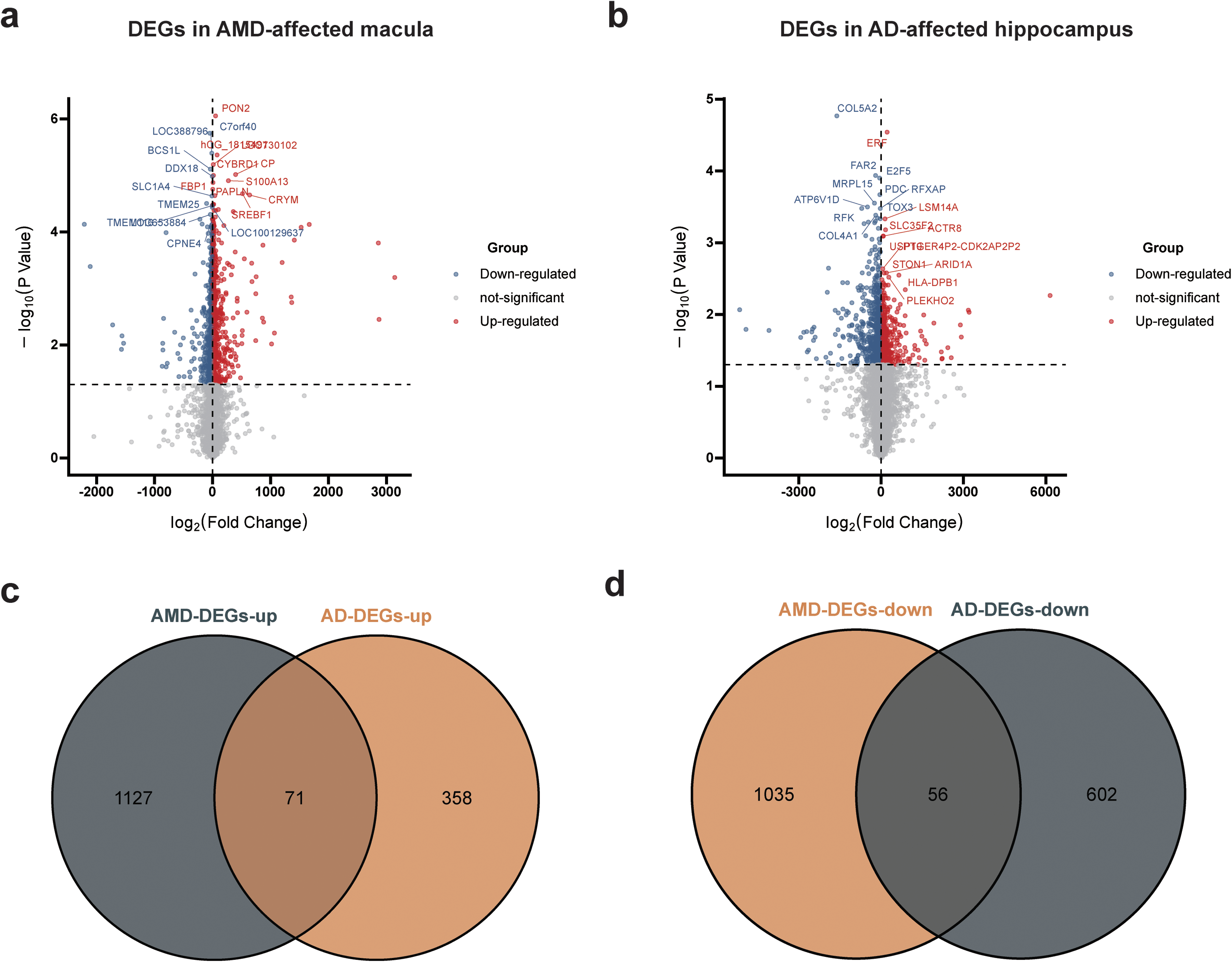
Identification of shared DEGs in AMD and AD. (a) The visualization of DEGs between control and AMD macula samples; (b) The visualization of DEGs between control and AD hippocampus; (c) Venn diagram showing the 71 shared upregulated DEGs in AMD and AD; (d) Venn diagram showing the 56 shared downregulated DEGs in AMD and AD. DEGs: differentially expressed genes; AMD: age-related macular degeneration; AD: Alzheimer's disease.
Functional enrichment analysis for DEGs
The shared upregulated DEGs (Supplemental Table 2) enriched in GO terms associated with cell morphologies and cellular responses to external stimuli (Figure 3(a)), and pathways associated with infection and inflammation (Figure 3(a)). The shared downregulated DEGs (Supplemental Table 2) enriched in GO terms and pathways involved energy production and mitochondrial function (Figure 3(b)).
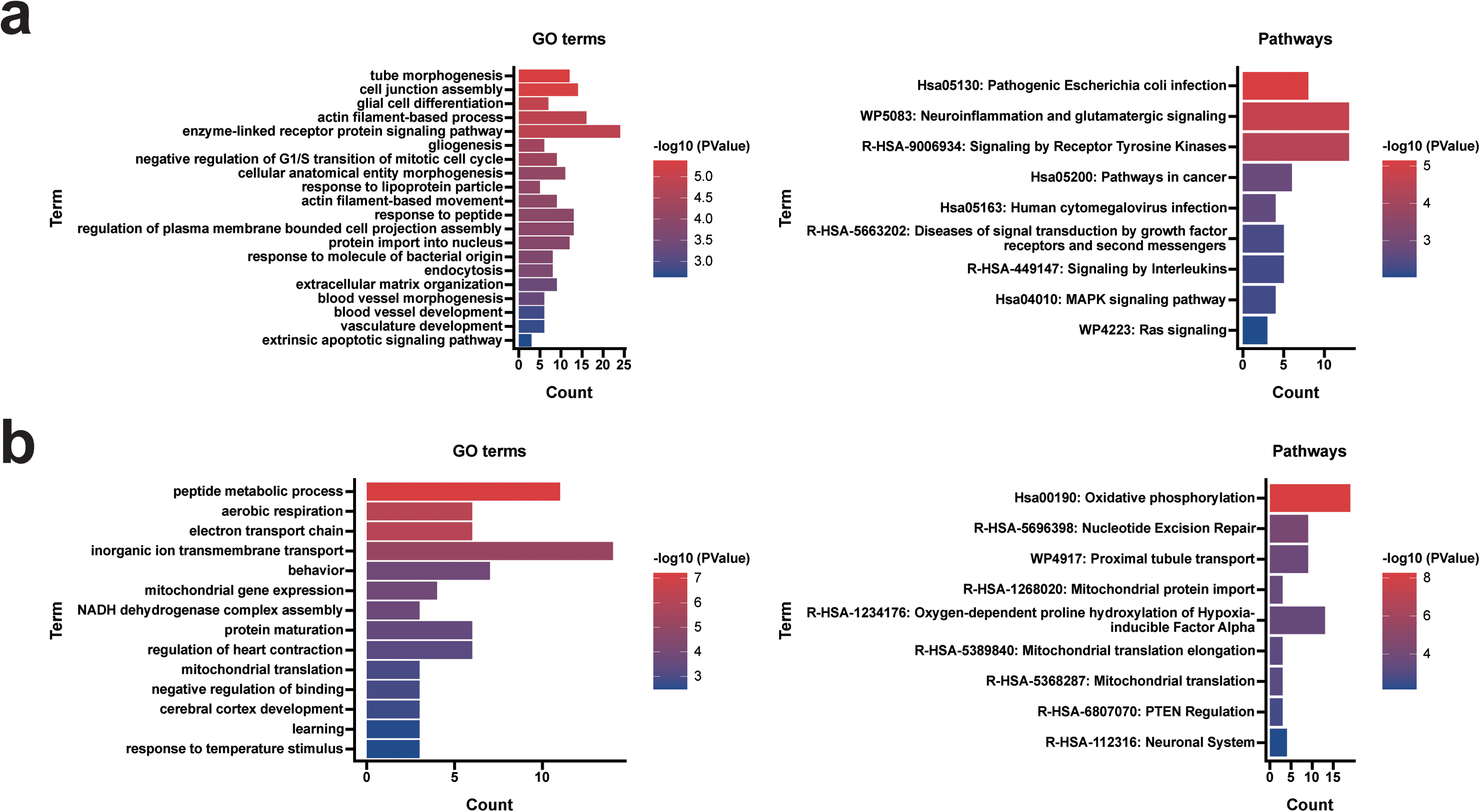
Functional enrichment analysis of the shared DEGs. (a) GO terms and pathways of 71 shared upregulated DEGs; (b) GO terms and pathways of 56 shared downregulated DEGs. DEGs: differentially expressed genes; GO: Gene Ontology.
PPI network and hub genes
The PPI network was established to show interactions between the 127 shared DEGs (Figure 4(a)). The MCC algorithm of the CytoHubba plugin in Cytoscape identified 10 hub genes in this network (Figure 4(b)), including: NDUFAB1, PSMD12, NDUFB5, NDUFB3, CYC1, CUL2, NFKBIA, COX7B, MRPS15, and TSFM (Table 1). Notably, only NFKBIA belonged to shared upregulated DEGs.

PPI network of shared DEGs and hub genes in this network. (a) PPI network of the 127 shared DEGs constructed via STRING; (b) Top 10 hub genes identified by Cytoscape software. PPI: protein-protein interaction; DEGs: differentially expressed genes.
Top 10 hub genes in DEGs network ranked by MCC method.
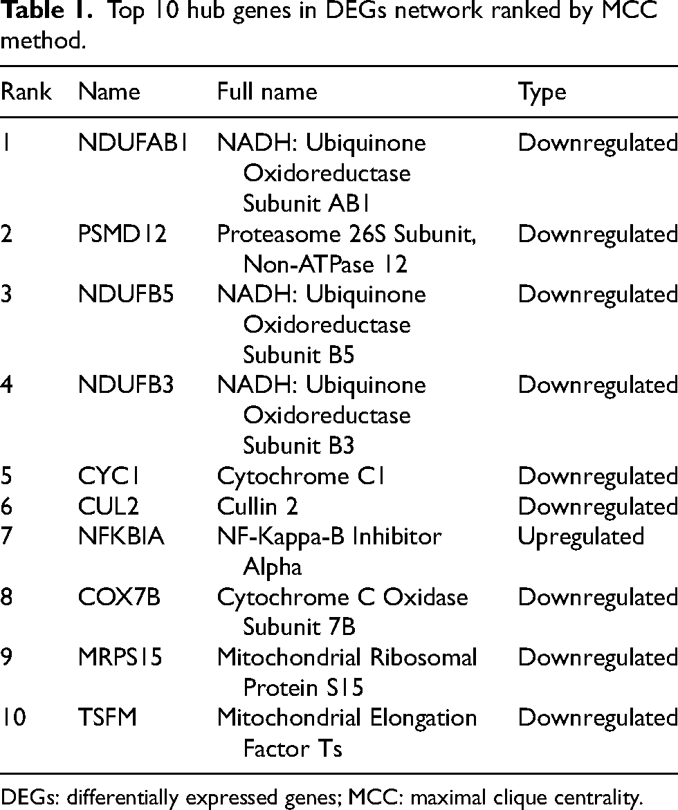
DEGs: differentially expressed genes; MCC: maximal clique centrality.
NDUFAB1 (NADH Ubiquinone Oxidoreductase Subunit AB1), NDUFB5 (NADH Ubiquinone Oxidoreductase Subunit B5), and NDUFB3 (NADH Ubiquinone Oxidoreductase Subunit B3) are the subunits of the NADH ubiquinone oxidoreductase (mitochondrial respiratory chain complex I). In addition, MRPS15 (Mitochondrial Ribosomal Protein S15) and TSFM (Mitochondrial Elongation Factor Ts) are also associated with mitochondrial functions. CYC1 (Cytochrome C1) is a subunit of the cytochrome bc1 complex, which plays an important role in the mitochondrial respiratory chain by transferring electrons from the iron-sulfur protein to cytochrome c. COX7B (Cytochrome C Oxidase Subunit 7B) is subunit of cytochrome c oxidase (COX), which is the terminal component of the mitochondrial respiratory chain, catalyzes the electron transfer from reduced cytochrome c to oxygen. The downregulation of these genes indicated the damaged mitochondrial functions in both AMD- and AD-affected tissues, which is consistent with previous studies suggesting the involvement of mitochondrial dysfunction in NDDs.34,35 Notably, recent data demonstrate that mitochondrial functions such as the ubiquitination of mitochondrial proteins and mitochondrial permeabilization is essential for the activation of NF-kB signaling. 36 Therefore, there might be a synergy between the downregulation of these genes and the upregulation of NFKBIA in inhibiting NF-kB pathway and contributing neuronal death in AMD and AD. PSMD12 (Proteasome 26S Subunit) is a component of the 26S proteasome, a multiprotein complex involved in the degradation of ubiquitinated proteins. CUL2 (Cullin 2) is also involved in ubiquitin-dependent protein catabolic process. These two genes play a key role in the maintenance of protein homeostasis and is associated with multiple cellular processes, including cell cycle progression and apoptosis. Therefore, the downregulation of these two genes could impair cellular functions and cell survival. Additionally, NF-kB pathway plays an important role in cell survival and the inhibition of NF-kB pathway by NFKBIA can also negatively impact cell survival. Collectively, through different cellular and molecular mechanisms, these hub genes may all significantly contribute to the pathophysiology of AMD and AD.
Value of hub genes
Hub genes expression in macula samples with different subtypes and stages of AMD were compared and the results showed that NFKBIA expression was significantly upregulated with the advance of AMD, especially higher in wet AMD (p < 0.05, Figure 5(a)). Similarly, NFKBIA expression was correlated with the advance of AD (Figure 5(b)). Subsequently, the diagnostic and discriminative value of the hub genes was examined, and the ROC curves showed that NFKBIA had the highest value of area under curve (AUC) in both AMD (AUC = 0.798, Figure 6(a)) and AD (AUC = 0.864, Figure 6(b)).
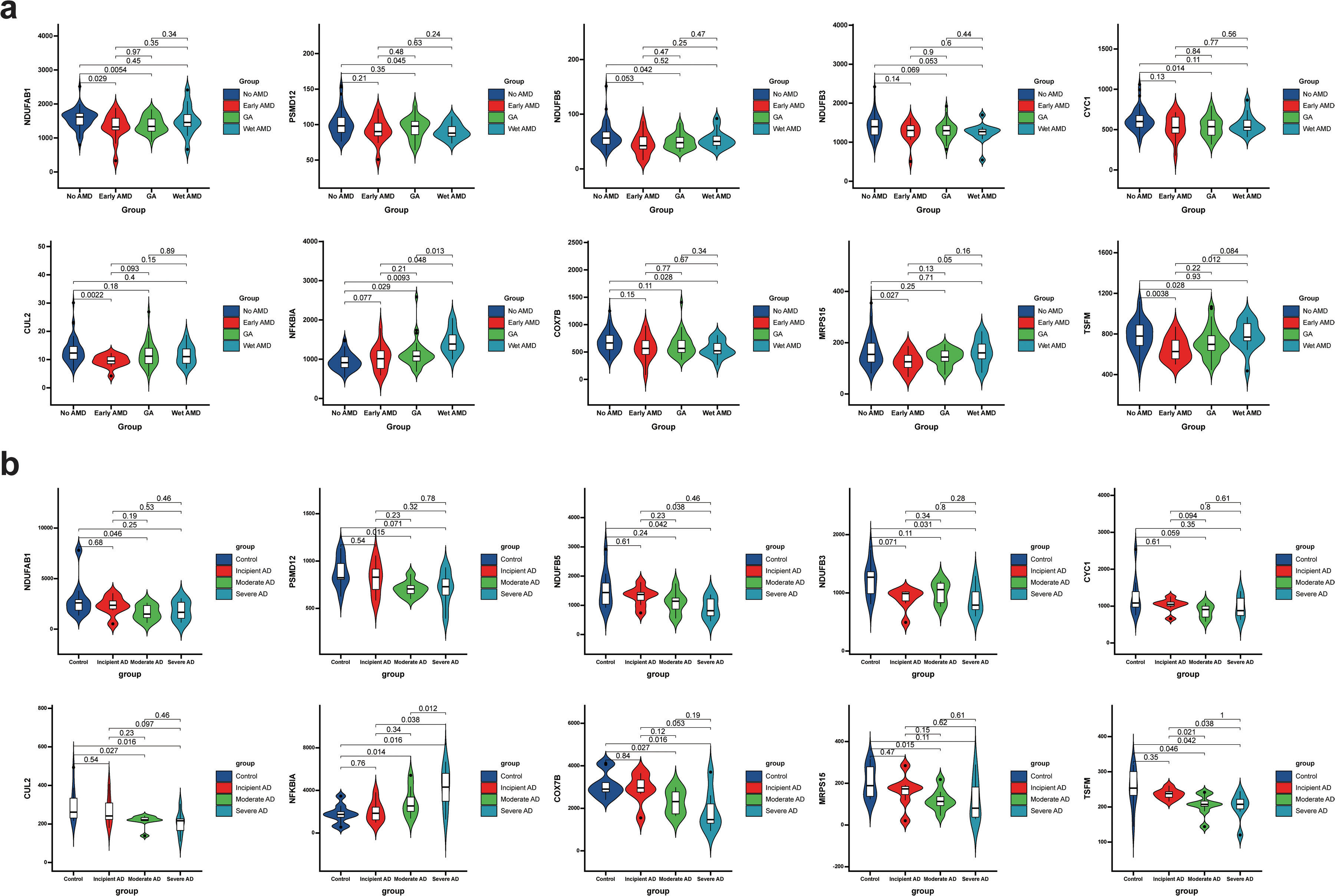
Gene expression-disease severity correlation analysis for hub genes. (a) The comparison of hub genes expression in macula samples with different AMD stages; (b) The comparison of hub genes expression in hippocampus samples with different AD stages. AMD: age-related macular degeneration; AD: Alzheimer's disease.

The diagnostic and discriminative value of the hub genes. (a) The ROC curve and AUC value of hub genes in AMD; (b) The ROC curve and AUC value of hub genes in AD. ROC: receiver operating characteristic; AUC: area under curve; AMD: age-related macular degeneration; AD: Alzheimer's disease.
Single-cell transcriptomic analysis
Nine types of cells were identified in AMD-affected macula samples: Amacrine cells; Bipolar cells; Cones; Endothelial cells; Horizontal cell; Macroglia (Müller glia and Astrocyte); Microglia; Retinal ganglion cells (RGCs); and Rods (Figure 7(a)). Single-cell RNA sequencing data showed that NFKBIA was the most predominantly expressed hub genes and highly expressed in microglia from AMD-affected macula samples (Figure 7(b)). Moreover, the heatmap showed that the expression of NFKBIA was higher in TREM2WT microglia than that in TREM2KO ones, especially when exposed to AD pathologies (Figure 8), which indicated that NFKBIA was associated with the pathogenesis of AD.
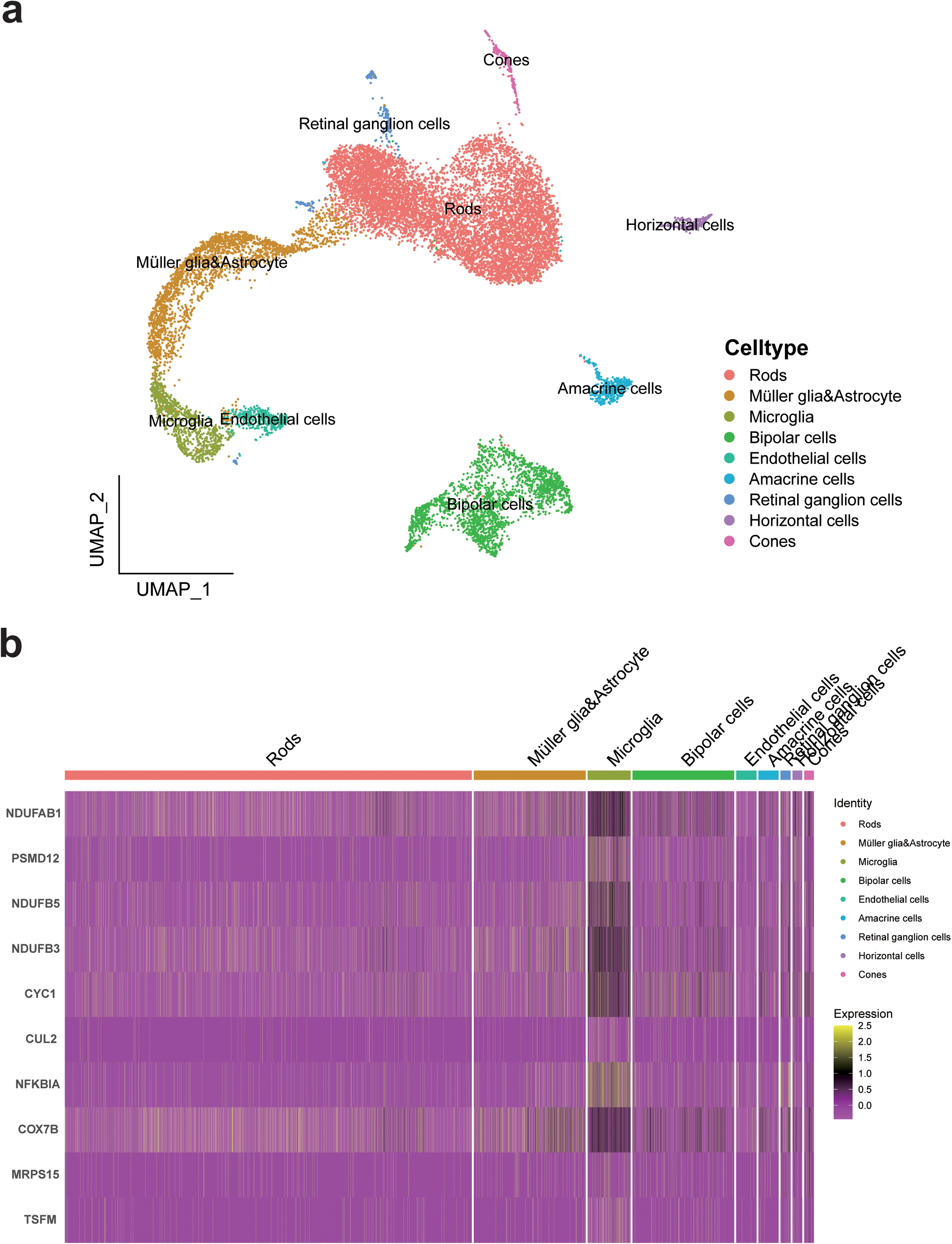
Cell atlas and gene expression analysis of hub genes in AMD macula samples. (a) The clustering and cell annotation of the single-cell transcriptomic data of macula samples with AMD; (b) Heatmap of hub genes expression in different retinal cell types. AMD: age-related macular degeneration.
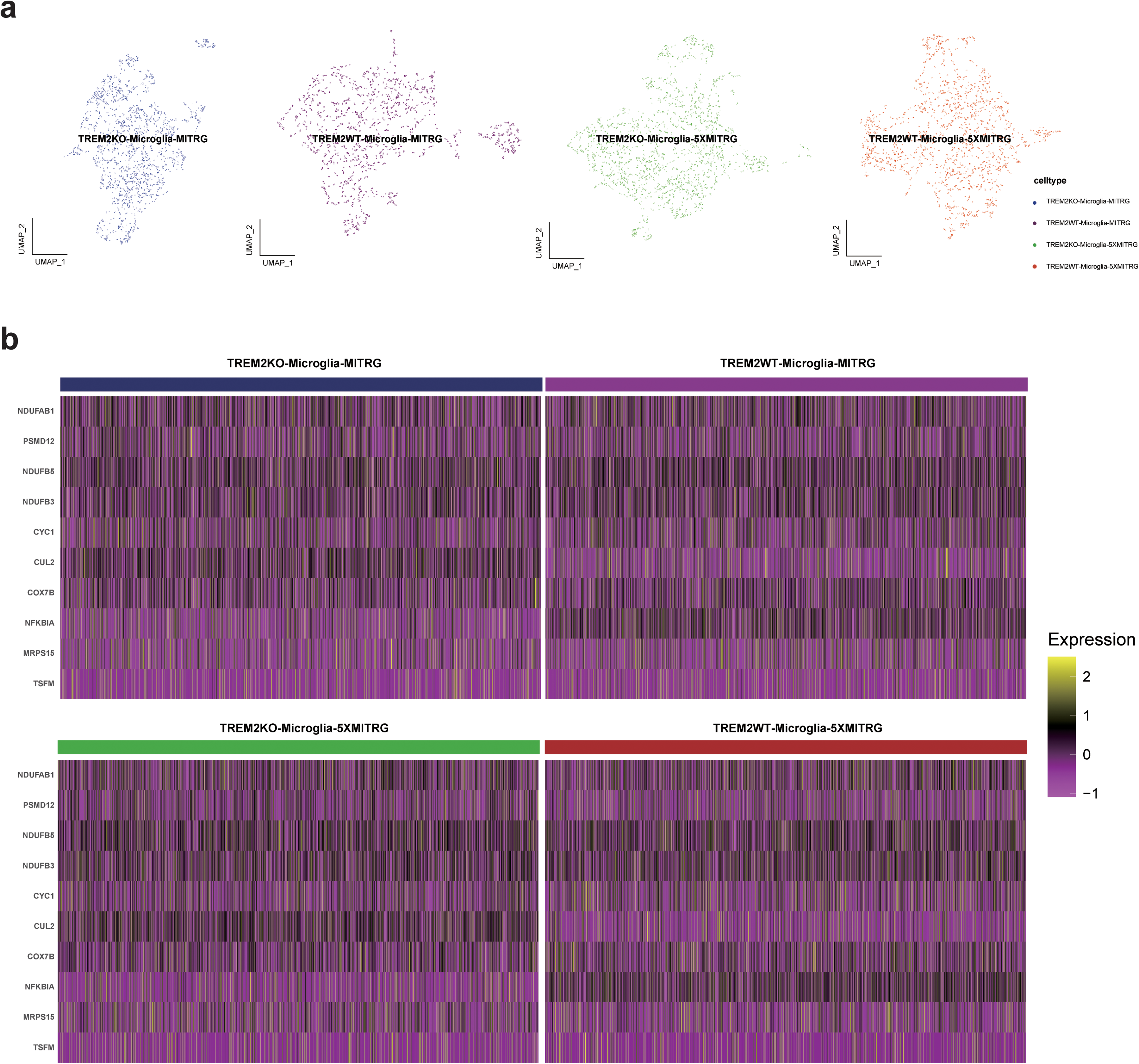
Gene expression analysis of hub genes in AD-associated microglia. (a) The clustering and visualization of AD-associated microglia; (b) Heatmap of hub genes expression in AD-associated microglia with different genotype and from different conditions. AD: Alzheimer's disease.
Discussion
Worldwide, age-related conditions have become major public concern with the aging of population. Among them, AD and AMD place considerable health burden on affected individuals and significant economic burden on society. Considering the potential health burden related to AD and AMD, the preventive and therapeutic strategies of these two diseases present critical unmet medical needs. The idea that they share common pathways such as mitochondrial dysfunction, dysregulation of proteome homeostasis, and inflammation in their pathogenesis has been proposed.14,37,38 Investigation in this area may pave the way for novel and effective therapeutic strategies for these two serious diseases. In this study, we applied single-cell and bulk transcriptomic analysis to explore the shared cellular and molecular mechanisms underlying the pathogenesis of AD and AMD. Importantly, the findings suggested that NFKBIA was a shared disease progression-related gene and NFKBIA + microglia might be important contributors to the progression of both AD and AMD. The findings may help us further understanding the etiology and pathophysiology of these two NDDs.
The important involvement of microglia in the pathogenesis of AMD and AD has been widely recognized. Microglial activation and migration are considered hallmarks of AMD pathogenesis.39–41 Activated microglia have two primary phenotypes, 42 among them, M1-phenotype microglia are neurotoxic. 43 M1 microglia promote the synthesis of inflammatory mediators such as tumor necrosis factor-alpha (TNF-α) and interleukin-1β (IL-1β), which can induce phagocytosis, chronic neuroinflammation, and neurodegeneration. 44 Importantly, phagocytotic effect of microglia has been implicated in the pathogenesis of NDDs. In AD, the phagocytic function of microglia plays a dual role: promotes the clearance of toxic Aβ peptides 45 and induces synaptic loss and neuron death. 46 Notably, in AMD, the loss of photoreceptors and synapse elimination has been observed. 47 In AMD, the injury of blood-retinal barrier allows leakage of complement components into the retina from the choriocapillaris. 48 Local complement activation leads to the recruitment of microglia into the lesion, which prune complement-coated synapses away from neurons, depriving neurons of trophic support, and cause neuroinflammation that adds to neuronal damage and loss. 49 DAMs, a special status of microglia, have been associated with neuroinflammatory and neurodegenerative conditions.25–30 DAMs show upregulation of genes involved in phagocytic and lipid metabolism pathways, such as APOE and TREM2, which are well-known risk factors for AD. 31 Furthermore, previous studies have suggested the potential synergy between TREM2-APOE signaling and complete system in inducing synaptic pruning of microglia and neuron loss in AD.32,33 However,this study first identified the potential role of microglia-derived NFKBIA in the pathogenesis of both AMD and AD.
The NF-kB transcription factor complex includes five members, including RelA, RelB, c-Rel, NF-κB1, and NF-κB2. These factors play an important role in the immune response and cell survival, mainly by targeting the regulation of expression of proinflammatory genes, antiapoptotic proteins, and angiogenesis regulators. NFKBIA (NF-kB Inhibitor Alpha) gene encodes a member of the NF-kB inhibitor family, with the biological power to inhibit NF-kB signaling. It was reported that the accumulation of NFKBIA protein downregulated NF-kB-mediated antiapoptotic pathway, thereby promote colonic cell death and lead to the onset of severe colitis. 16 In addition, the association between NFKBIA and inflammation has been noticed. For instance, NFKBIA was identified as a key gene contributing inflammatory response in allergic rhinitis (AR) and significantly associated with the progression of AR. 17 Moreover, a previous study found that the expression of NFKBIA was upregulated in microglia and related to IL-1 inflammatory pathway in traumatic brain injury. 50 Collectively, NFKBIA derived from microglia might contribute to the pathogenesis of AMD and AD through two potential pathways, including: 1) trigger inflammation; and 2) inhibit NF-kB pathways, thereby lowering the capacity of neurons to survive and leading to ultimate neuronal death (Figure 9).

The diagram summarizing the potential role of NFKBIA + microglia in the pathophysiology of AD and AMD. AD: Alzheimer's disease; AMD: age-related macular degeneration.
Additionally, the results showed that shared upregulated genes in AD- and AMD-affected tissues enriched in pathways such as response to molecule of bacterial origin, pathogenic E. coli infection, and human cytomegalovirus infection. These functional enrichment results indicated that bacterial or viral infection might play a role in the development of AMD and AD, which has been mentioned in previous studies. 51 Moreover, among the identified top 10 hub genes, there are 5 genes associated with mitochondrial functions, including NDUFAB1, NDUFB5, NDUFB3, MRPS15, and TSFM. Notably, they were all downregulated in AD- and AMD-affected tissue. The role of mitochondrial dysfunction in the development of NDDs has been reported.34,35 Therefore, this study highlighted the value of further investigation of mitochondrial functions in AMD and AD.
There were some limitations. First, this study only analyzed publicly available transcriptomic data without validating the results in local datasets, which limited the applicability of the findings in this study. Second, no clinical data was presented in this study, which limited the depth of the findings.
Conclusion
In conclusion, by single-cell and bulk tissue transcriptomic analysis, the findings in this study suggested that NFKBIA derived from microglia might be an important contributor to the pathogenesis of both AD and AMD. Targeting NFKBIA or NFKBIA + microglia might be a promising therapeutic strategy for the treatment of AD and AMD.
Supplemental Material
sj-xlsx-1-alz-10.1177_13872877251326267 - Supplemental material for Identifying microglia-derived NFKBIA as a potential contributor to the pathogenesis of Alzheimer’s disease and age-related macular degeneration
Supplemental material, sj-xlsx-1-alz-10.1177_13872877251326267 for Identifying microglia-derived NFKBIA as a potential contributor to the pathogenesis of Alzheimer’s disease and age-related macular degeneration by Shizhen Lei and Yani Liu in Journal of Alzheimer's Disease
Footnotes
Acknowledgments
The authors have no acknowledgments to report.

Ethical considerations
This study used only publicly available datasets and the Research Ethics Committee of Wuhan No. 1 Hospital has confirmed that no ethical approval is required. Our research adhered to the tenets of the Declaration of Helsinki.
Consent to participate
Not applicable.
Consent for publication
Not applicable.
Author contributions
Shizhen Lei (Conceptualization; Data curation; Formal analysis; Funding acquisition; Investigation; Methodology; Project administration; Resources; Software; Supervision; Validation; Visualization; Writing – original draft; Writing – review & editing); Yani Liu (Formal analysis; Investigation; Visualization; Writing – original draft).
Funding
The authors disclosed receipt of the following financial support for the research, authorship, and/or publication of this article: This study was supported by The Funding for Scientific Research Projects from Wuhan Municipal Health Commission (grant number: WX23Z33).
Declaration of conflicting interests
The authors declared no potential conflicts of interest with respect to the research, authorship, and/or publication of this article.
Data availability
All data generated during the current study are available from the corresponding author upon reasonable request.
Supplemental material
Supplemental material for this article is available online.
References
Supplementary Material
Please find the following supplemental material available below.
For Open Access articles published under a Creative Commons License, all supplemental material carries the same license as the article it is associated with.
For non-Open Access articles published, all supplemental material carries a non-exclusive license, and permission requests for re-use of supplemental material or any part of supplemental material shall be sent directly to the copyright owner as specified in the copyright notice associated with the article.
