



Abstract
Background
Deficits in Reelin expression play a significant role in the pathogenesis of various neurological disorders, including schizophrenia and Alzheimer's disease (AD). Notably, Reelin-expressing neurons of the entorhinal cortex layer II are among the first to be affected in AD.
Objective
Strategies aimed at correcting deficits in Reelin might provide a novel therapeutic approach for AD.
Methods
Here, we examined the effects of the whey protein supplement and glutathione (GSH) precursor, Immunocal®, on Reelin expression both in vitro in hippocampal-entorhinal cortex slices from rat brain and in vivo in the hAβPPSweInd (J20) mouse model of AD.
Results
Incubation of brain slices with Immunocal® increased Reelin expression at the mRNA and protein levels. Oral treatment with Immunocal®, given ad libitum in drinking water beginning at 3 months of age, corrected a deficit in cortical GSH levels observed in untreated mice and preserved Reelin expression in the hippocampal-entorhinal cortex sub-region of 5-month-old J20 mice. We also assessed the long-term effects of Immunocal® by treating J20 mice from 3 months old to 12 months old. Long-term Immunocal® treatment preserved brain GSH and rescued Reelin mRNA and protein expression, while significantly reducing amyloid plaque formation in the entorhinal cortex and hippocampus of AD mice.
Conclusions
These findings suggest that Immunocal® promotes Reelin expression in vitro and sustains brain GSH and Reelin expression while diminishing amyloid plaque load in the entorhinal cortex and hippocampus of J20 mice. Thus, Immunocal® offers a promising therapeutic approach to enhance Reelin expression and curtail amyloid deposition in AD.
Introduction
Alzheimer's disease (AD) is the leading cause of dementia and cognitive decline with approximately 6.7 million patients currently diagnosed in the United States and over 500,000 new cases each year. Only about 5% of AD cases are familial in origin with ageing being the most significant risk factor for developing sporadic AD. Given the demographic shift to advancing age in our population, there is predicted to be a very significant increase in the number of people diagnosed with AD in the next several decades. By 2060, the number of people in the United States age 65 and older diagnosed with AD is predicted to be approximately 13.8 million (Alzheimer's Association, 2023 Alzheimer's Disease Facts and Figures). 1 Research focused on identifying new molecular targets that underlie the pathogenesis of AD is critically needed to develop novel therapeutic strategies for this devastating disorder.
Reelin is a large glycoprotein secreted by specific cells within the central nervous system (CNS) that plays a key role in patterning and layering of the cerebral cortex and other regions of the brain during development. In adults, Reelin plays a central role in processes that influence synapse formation and neuronal plasticity required for learning and memory, such as the regulation of dendritic spine architecture and the maintenance of long-term potentiation.2–6 In particular, Reelin-expressing neurons of the entorhinal cortex project to the hippocampus and are involved in declarative memory formation and consolidation.7–10
Entorhinal cortex layer II neurons provide the principal excitatory input to the dentate gyrus of the hippocampus and are also one of the first neuronal populations to show significant pathology in AD, resulting in a severe loss of these synaptic contacts.11–13 These Reelin-expressing cells are significantly reduced in the brains of human AβPP transgenic mice expressing the Swedish and Indiana mutant form of the hAβPP gene (J20 strain). In accordance with the loss of these Reelin-expressing entorhinal cortex layer II neurons, Reelin levels in the hippocampus of J20 mice are also significantly reduced, compared to non-transgenic controls. 14 Similar loss of Reelin-expressing entorhinal cortex layer II neurons is also observed in the brains of patients with AD.14,15 Finally, in a transgenic rat model of AD (McGill-R-Thy1-AβPP strain), Reelin-expressing neurons of the entorhinal cortex layer II were found to selectively express increased levels of soluble intracellular Aβ early in disease, prior to the deposition of amyloid plaques. 16 Collectively, these studies suggest that Reelin-expressing neurons of entorhinal cortex layer II play a central role in the early pathogenic changes in AD and the loss of these Reelin-expressing cells and their synaptic projections to the hippocampus are early markers of disease. 17 Thus, deficits in Reelin signaling to the hippocampus likely underlie some of the cognitive deficits observed in patients with AD.18,19
Several studies suggest that modulating Reelin expression has marked effects on AD progression. For example, further reducing Reelin in J20 AD mice by crossing them with heterozygous reeler mice accelerates amyloid plaque formation and tau pathology. 20 On the other hand, Reelin overexpression in J20 AD model mice significantly delays Aβ fibril formation and rescues cognitive deficits in these mice. 21 Thus, J20 mice are an established model of AD and the disease course of these mice is significantly impacted by alterations in Reelin expression, making this an excellent model system to investigate the effects of modulators of Reelin expression and/or signaling on AD progression.
The whey protein supplement, Immunocal®, is a rich source of the glutathione precursor, cysteine, and is known to boost antioxidant levels both in vitro and in vivo.22,23 In addition, in a schizophrenia mouse model characterized by low Reelin levels in the prefrontal cortex, Immunocal® was shown to elevate Reelin in the brain. 24 Given that a similar loss of Reelin has been observed in entorhinal cortex layer II neurons in the brains of AD patients and in animal models of AD, determining whether Immunocal® preserves Reelin expression in the entorhinal cortex-hippocampus of J20 mice and mitigates amyloid plaque deposition in these animals, will provide key preclinical data to support future clinical trials of Immunocal® in patients with AD.
Methods
Reagents
Immunocal® was provided by Immunotec, Inc. (Vaudreuil-Dorion, QC, CA). Gey's balanced salt solution and ProLong Gold were purchased from Invitrogen (Carlsbad, CA, USA). Primary antibodies for Reelin (ab78540), NeuN (ab104225), DRAQ7™, and β-actin (ab 115777) were purchased from Abcam (Cambridge, UK). Secondary antibodies for immunofluorescence were purchased from Jackson Immunoresearch (Westgrove, PA, USA). Horseradish peroxidase (HRP)-conjugated secondary antibodies for western blotting and all probes and reagents for Quantitative real-time polymerase chain reaction (qPCR) were purchased from Bio-Rad (Hercules, CA, USA). All probes (RNAscope® Probe Mm-Reln-C3; catalogue no. 319361-C3) and reagents (RNAscope® fluorescent multiplex kit; catalogue no. 320850) for in situ hybridization were purchased from Advanced Cell Diagnostics (Newark, CA, USA). Amylo-Glo was purchased from Biosensis (Thebarton, SA, AU).
Experimental models
Rat brain slice cultures. To investigate the effects of Immunocal® on Reelin expression in vitro, we utilized organotypic hippocampal-entorhinal cortex slices prepared from postnatal day 25 Sprague-Dawley male rats. Animals were euthanized by deep isoflurane inhalation to prevent pain or discomfort, followed by decapitation, prior to removal of brain tissue. Brain slices were sliced using a vibrating microtome essentially as described by Leutgeb et al. 25 Animal studies were conducted in accordance with a protocol approved by the University of Denver Institutional Animal Care and Use Committee (Approval #983340-4).
J20 AD mouse model studies. To investigate the effects of Immunocal® on Reelin expression in vivo, we utilized the hAβPP(Swe/Ind) mutant transgenic mouse model of AD (J20 strain). This mouse model was obtained from Jackson Laboratories (Bar Harbor, ME, USA; strain B6.Cg-Tg(PDGFB-AβPPSwInd) 20Lms/2Mmjax) and displays significant brain pathology, amyloid plaques, and cognitive deficits that recapitulates multiple aspects of AD in humans.26–28 According to previous reports, these transgenic mice typically show significant cognitive deficits, diminished numbers of Reelin-positive entorhinal cortex (EC) layer II neurons, and decreased Reelin expression in the hippocampus by approximately 4–5 months of age. As these mice age, amyloid plaques (a hallmark of AD pathology) are visible in the brain, particularly the hippocampus, by around 12 months of age. All animal studies were conducted in accordance with a protocol approved by the University of Denver Institutional Animal Care and Use Committee (Approval #927465-8).
Immunocal® treatment of brain slices
Approximately 400 micron hippocampal-entorhinal cortex slices were cultured in a humidified incubator in tissue culture medium with 5% CO2 at 37°C. After equilibration overnight, slices were subsequently treated with either culture medium alone or containing Immunocal® (3.3% w/v) for 24 h, followed by subsequent experimental analyses. The dose of Immunocal® used was identical to that used in our prior in vitro neuroprotection studies in neuronal culture systems. 23
Immunoprecipitation
Following incubation in either culture medium alone or containing recombinant Reelin, brain slices were lysed using a Dounce homogenizer in standard RIPA lysis buffer supplemented with aprotinin and leupeptin. Lysates were cleared by centrifugation, protein measured using a BCA protein assay, and immunoprecipitated with an antibody to Dab-1 followed by protein A/G-sepharose beads. Immune complexes were resolved by SDS-PAGE, transferred to polyvinylidene difluoride (PVDF) membranes and immunoblotted for phosphotyrosine. Membranes were then stripped and reprobed for Dab-1.
Quantitative real-time polymerase chain reaction (qPCR)
Following incubation in either culture medium alone or containing Immunocal®, recombinant Reelin, or the combination, brain slices were lysed using a Dounce homogenizer and RNA was isolated using a miRNeasy kit purchased from Qiagen (Valencia, CA, USA). RNA was analyzed by qPCR on an iQ5 Real Time PCR System (Bio-Rad, Hercules, CA, USA) using an iTAq Universal SYBR Green One-step kit (BioRad, Hercules, CA, USA). Primers for rat Reelin (PrimePCR SYBR Green Assay, Reln, rat, qRnoCED0008838) and rat GAPDH (PrimePCR SYBR Green Assay, Gapdh, rat, qRnoCID0057018) were also purchased from Biorad. PCR was performed using the following conditions: 95 °C for 5 min followed by 40 cycles of 95 °C for 15 s, 60 °C for 30 s, and 68 °C for 1 min. Threshold cycle (Ct) values for each sample were selected by the iQ5 software. The analysis of differential fold change was done using the Livak (ΔΔCt) method. 29
Immunofluorescence staining
Approximately 200-micron thick brain slices were harvested in ice cold Gey's balanced salt solution such that the hippocampal entorhinal cortex region was preserved, then added to a 6-well plate (1 slice/well) containing ice cold 4% paraformaldehyde for 90 min. Fixative was removed from the wells and the slices were washed three times for 5 min in PBS-T. After the final wash, PBS-T was removed and 1 mL of Blocking Solution (5% BSA, 1× PBS, 0.05% Tween-20, pH 7.4) was added to the wells and incubated at 25 °C for 90 min. Blocking solution was replaced with primary antibody in blocking solution at the necessary dilutions and incubated overnight at 4 °C. The following day, primary antibody was removed, and the tissue was washed 3 times for 5 min in PBS-T. Slices were then incubated with the secondary antibody diluted in blocking solution at the indicated concentrations for 90 min at 25 °C. Then, secondary antibody solution was removed and immediately replaced with 1 mL of DRAQ7™ diluted in PBS-T and incubated for 20 min at 25 °C. The tissue was carefully transferred to a SuperFrost Plus Slide and excess PBS-T was removed using a Kimwipe. Prolong Gold was added onto the tissue and a coverslip was placed over it.
Immunocal® treatment of mice
For the in vivo study, we used three groups of mice: hAβPP(Swe/Ind) mutant hemizygous mice (J20 strain) treated with Immunocal® (3.3% w/v in drinking water ad libitum), as previously described by Ross et al., 30 untreated hemizygous J20 mice, and untreated non-transgenic (non-carrier) control mice. For 5-month studies, Immunocal® treatment was initiated at 3 months old and continued until the mice were 5 months old. For 12-month studies, Immunocal® treatment was initiated at 3 months old and continued until the mice were 12 months old.
Tissue processing
Prior to removal of brain tissue, animals were euthanized by deep isoflurane inhalation, followed by decapitation. Mice were euthanized and biochemical and immunohistochemical analysis was conducted on brain tissue to evaluate Reelin-expressing neurons in the entorhinal cortex and Reelin staining in the dentate gyrus, CA1, and CA3 of the hippocampus. For western blotting, brains were micro-dissected to obtain tissue samples enriched for the hippocampal-entorhinal cortex architecture. For immunofluorescence staining of brain tissue, slices were prepared using a vibrating microtome essentially as described by Leutgeb et al. before fixing with PFA for staining. 25
Cortical tissue was obtained from mice and immediately frozen in liquid nitrogen. For HPLC-ECD analysis, 2.5 M perchloric acid was added and the brains were roughly chopped using pointed surgical scissors. Samples were then sonicated 3 times for 15 s intervals. Samples were then centrifuged for 5 min at 13,000 rpm and the supernatant was removed. An aliquot of the original brain tissue was used for a BCA protein assay. The supernatant was neutralized with 500 µL of 4 M KOH and vortexed thoroughly. Samples were then centrifuged for 15 min at 13,000 rpm, and stored at −80 °C until separation and analysis by HPLC-ECD.
Analysis of brain GSH and GSSG by HPLC with electrochemical detection (HPLC-ECD)
Glutathione (GSH) in samples and known standards were separated by reversed-phase HPLC on a C18 bonded silica column at 35 °C (5 µm, 4.6 mmID × 25 cm) from Tosoh Bioscience. (Grove City, OH, USA). Analytes were detected using a CoulArray® detector (model 5600, ESA) on three coulometric array cells in series; electrochemical detectors were set between 0 and 900 mV at increments of 75 mV. Concentrations were determined with a standard curve of each identified analyte. Mobile phase consisted of 50 mM lithium acetate and 1% acetonitrile in water, pH 3.8. The flow rate was set to 0.4 mL/min for all samples. CoulArray® software was used for baseline correction and peak analysis.
Western blotting
Mouse brains were rapidly dissected and one hemisphere was placed into ice cold Gey's balanced salt solution for slicing while the other half was flash frozen in liquid nitrogen and stored at −80 °C. Briefly, the flash frozen halves were thawed, then brain slices were lysed using a Dounce homogenizer and lysis buffer supplemented with aprotinin and leupeptin. The samples were diluted 1:100 for a BCA protein assay. Western blotting was done to immunochemically detect proteins immobilized on PVDF membranes. Protein samples (50 µg/lane) were resolved by SDS-PAGE and proteins were then transferred to PVDF membranes. Nonspecific binding sites were blocked using 1% BSA in PBS (pH 7.4) containing 0.1% Tween-20 (PBS-T) for 1 h at 25 °C. The blocking buffer was drained, and the membrane was allowed to incubate in primary antibody diluted in blocking buffer per manufacturer's recommendations overnight at 4 °C. The membrane was washed 3 times for 15 min in PBS-T and was then incubated with the secondary antibody (1:5000) for 1.5 h at 25 °C. The secondary was then removed, and the membrane was washed again in PBS-T, 3 times for 15 min. Immunoreactive proteins were detected using enhanced chemiluminescence (GE Healthcare; Pittsburgh, PA, USA) and films were developed using a ChemiDoc developer (BioRad, Hercules, CA, USA). Re-probing of blots was performed by stripping in 0.1 M Tris-HCl (pH 8.0), 2% SDS, and 100 mM β-mercaptoethanol for 30 min at 52 °C. The blots were rinsed twice in PBS-T and processed as above with a different primary antibody. Relative intensities of the protein bands were quantified by scanning densitometry using ImageJ.
In situ mRNA hybridization
Samples were processed according to ACD RNAscope® Fluorescent Multiplex Assay manual (Newark, CA, US). Using a vibratome (Leica), brain tissue was freshly sliced in ice cold Gey's balanced salt solution at approximately 500 microns thickness such that the hippocampal entorhinal cortex region was preserved, then the slices were embedded in OCT and frozen in liquid nitrogen. Brains were then sliced at 30 microns thickness with a cryostat (Leica CM 1950) and adhered onto SuperFrost Plus Slides and stored at −80 °C until use. Sections were fixed and permeabilized using 4% paraformaldehyde and incubated for 15 min. Slides were rinsed twice with 1X PBS (pH 7.4) buffer. Tissues were dehydrated consecutively in 50%, 70% and 100% ethanol for 2 min each. A barrier was created around the tissue using a hydrophobic pen. Tissues were then incubated with protease inhibitor for 30 min at 25 °C in HybEZ™ Humidity Control Tray. About 4–6 drops of Reelin or NeuN (positive control) probe was added to tissues. The slides were inserted in a slide holder and placed in HybEZ™ Humidity Control Tray and incubated for 2 h at 40 °C in HybEZ™ Oven. After washing in 1X PBS, samples were hybridized by adding 4–6 drops with Amp1, Amp2 and Amp3 for 30 min, 15 min, and 30 min respectively at 40 °C. Samples were incubated with FITC dye for 15 min at 25 °C and counter stain DRAQ7 was added for 15 min.
Quantification of Reelin staining and amyloid plaques
Images were collected using an Olympus FLUOVIEW FV3000 confocal laser scanning microscope using an Olympus 20X PLN objective. Confocal z-series were captured at equal exposure times across all samples (n = 4 to 7 in each group for protein staining and n = 7 to 9 in each group for in situ hybridization). Each region (EC, dentate gyrus (DG), CA3, CA1) was captured in triplicate. Total fluorescent intensity was analyzed in Adobe Photoshop for protein staining and in situ hybridization. Any adjustments made in the analysis step were equally applied to all groups.
For amyloid plaque quantification by image J software, the color images were converted into HSV format and 8-bit channels. Plaques were quantified in an unbiased manner by an investigator blind to the treatment group assignments of the samples. Plaque number was calculated from each area of hippocampus, whereas plaque load was calculated as the area occupied by the plaques divided by the area of the total brain region analyzed.
Behavioral cognitive tests
Barnes maze. Barnes maze (ANY-maze, Wood Dale, IL) testing was performed in female mice at 12 months old in the week prior to euthanasia. The first 6 days of testing comprised the acquisition phase, followed by a single probe/test day. The circular maze was divided into quadrants with an arrow on the wall used as a visual cue to identify the location of the escape pod. During the acquisition phase, mice were placed in each quadrant and allowed 90 s to find the escape pod. If the mice were unable to find the pod after the allotted time, they were directed to it and remained in the pod for 30 s. If they found the pod and entered on their own, the pod was then covered and they remained there for 30 s. Videos were reviewed and latency times to find the escape pod were recorded. On the probe day, the pod was blocked so that mice could not enter. Mice were placed in the middle of the maze and allowed to search the maze for 60 s. Videos were reviewed and latency times to the escape pod zone (encompassing the escape pod and either pod directly adjacent to it) were recorded.
Novel object recognition (NOR) test. NOR testing was performed in female mice at 12 months old in the week prior to euthanasia. Following habituation, the mice were exposed to two identical objects for 5 min. Ninety min after training, a second 5 min testing phase occurred, in which the mice were presented with one old (familiar) object and one new (novel) object. The time spent exploring each of the objects (time spent exploring the familiar object = F; time spent exploring the novel object = N) was recorded by a video tracking system and by a researcher present in the room. The discrimination index was then calculated for each mouse by the equation: (N − F)/(N + F).
Statistical analysis
All data collected were analyzed with GraphPad Prism 5 software using one-way analysis of variance (ANOVA) followed by a post-hoc Tukey's test. Descriptive statistics are displayed as an expressed mean ± S.E.M. Quantitative differences were deemed significant when p < 0.05.
Results
Immunocal® increases expression of Reelin in vitro
To demonstrate the overall health of our brain slice preparation, we first examined Reelin signaling in vitro by incubating rat hippocampal-entorhinal cortex slices with recombinant Reelin which resulted in a marked increase in the tyrosine phosphorylation of the adapter protein DAB1 (Figure 1(a)). Next, we incubated slices in either tissue culture medium alone or containing Immunocal®. Immunocal® treatment induced an increase in the full length Reelin protein and in the most prominent Reelin cleavage product (180 kDa) (Figure 1(b)). 31 We also measured the Reelin transcript by qPCR after incubation of brain slices in control medium, medium containing Immunocal®, recombinant Reelin, or a combination of the two. Incubation with Immunocal® induced a statistically significant increase in Reelin mRNA transcript levels in either the absence or presence of recombinant Reelin protein (Figure 1(c)). Incubation with recombinant Reelin protein alone had no significant effect on the amount of Reelin transcript detected in the slices, although a slight trend towards decreased Reelin mRNA levels was observed under these conditions. Next, we assessed the effects of Immunocal® treatment on Reelin expression in hippocampal-entorhinal cortex slices by co-staining for Reelin and NeuN using specific antibodies and immunofluorescence microscopy. Incubation of brain slices with Immunocal® induced a striking increase in Reelin immunoreactivity in the entorhinal cortex, dentate gyrus, and CA1 region of the hippocampus (Figure 2).

Immunocal® treatment increases Reelin expression in vitro in hippocampal-entorhinal cortex slices. (a) Brain slices were incubated for 24 h in either control medium alone (Con) or containing recombinant Reelin (recRln). Following incubation, slices were lysed and DAB1 was immunoprecipitated (IP). The immune complexes were resolved by SDS-PAGE and immunoblotted (IB) for phospho-tyrosine (PY) followed by stripping and re-probing for DAB1. (b) Brain slices were incubated for 24 h in either Con medium or containing Immunocal® (ICAL). Protein lysates were IB for Reelin. (c) Brain slices were incubated for 24 h in either control medium (Con), medium containing Immunocal® (ICAL), recombinant Reelin protein (Rln), or a combination of the two (Rln + ICAL). Total RNA was extracted from the slices, cDNA was prepared and subjected to qPCR using primers and probes for rat Reelin and GAPDH. Data are expressed as the mean ± SEM ΔΔCt levels of the Reelin transcript normalized to GAPDH, n = 5 independent preparations. **p < 0.01 compared to Con; #p < 0.05 versus Rln alone, as determined using a one-way ANOVA with a post hoc Tukey's test.
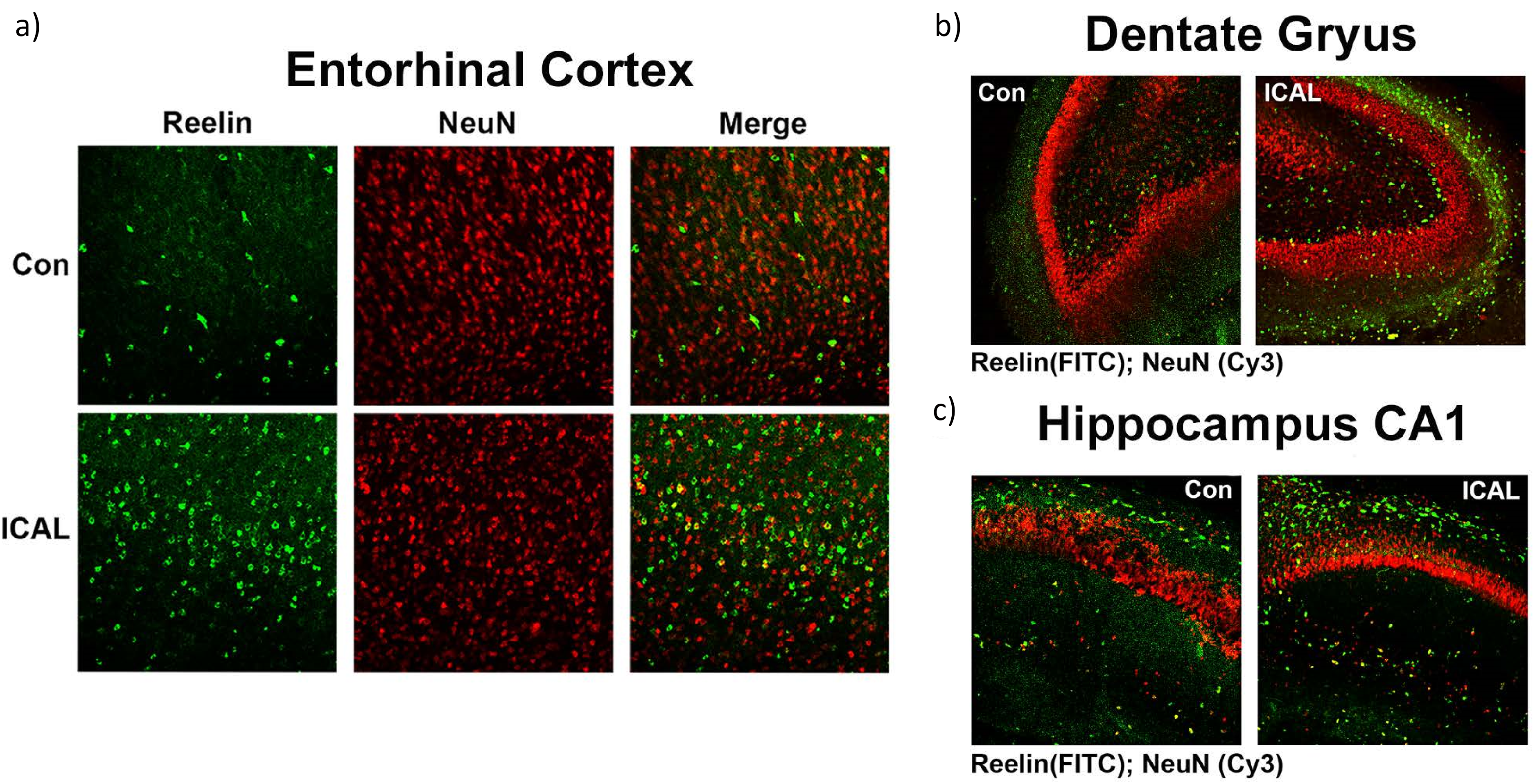
Immunofluorescence staining of hippocampal-entorhinal cortex slices for Reelin. Cortex brain slices were incubated for 24 h in either control medium alone (Con) or Immunocal ® (ICAL) then fixed in formalin and stained with antibodies for Reelin (green) and NeuN (red) then imaged in specific regions including the EC (a), DG (b), and CA1 of the hippocampus (c) (colors are visible in the online version).
Immunocal® preserves the GSH/GSSG ratio in 5-month-old AD model mice
Next, we analyzed the effects of Immunocal® treatment in vivo utilizing J20 AD model mice which were given Immunocal® supplementation ad libitum from 3 to 5 months old. We measured the brain levels of reduced GSH and oxidized glutathione disulfide (GSSG) in cortical tissue from the mice using HPLC-ECD. Untreated hemizygous J20 mice displayed a statistically significant decrease in cortical GSH and coinciding increase in GSSG at 5 months old when compared to non-carrier control mice (Figure 3(a) and (b)). However, hemizygous J20 mice treated with Immunocal® showed a complete preservation of cortical GSH and a reduction in GSSG, which was statistically significantly different than the levels observed in untreated hemizygous mice (Figure 3(a) and (b)). Furthermore, the GSH/GSSG ratio, a measure of oxidative stress, showed a statistically significant reduction in the untreated hemizygous mice when compared to the non-carrier control mice, and this effect was completely prevented by treatment with Immunocal® (Figure 3(c)).
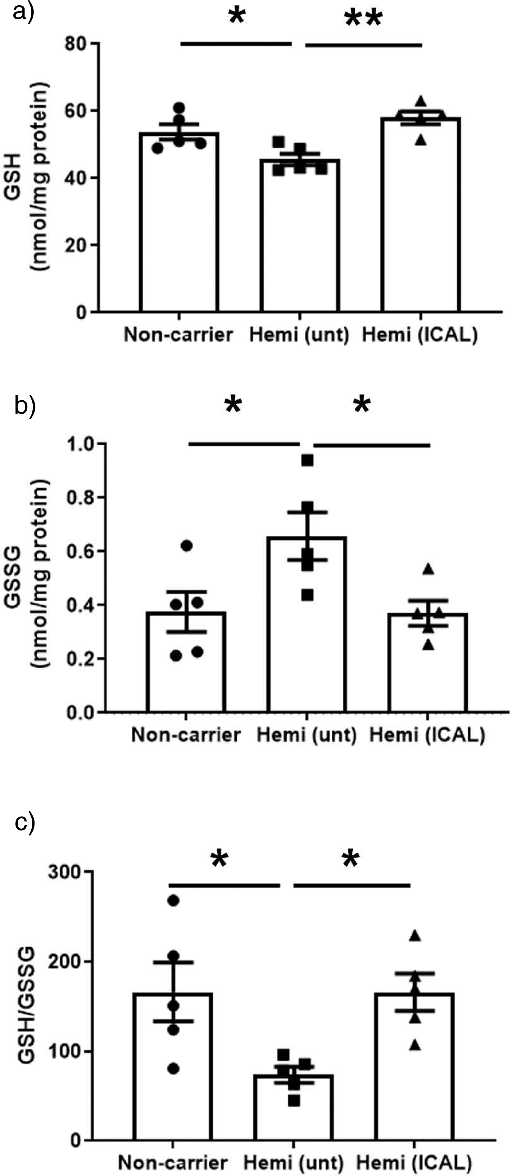
Immunocal® treatment preserves the brain GSH/GSSG ratio in 5-month-old J20 AD model mice. Cortical brain tissue was harvested from 5-month-old non-carrier control mice (Non-carrier) and hemizygous J20 AD mice (Hemi) either untreated (unt) or treated with 3.3% w/v Immunocal® in drinking water ad libitum for 2 months (ICAL). Tissue was extracted then GSH (a) and GSSG (b) and GSH:GSSG ratio (c) were measured by HPLC with electrochemical detection. Values are normalized to protein content represented as mean ± SEM, n = 5 mice per group. *p < 0.05, **p < 0.01 as determined using a one-way ANOVA with a post hoc Tukey's test.
Immunocal® rescues Reelin expression in 5-month-old AD model mice
We used immunofluorescence microscopy to evaluate Reelin expression in the entorhinal cortex, dentate gyrus, and CA1/CA3 regions of the hippocampus in J20 AD model mice (Figure 4). The entorhinal cortex of non-carrier control mice showed significant Reelin immunoreactivity, particularly in layer II, the area demarcated in the Figure (Figure 4(a), see lower panels). In comparison to the non-carrier control, untreated hemizygous J20 mice displayed a marked reduction in Reelin immunoreactivity in layer II of entorhinal cortex, which was increased upon Immunocal® treatment (Figure 4(a)).
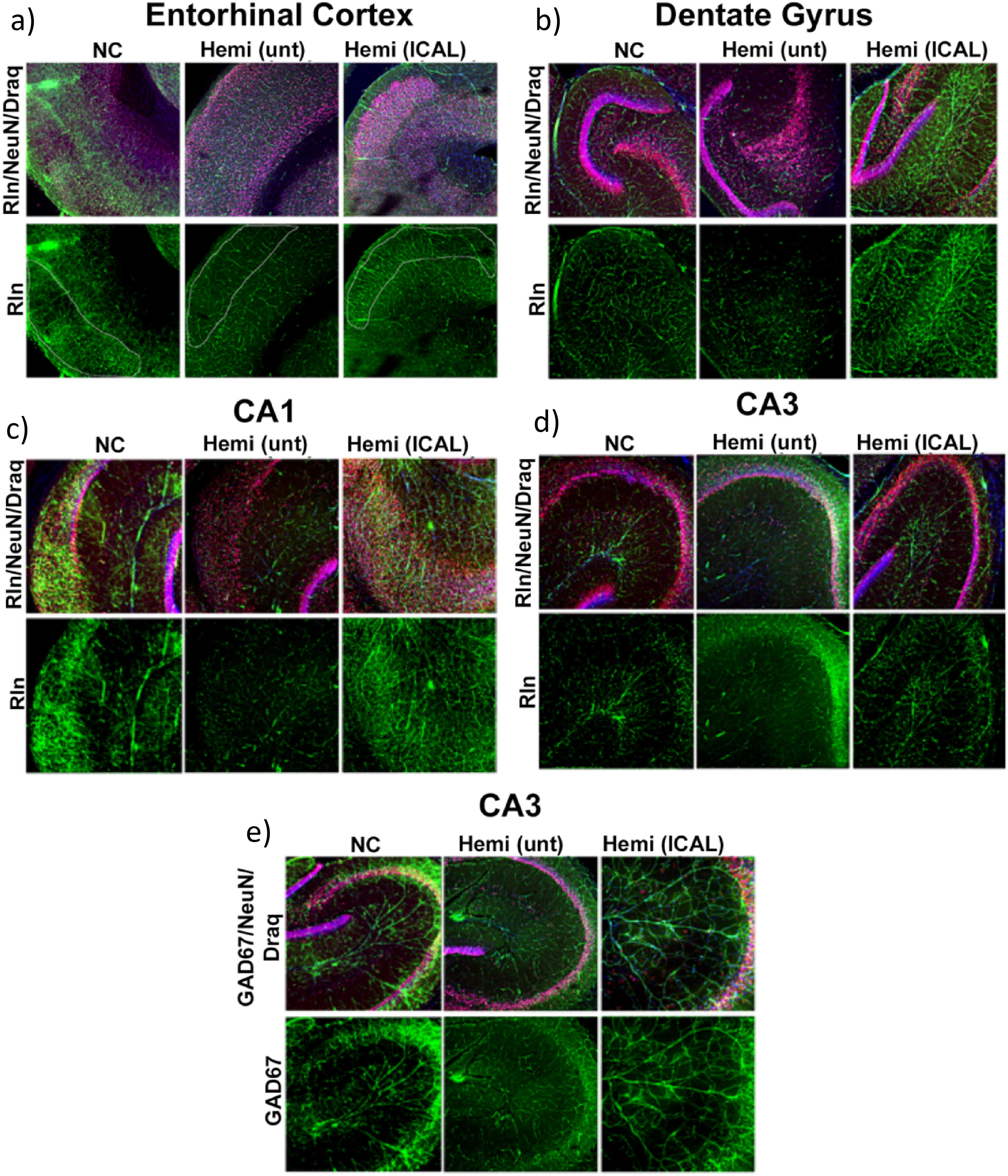
Immunofluorescence staining of hippocampal-entorhinal cortex regions in 5-month-old J20 AD model mice for Reelin and GAD67. Brain slices from the hippocampal-entorhinal cortex region were harvested from 5-month-old non-carrier control mice (NC) and hemizygous J20 AD mice (Hemi) either untreated (unt) or treated with 3.3% w/v Immunocal® in drinking water ad libitum for 2 months (ICAL). (a-d) Sliced sections were fixed in formalin and stained with antibodies for Reelin (Rln; green), NeuN (red), and Draq nuclear stain (blue). Specific regions were imaged in the hippocampal-entorhinal cortex region including the EC (A), DG (b), CA1 of the hippocampus (c), and CA3 of the hippocampus (d). (e) Sliced sections were fixed in formalin and stained with antibodies for GAD67 (green), NeuN (red), and Draq nuclear stain (blue) and then imaged in the CA3 region (colors are visible in the online version).
Next, we assessed Reelin staining in the dentate gyrus and CA1/CA3 regions of the hippocampus in J20 mice. In the dentate gyrus, Reelin expression was diffuse and moderate in intensity in non-carrier control mice (Figure 4(b)). Untreated hemizygous J20 mice showed a slight overall reduction in Reelin staining in the dentate gyrus, whereas Immunocal®-treated hemizygous J20 mice displayed a striking increase in the intensity of Reelin staining in this brain region (Figure 4(b)). In the CA1 region of the hippocampus, Reelin expression was very high in non-carrier control mice and was markedly reduced in untreated hemizygous J20 AD mice (Figure 4(c)). Notably, this deficit in Reelin staining was essentially prevented by treatment of hemizygous J20 mice with Immunocal® for 2 months (Figure 4(c)). A very similar effect on Reelin expression was observed in the CA3 region of the hippocampus. In particular, Reelin-expressing neurons in the middle section of this sub-region were particularly evident in non-carrier control mice, essentially absent in untreated hemizygous J20 mice, and largely preserved in Immunocal®-treated J20 mice (Figure 4(d)).
Finally, we evaluated GAD67 staining, a marker for inhibitory GABAergic interneurons, in the CA3 region of the hippocampus
Immunocal® preserves the GSH/GSSG ratio in 12-month-old AD model mice
Next, we measured the effects of long-term Immunocal® treatment in vivo utilizing J20 AD model mice which were given Immunocal® supplementation ad libitum from 3 to 12 months of age. First, we measured brain levels of GSH and GSSG in cortical tissue from the mice using HPLC-ECD as in the 5-month-old mice. Untreated hemizygous J20 mice displayed a significant reduction in cortical GSH and a marked increase in GSSG at 12 months old when compared to non-carrier control mice (Figure 5(a) and (b)). Hemizygous J20 mice treated with Immunocal® from 3 months old to 12 months old showed a complete preservation of cortical GSH and a reduction in GSSG, both of which were statistically significantly different from the levels observed in untreated hemizygous mice (Figure 5(a) and (b)). Finally, the GSH/GSSG ratio showed a statistically significant reduction in the untreated hemizygous mice when compared to the non-carrier control mice; an effect that was completely blunted by treatment with Immunocal® (Figure 5(c)).
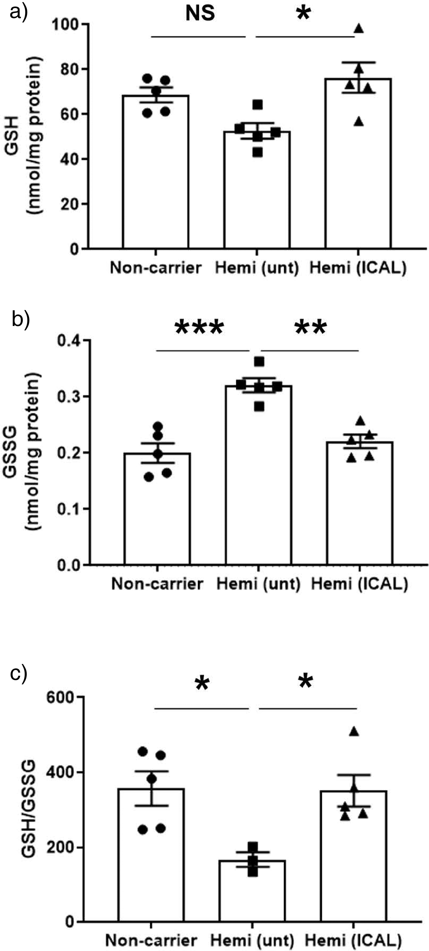
Immunocal® treatment preserves the brain GSH/GSSG ratio in 12-month-old J20 AD model mice. Cortical brain tissue was harvested from 12-month-old non-carrier control mice (Non-carrier) and hemizygous J20 AD mice (Hemi) either untreated (unt) or treated with 3.3% w/v Immunocal® in drinking water ad libitum for 9 months (ICAL). Tissue was extracted, then GSH (a) and GSSG (b) and GSH/GSSG ratio (c) were measured by HPLC with electrochemical detection. Values are normalized to protein content and represented as mean ± SEM, n = 5 mice per group. *p < 0.05, **p < 0.01, ***p < 0.001, NS indicates no significant difference, as determined using a one-way ANOVA with a post hoc Tukey's test.
Immunocal® sustains Reelin mRNA expression in 12-month-old AD model mice
To analyze mRNA expression of Reelin in response to Immunocal® treatment, we used in situ hybridization to analyze regions of hippocampus and entorhinal cortex that express Reelin transcripts. In particular, we measured these levels in cells which produce Reelin including the entorhinal cortex layer 2 and interneurons of the CA3 region. We found mRNA expression for Reelin mostly concentrated in the regions of hilus layer in dentate gyrus, CA3 of hippocampus and entorhinal cortex (Figure 6(a)). We quantified regions of entorhinal cortex and CA3 of the treatment groups and found there was a significant decrease in Reelin transcript levels detected in CA3 and entorhinal cortex regions of hemizygous untreated mice when compared to non-carrier controls (Figure 6(b) and (c)). Further, Immunocal® treatment significantly sustained Reelin transcript expression, but only in the entorhinal cortex (Figure 6(c)).
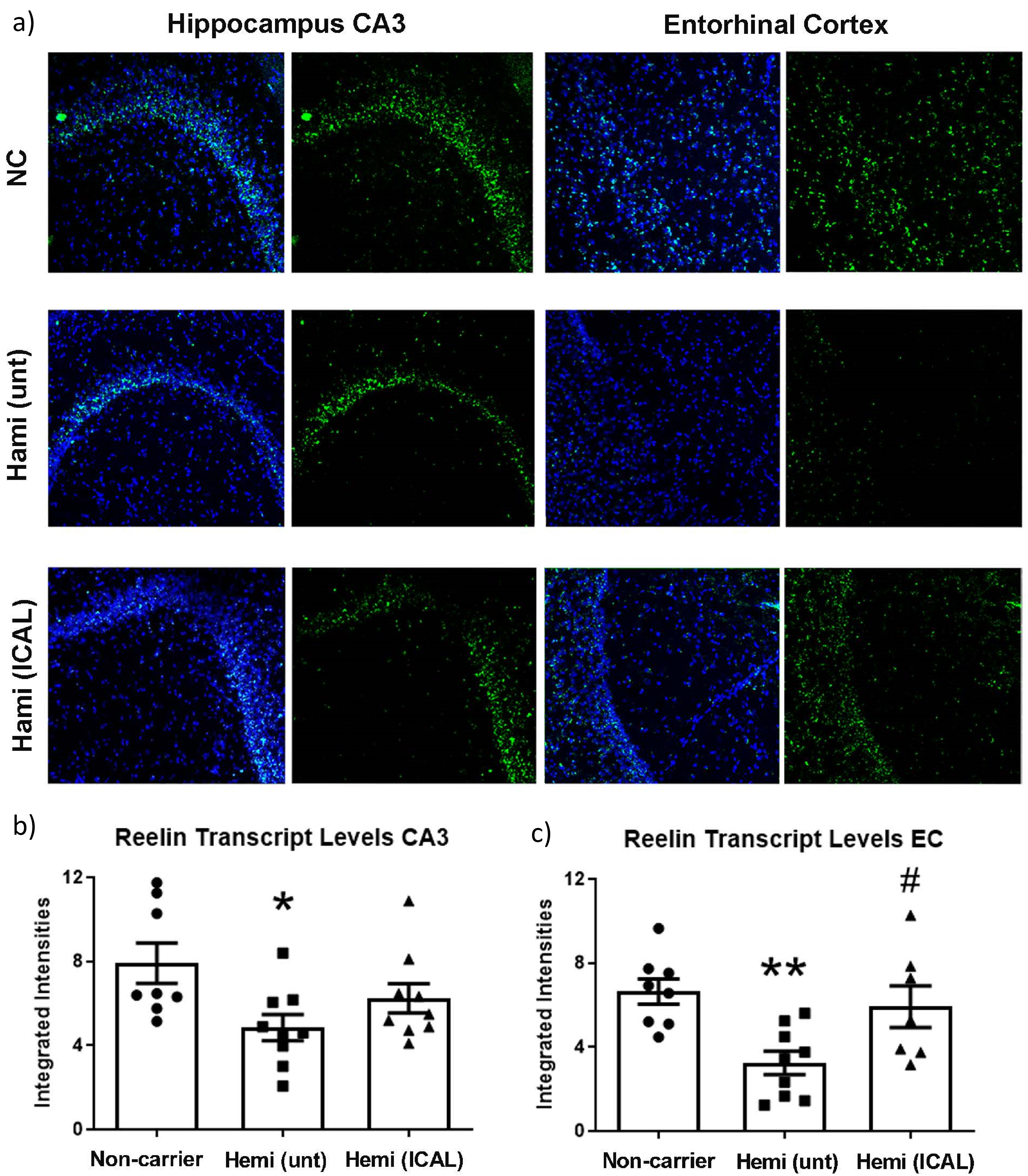
In situ mRNA hybridization for Reelin transcript in the hippocampal-entorhinal cortex region of 12-month-old J20 AD model mice. (a) Brain slices from the hippocampal-entorhinal cortex region were harvested from 12-month-old non-carrier control mice (Non-carrier) and hemizygous J20 AD mice (Hemi) either untreated (unt) or treated with 3.3% w/v Immunocal® in drinking water ad libitum for 9 months (ICAL). Sliced sections were fixed in formalin and hybridized with antisense Reelin transcript (Rln; green) along with Draq nuclear stain (blue). (b) Total fluorescent intensities in the CA3 region of the hippocampus from (A) represented as the mean ± SEM, n = 8–9 mice per group. *p < 0.05 for Hemi (unt) as compared to Non-carrier. (c) Total fluorescent intensities in the entorhinal cortex from (a) represented as mean ± SEM, n = 7–9 mice per group. **p < 0.01 as compared to Non-carrier, #p < 0.05 as compared to Hemi (unt), as determined using a one-way ANOVA with a post hoc Tukey's test (colors are visible in the online version).
Immunocal® rescues Reelin protein expression in 12-month-old AD model mice
We next evaluated the expression of Reelin protein in the 12-month-old J20 AD mice by western blotting brain tissues enriched for the hippocampal-entorhinal cortex region. Reelin expression was reduced in untreated hemizygous J20 mice compared to non-carrier controls and Immunocal® treatment largely corrected this deficiency (Figure 7(a)). Using quantitative densitometric analysis of the 180 kDa Reelin band revealed an approximately 20% mean reduction in Reelin protein expression in hemizygous untreated AD mice compared to non-carrier controls (not statistically significant), and a complete rescue of Reelin expression in Immunocal®-treated mice (Figure 7(b)).
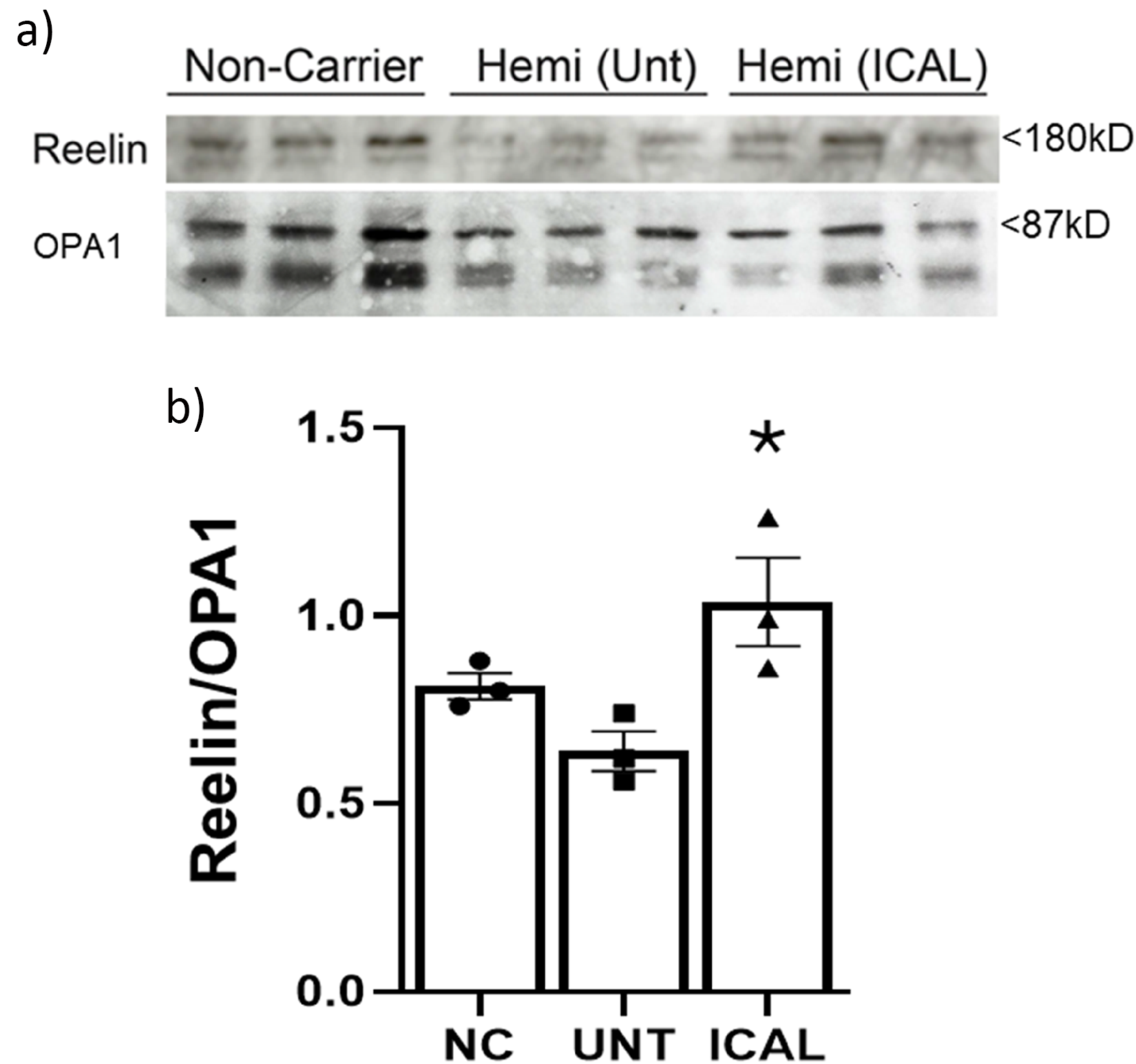
Immunocal® treatment rescues Reelin expression in 12-month-old J20 AD model mice. (a) Western blot analysis using antibodies specific for Reelin (top) and OPA1 (bottom) in tissue harvested from 12-month-old non-carrier control mice (Non-carrier; NC) and hemizygous J20 AD mice (Hemi) either untreated (Unt) or treated with 3.3% w/v Immunocal® in water ad libitum for 9 months (ICAL). Tissue was micro-dissected to enrich for the hippocampal-entorhinal cortex sub-region. (b) Densitometric quantification of (a) was calculated by dividing the Reelin 180 kD band by the full length OPA1 87 kD band and represented as the mean ± SEM, n = 3 mice per group. *p < 0.05 as compared to Hemi (Unt), as determined using a one-way ANOVA with a post hoc Tukey's test.
Next, we performed immunofluorescence microscopy to evaluate Reelin expression in the entorhinal cortex, dentate gyrus, and CA1/CA3 regions of the hippocampus in 12 month-old J20 mice. The entorhinal cortex of non-carrier control mice showed significant Reelin immunoreactivity (Figure 8(a)), particularly in layer II, similar to the pattern observed in J20 mice analyzed at 5 months old. In comparison to the non-carrier control, untreated hemizygous J20 mice displayed a marked reduction in Reelin immunoreactivity in layer II of entorhinal cortex. Intriguingly, treatment with Immunocal® from 3 months old to 12 months old essentially rescued this deficit in Reelin expression within layer II of the entorhinal cortex of J20 mice (Figure 8(a)). Next, we assessed Reelin staining in the dentate gyrus and CA1/CA3 regions of the hippocampus in J20 mice
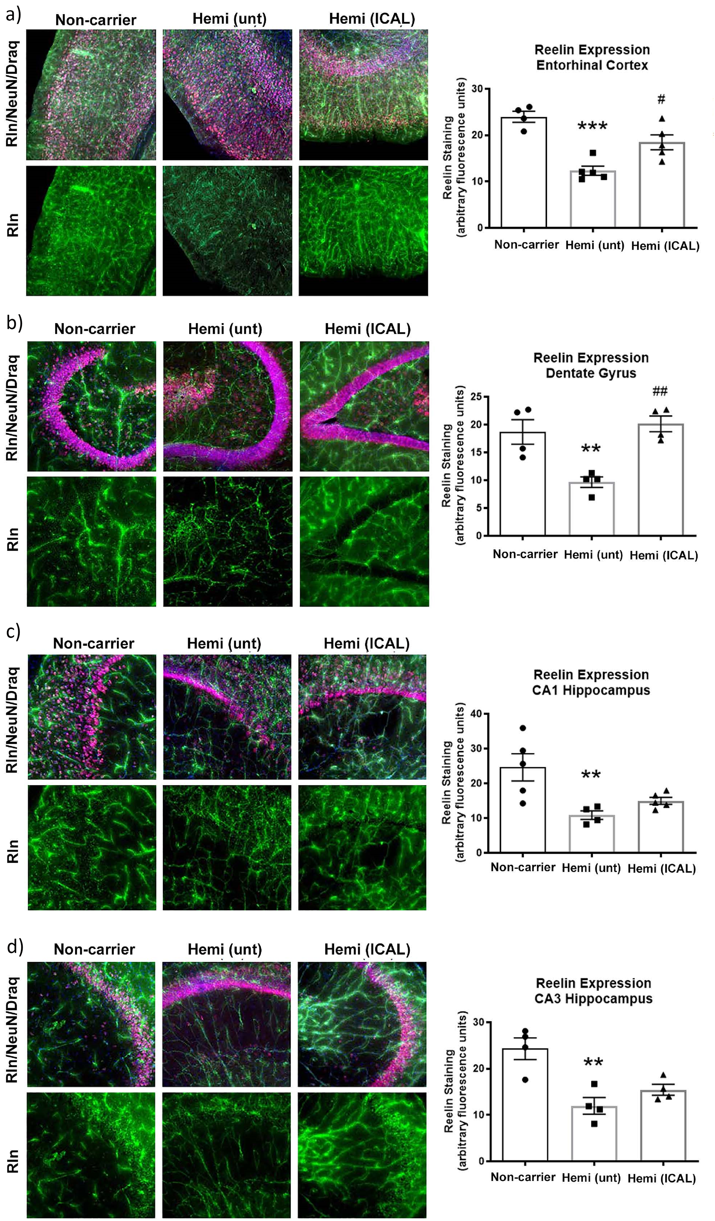
Immunofluorescence staining of hippocampal-entorhinal cortex regions in 12-month-old J20 AD model mice for Reelin. Brain slices from the hippocampal-entorhinal cortex region were harvested from 12-month-old non-carrier control mice (Non-carrier) and hemizygous J20 AD mice (Hemi) either untreated (unt) or treated with 3.3% w/v Immunocal® in drinking water ad libitum for 9 months (ICAL). Sliced sections were fixed in formalin and stained with antibodies for Reelin (Rln; green), NeuN (red), and Draq nuclear stain (blue). (a) Images taken in the EC and quantification of total fluorescent intensities represented as the mean ± SEM, n = 4 for Non-carrier, n = 5 for Hemi (unt) and Hemi (ICAL) mice per group. ***p < 0.001 as compared to Non-carrier, #p < 0.05 as compared to Hemi (unt). (b) Images taken in the DG and quantification of total fluorescent intensities represented as the mean ± SEM, n = 4 mice per group. ** p < 0.01 as compared to Non-carrier, ##p < 0.01 as compared to Hemi (unt). (c) Images taken in the CA1 region of the hippocampus and quantification of total fluorescent intensities represented as the mean ± SEM, n = 5 for Non-carrier and Hemi (ICAL), n = 4 for Hemi (unt) mice per group. **p < 0.01 as compared to Non-carrier. (d) Images taken in the CA3 region of the hippocampus and quantification of total fluorescent intensities represented as the mean ± SEM, n = 4 mice per group. **p < 0.01 as compared to Non-carrier. All statistical analyses were determined using a one-way ANOVA with a post hoc Tukey's test (colors are visible in the online version).
Immunocal® reduces amyloid plaque burden in 12-month-old AD model mice
The J20 mice have been shown to display extensive Aβ plaque pathology in the hippocampal-entorhinal cortex formation by 12 months of age. To evaluate the effects of Immunocal® treatment on the deposition of amyloid plaques, we used Amylo-glo, an amyloid peptide aggregate-specific stain used to mark amyloid plaques. After treating the mice from 3 months old to 12 months old with Immunocal®, there was a dramatic reduction in both the plaque number and plaque load in the entorhinal cortex (Figure 9(a)). Further, in the dentate gyrus, CA3, and CA1 hippocampal regions of untreated J20 mice there was a significant plaque load throughout with distinct dense formations (Figure 9(b)–(d)). However, treatment with Immunocal® for 9 months dramatically diminished both the plaque number and plaque load in each of these regions in comparison to the untreated J20 mice (Figure 9(b)–(d)).
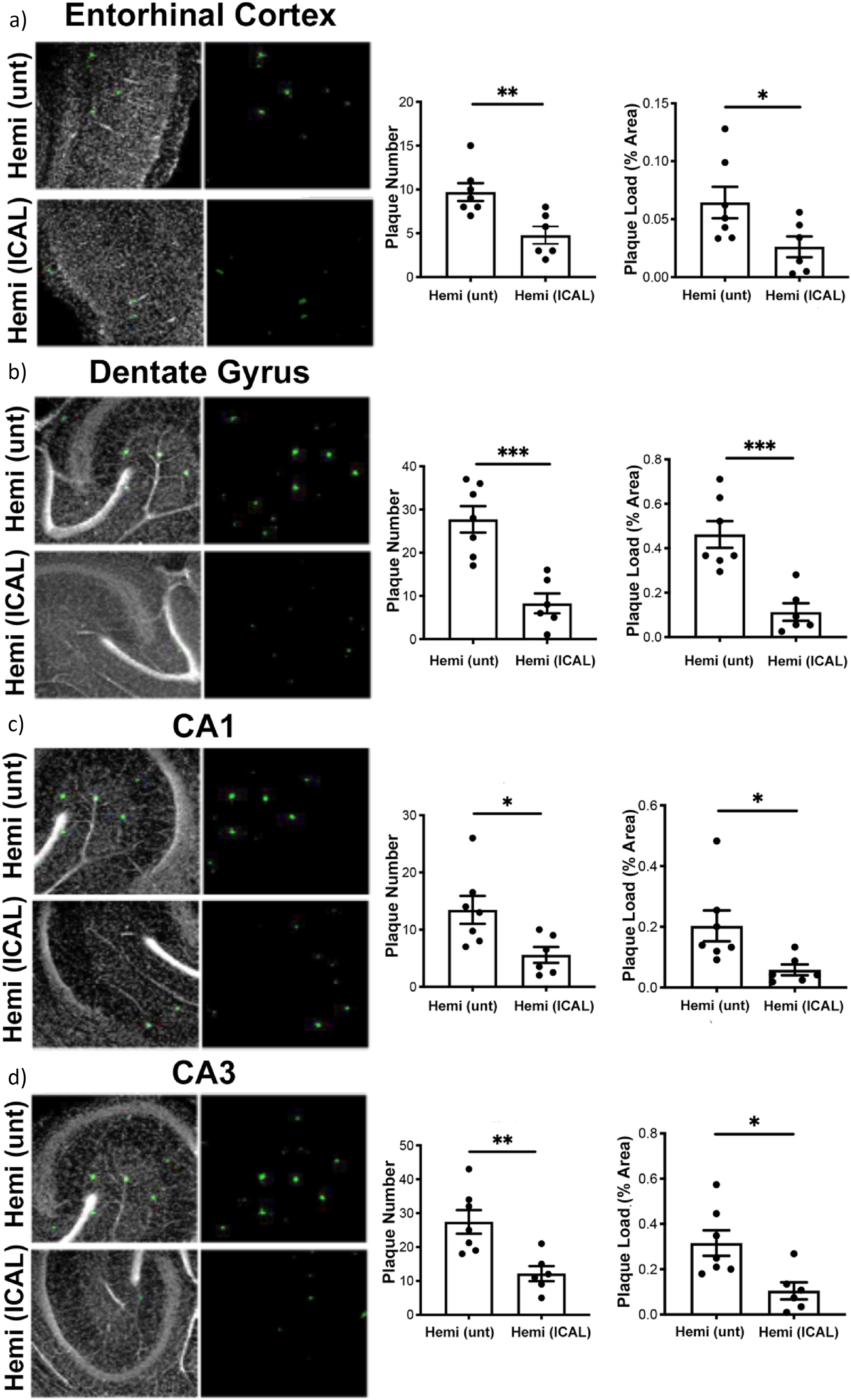
Immunocal® treatment reduces amyloid plaque load and number in the hippocampal-entorhinal cortex region of 12-month-old J20 AD model mice. Brain slices from the hippocampal-entorhinal cortex region were harvested from 12-month-old hemizygous J20 AD mice (Hemi) either untreated (unt) or treated with 3.3% w/v Immunocal® in drinking water ad libitum for 9 months (ICAL). Sections were fixed in formalin and stained with Amylo-glo (green) and Draq nuclear stain (grayscale) then imaged in the EC (a), DG (b), CA1 of the hippocampus (c), and CA3 region of the hippocampus (d). The number of the plaques and plaque load (% area) were quantified and shown as the mean ± SEM, n = 7 for Hemi (unt), n = 6 for Hemi (ICAL) mice per group. *p < 0.05, **p < 0.01, ***p < 0.001 as compared to Hemi (unt) as determined using a one-way ANOVA with a post hoc Tukey's test (colors are visible in the online version).
Immunocal treatment modestly improves performance on cognitive behavioral tests
Finally, we evaluated the effects of Immunocal® treatment on cognitive function in 12-month-old female J20 mice. Data for male mice are not shown as the study was under powered for the number of 12-month-old male J20 mice necessary to document statistically significant changes in cognitive function between any of the groups. We first used the Barnes maze to assess spatial learning and memory. When data were averaged for days 5 and 6 of the six-day acquisition phase of the Barnes maze test, untreated hemizygous J20 female mice showed a statistically significant increase in the delay time to find the escape pod, when compared to non-carrier control mice, and this deficit was largely corrected by treatment with Immunocal® (Figure 10(a)). In the probe phase of the Barnes maze test, untreated hemizygous J20 female mice showed a statistically significant increase in the delay time to find the escape pod zone, when compared to non-carrier control mice. However, treatment with Immunocal® did not correct this deficit as there was substantial variability in the response of Immunocal®-treated J20 mice during the probe phase of the Barnes maze test (Figure 10(b)). Next, we evaluated recognition memory using the NOR test. Untreated hemizygous J20 female mice showed a statistically significant decrease in the calculated discrimination index, when compared to non-carrier control mice, and this deficit was largely corrected by treatment with Immunocal® (Figure 10(c)). Collectively, these data suggest that Immunocal® treatment has some moderate beneficial behavioral effects on cognitive function in the J20 mouse model of AD.
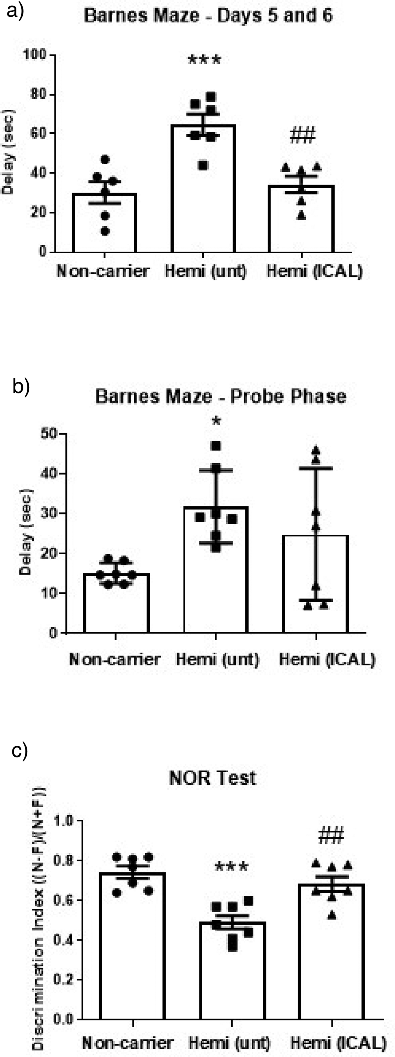
Immunocal® treatment has some beneficial effects on cognitive behavioral tasks in 12-month-old J20 AD model mice. Female J20 mice were subjected to the Barnes maze test for spatial learning and memory and the novel object recognition (NOR) test for recognition memory. (a) Data for days 5 and 6 of the acquisition phase of the Barnes maze test were averaged and are shown as the mean ± SEM for the delay times observed (n = 6 mice per group). ***p < 0.001 compared to Non-carrier, ##p < 0.01 compared to Hemi (unt), as determined using a one-way ANOVA with a posthoc Tukey's test. (b) Delay times to find the escape pod zone for the probe phase of the Barnes maze test are shown as the mean ± SEM for each treatment group (n = 7 mice per group). *p < 0.05 compared to Non-carrier, as determined using a one-way ANOVA with a posthoc Tukey's test. (c) The discrimination index for the NOR test is plotted as the mean ± SEM (n = 7 mice per group). ***p < 0.001 compared to Non-carrier, ##p < 0.01 compared to Hemi (unt), as determined using a one-way ANOVA with a posthoc Tukey's test.
Discussion
The amyloid cascade hypothesis has dominated the field of AD research for nearly three decades and is founded on the premise that deposition of Aβ peptide is the initiating event in disease pathogenesis, ultimately leading to neurofibrillary tangle formation, synaptic loss, and neuronal cell demise. 32 For more than two decades, the amyloidogenic processing of AβPP to form Aβ through the sequential cleavage of AβPP by β-secretase (BACE) and γ-secretase has been recognized as a viable molecular target for AD therapeutic development.33,34 Due to this, selective inhibitors of BACE and γ-secretase were developed as drugs to decrease the tissue load of Aβ in AD brains.35,36 Overall, these drugs met with clinical failure.
More recently, the Food and Drug Administration (FDA) granted accelerated approval for Biogen's anti-amyloid antibody, aducanumab (2021), and for the Eisai/Biogen anti-amyloid antibody, lecanemab (2023), for AD. Another anti-amyloid antibody, donanemab, also recently demonstrated some apparent therapeutic benefit in a phase III trial and was most recently approved by the FDA (2024). 37 Despite these recent positive clinical developments, considerable skepticism remains surrounding the therapeutic efficacy of anti-amyloid strategies for AD.38–40 Additionally, the observation that brain amyloid load does not necessarily correlate with the severity of cognitive deficits in AD has led some to question whether the amyloid cascade hypothesis is sufficient to explain the underlying pathogenesis of late onset, sporadic AD.41–48
In the search for new therapeutic approaches in AD, Reelin deficiency has emerged as a possible molecular target.49,50 Recently, a genome-wide association study linked the Reelin-DAB1 signaling pathway with AD risk in a cohort of APOE ε4 homozygous (ε4/ε4) individuals. 51 Further implicating Reelin as a protective molecule against AD, Lopera et al. recently reported the case history of a man who displayed dramatic resistance to developing autosomal dominant AD despite carrying a PSEN1-E280A mutation. Genetic testing showed that he was heterozygous for a rare variant in RELN (H3447R, termed COLBOS after the Colombia-Boston biomarker research study), a gain-of-function variant showing enhanced activation of its downstream signaling target DAB1. 52 Thus, enhanced activation of the Reelin-DAB1 signaling pathway appears to increase resilience to AD. These studies suggest that strategies aimed at correcting Reelin deficits might provide a novel therapeutic approach to AD.
In addition to deficiencies in Reelin expression and/or signaling, depletion of the critical antioxidant GSH has also been implicated in the pathogenesis of AD. Using magnetic resonance spectroscopy methods, investigators have shown that brain GSH (particularly in the hippocampus) is depleted in patients with AD and this decrement correlates with the level of cognitive dysfunction.53,54 Studies in mouse models have demonstrated that depletion of GSH occurs prior to the deposition of intracellular Aβ aggregates or extracellular amyloid plaques. 55 Moreover, administration of the GSH precursor, N-acetylcysteine-amide, shows neuroprotective activity against Aβ-induced AD-like pathology in rats. 56 Collectively, these findings suggest that supplementation with GSH precursors may be a novel therapeutic strategy for AD.57,58
The cysteine-rich whey protein and GSH precursor, Immunocal®, has previously been shown to be neuroprotective in vitro in diverse models of oxidative stress and neurotoxicity. 23 This GSH precursor has also been demonstrated to elevate GSH in the brain and spinal cord and is therapeutically efficacious in mouse models of schizophrenia, amyotrophic lateral sclerosis, and traumatic brain injury.24,30,59 In addition, Immunocal® treatment has been shown to rescue Reelin expression at the mRNA and protein level in the prefrontal cortex of a mouse model of schizophrenia. 24 The purpose of the present study was to determine if supplementation with Immunocal® increases Reelin expression in vitro in a hippocampal-entorhinal cortex slice model and in vivo within the brain of an AD mouse model (J20 strain). In addition, we assessed whether Immunocal® administration to J20 AD model mice resulted in preserved brain GSH and diminished brain amyloid load when compared to untreated mice.
We found that incubation with Immunocal® significantly increased Reelin expression in vitro in hippocampal-entorhinal cortex slices. We also show that Immunocal® treatment of J20 AD model mice was effective at sustaining brain GSH levels, rescuing Reelin expression and diminishing amyloid plaque pathology in vivo. Immunocal® treatment also modestly improved performance on some behavioral cognitive tests, Barnes maze and NOR test; however, the rigor of these data are limited by the fact that we were only able to analyze behavior in female 12-month-old J20 AD mice.
Together, our findings demonstrate that Immunocal® treatment produces marked beneficial effects on antioxidant status, Reelin expression and amyloid pathology in the J20 mouse model of AD. This is significant since the neuronal population of Reelin-secreting cells in the entorhinal cortex, and the hippocampal neurons to which they project, are some of the earliest populations most vulnerable to neurodegeneration in AD. Furthermore, our data are consistent with previous studies showing that genetic manipulation of Reelin expression in J20 mice has profound effects on AD pathology.20,21 Finally, our findings support the hypothesis that GSH depletion is a key pathogenetic mechanism underlying AD and therefore, supplementation with GSH precursors may be a novel therapeutic approach to treat this devastating disorder.
We conclude that Immunocal® represents a novel therapeutic approach to preserve brain GSH levels, rescue Reelin expression, and decrease amyloid deposition in AD. Recent studies place Reelin as a central player in AD etiology linking APOE4, tau, and Aβ pathogenetic mechanisms. 60 Given its capacity to rescue Reelin in an AD mouse model based largely on amyloid pathology (the J20 mouse model), Immunocal® should be evaluated in additional preclinical models of AD. These models should include those exhibiting prominent tau pathology and enhanced risk associated with APOE4 (ε4/ε4) expression to determine if the therapeutic effects of this GSH precursor extend to mitigating tau pathology and ameliorating the cognitive deficits associated with AD progression.
Footnotes

Ethical considerations
All animal housing and experiments were conducted in strict accordance with the institutional Guidelines for Care and Use of Laboratory Animals at the University of Denver. Animal studies using rats were conducted in accordance with a protocol approved by the University of Denver Institutional Animal Care and Use Committee (Approval #983340-4). All animal studies using mice were conducted in accordance with a protocol approved by the University of Denver Institutional Animal Care and Use Committee (Approval #927465-8).
Author contributions
Alexandra Sandberg: Formal analysis; Investigation; Methodology; Writing - original draft.
Srivalli Puttagunta: Formal analysis; Investigation; Methodology; Writing - original draft.
Nathan Duval: Formal analysis; Investigation; Methodology.
Holly Fleming: Investigation; Methodology.
Lilia Koza: Investigation; Methodology.
Kade Hieber: Investigation; Methodology.
Jessica Holsopple: Investigation; Methodology.
Michael Reyna: Formal analysis; Investigation; Methodology.
Daniel Paredes: Project administration; Writing - review & editing.
Daniel A Linseman: Conceptualization; Project administration; Supervision; Writing - review & editing.
Funding
The authors disclosed receipt of the following financial support for the research, authorship, and/or publication of this article: Funding was provided by Immunotec, Inc. (Quebec, Canada), the manufacturer of Immunocal®.
Declaration of conflicting interests
The authors declared the following potential conflicts of interest with respect to the research, authorship, and/or publication of this article: Daniel Linseman is a member of the Scientific Advisory Board for Immunotec, Inc. (Quebec, Canada), the manufacturer of Immunocal®.
Data availability statement
The data supporting the findings of this study are available within the article.