



Abstract
Background
Alzheimer's disease (AD) is a neurodegenerative disorder characterized by pathogenesis involving numerous factors. Recent research has highlighted the significant role of autoimmunity in the initiation and progression of AD, with autoantibodies emerging as a pivotal area of investigation. Nevertheless, the influence of autoantibodies in AD is marked by substantial heterogeneity, they may either mitigate disease progression by clearing pathogenic protein aggregates or exacerbate the pathological process through mechanisms such as the activation of inflammatory responses or the induction of neuronal damage.
Objective
This review aims to synthesize the various roles of autoantibodies in AD, examine the factors that influence their functions, and assess their potential application in precision immunotherapy.
Methods
PubMed and Web of Science databases were searched for English-language papers (2015–2025). Peer-reviewed human, animal and cell studies, systematic reviews and meta-analyses were screened independently by two reviewers.
Results
A total of 87 studies were selected for inclusion, spanning human, animal, and cellular research. The findings indicated that certain autoantibodies, such as those targeting amyloid-β, tau, or 4-hydroxynonenal, may confer neuroprotective effects. Conversely, other autoantibodies, including those against BACE1, aquaporin-4, or HuD, may exacerbate AD pathology. Importantly, some autoantibodies were found to exhibit dual roles, contingent upon their specific modifications or the context of the disease.
Conclusions
Autoantibodies constitute a double-edged immune axis in AD. Their impact hinges on antigen class, disease stage, isotype affinity and glycosylation. Precision strategies—like CAAR-T cell therapy, glycosylation modulation, and affinity optimization—offer therapeutic promise but require further validation.
Introduction
Alzheimer's disease (AD) is an irreversible, age-associated neurodegenerative disorder marked by progressive memory impairment and cognitive deterioration. 1 As the predominant cause of dementia among the elderly, AD significantly diminishes patients’ quality of life while imposing substantial caregiving demands and financial burdens on families and society. Projections suggest a considerable increase in AD-related mortality in the forthcoming decades. 2 The global estimates of cost for AD were US $9.12 trillion by 2050. 3
The pathogenesis of AD is highly intricate, encompassing a multitude of factors such as the amyloid-β (Aβ) hypothesis, the tau protein hypothesis, environmental influences, metabolic irregularities, immune system dysregulation, and genetic predispositions, notably Apolipoprotein E abnormalities which contribute to Aβ and tau protein accumulation. 4 Despite the diverse hypotheses and research trajectories, the precise mechanisms underlying AD remain inadequately understood.
Recent studies increasingly suggest that the autoimmunity may significantly contribute to the pathogenesis and progression of AD, with particular emphasis on the role of autoantibodies as a key area. 5 The immune system of the central nervous system (CNS) is sustained through a synergistic interaction between intrinsic innate immune cells, such as microglia and astrocytes, and peripherally derived adaptive immune cells, which continuously monitor the neurological health.6,7 Upon experiencing damage, these immune cells become activated to facilitate repair. However, dysfunction within the immune system can lead to erroneous attacks on healthy tissues, thereby initiating an autoimmune response. In the initial stages of AD, microglia exhibit neuroprotective properties by phagocytosing Aβ plaques and establishing a protective barrier. 8 As the disease advances, the overactivation of microglia may lead to an increased release of proinflammatory cytokines and an accumulation of Aβ proteins. 9 These observations indicate that targeting immunoregulatory pathways could emerge as a crucial strategy for the treatment of AD. Furthermore, the study highlights that the pathological alterations associated with AD, along with the compromised integrity of the blood-brain barrier, create distinct opportunities for the immune system to interact with CNS proteins, potentially resulting in the production of autoantibodies. 10 In fact, various types of autoantibodies have been identified in patients with AD, and ongoing research increasingly indicates that these autoantibodies may influence the disease's progression through multiple mechanisms. For instance, certain autoantibodies have been shown to promote Aβ deposition and tau protein phosphorylation in amyloid precursor protein/presenilin 1 mouse models, thereby inducing neuroinflammation and neurodegeneration. 11 Furthermore, autoantibodies may impair learning and memory functions by activating the complement system and inhibiting neuronal plasticity. 12 Conversely, other studies suggest that autoantibodies can mitigate Aβ pathology formation, reduce oxidative stress, and potentially serve as part of the body's defense mechanism. 13 Tau transgenic mice lacking autoantibodies exhibit spatial learning deficits, implying that autoantibodies may confer some cognitive protection. 14 The discrepancies among these findings indicate that the role of autoantibodies in AD is heterogeneous and may be modulated by additional factors. 15 This review aims to examine the multifaceted role of autoantibodies in AD, investigate the influencing factors, and evaluate related immunotherapeutic strategies to offer new insights for future clinical applications.
Methods
A thorough literature review was performed utilizing the PubMed and Web of Science databases to identify studies related to AD and autoantibodies, encompassing publications up to February 2025. The search strategy employed combinations of keywords such as “Alzheimer's disease”, “autoantibodies”, “autoantibody”, “antibodies”, “antibody”, and “IgG”.
Redundant records were eliminated utilizing the duplicate literature screening feature in EndNote. Subsequently, the search results were refined to include only English-language literature, with the publication period restricted to 2015 to 2025, as determined by the database's screening capabilities. The inclusion criteria encompassed studies involving human, animal, and cellular models, including original research articles, review papers, systematic reviews, meta-analyses, and research related to AD and autoantibodies. The exclusion criteria comprised studies unrelated to AD or autoantibodies, non-English language literature, studies involving traumatic brain injury or non-AD dementia, and other types of literature such as conference abstracts, editorials or letters. All selected literature was independently screened and evaluated by two researchers to ensure relevance and quality. A Preferred Reporting Items for Systematic Reviews and Meta-Analyses (PRISMA)-style flowchart was employed to illustrate the comprehensive process of literature screening and inclusion.
Results
A comprehensive search of databases yielded 2932 records, comprising 1522 entries from PubMed and 1410 from Web of Science. Following the removal of 694 duplicate entries, 2238 records were subjected to title and abstract screening. Of these, 1996 records were excluded due to their irrelevance to the study's objectives. Subsequently, full-text retrieval was attempted for 242 articles, with 5 articles being unobtainable. Consequently, 237 reports underwent eligibility assessment, resulting in the exclusion of 150 reports for the following reasons: lack of relevance to AD or autoantibodies (n = 48), classification as conference abstracts, editorials, or letters (n = 35), inadequate experimental design or data (n = 42), traumatic brain injury or non-AD dementia (n = 20), or cohort overlap/duplicate reporting (n = 5). Ultimately, 87 studies satisfied the inclusion criteria and were incorporated into the review (Figure 1).
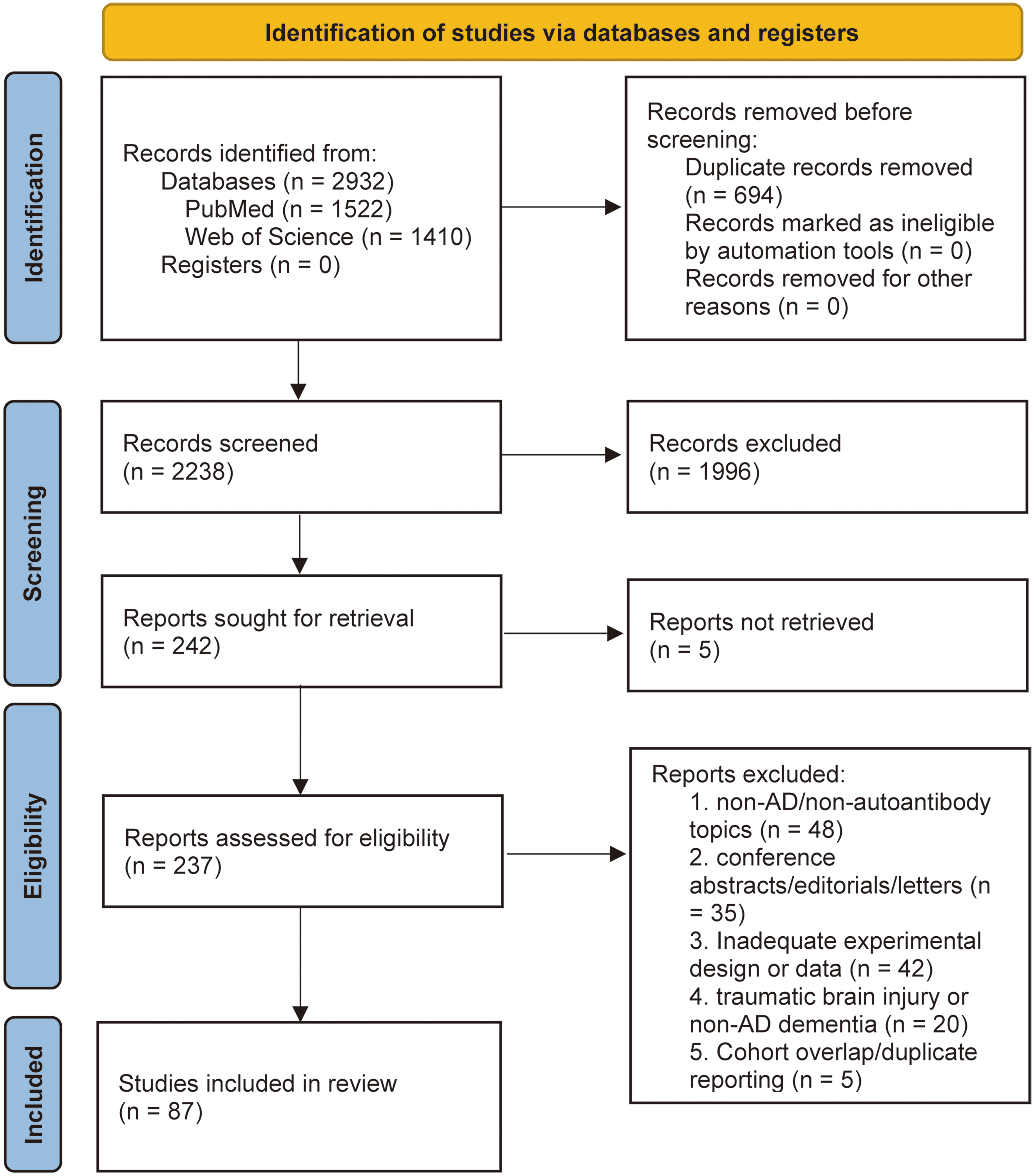
Flowchart illustrating inclusion process.
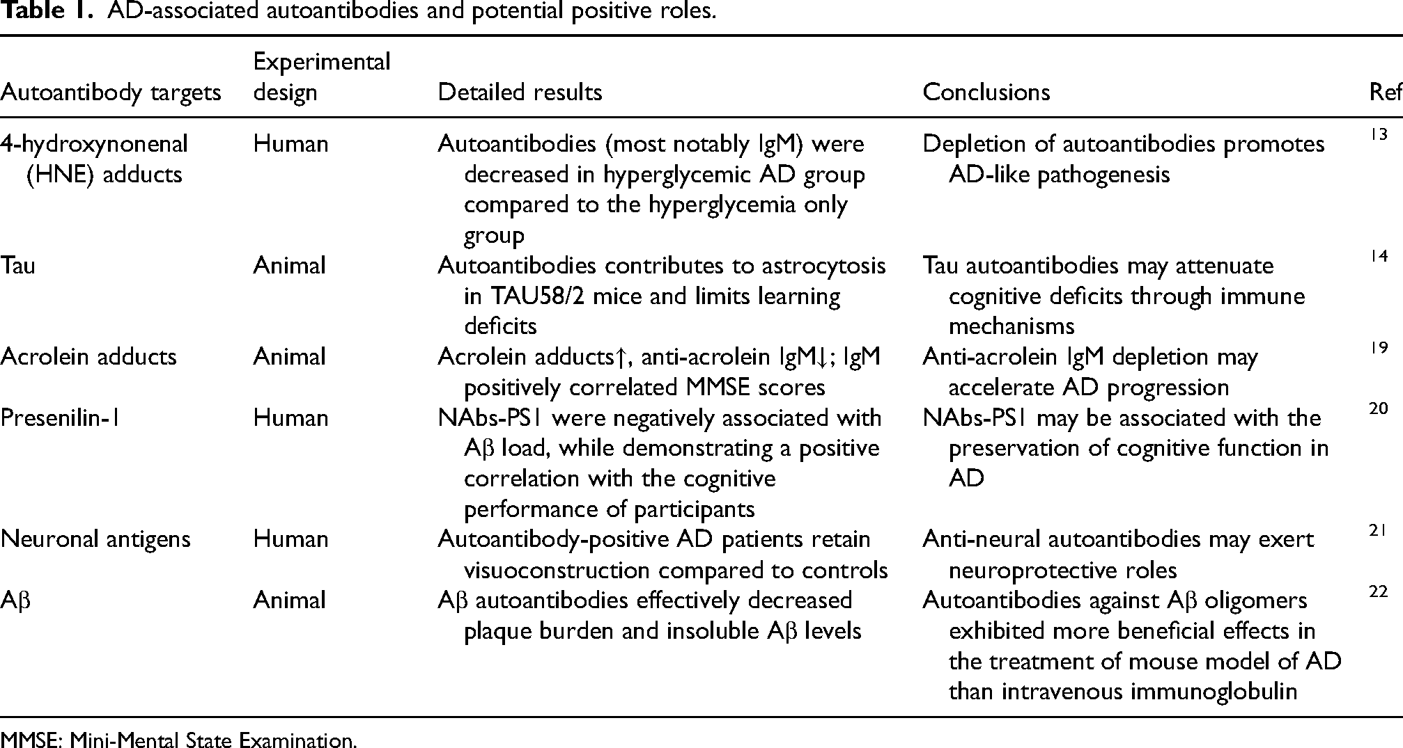
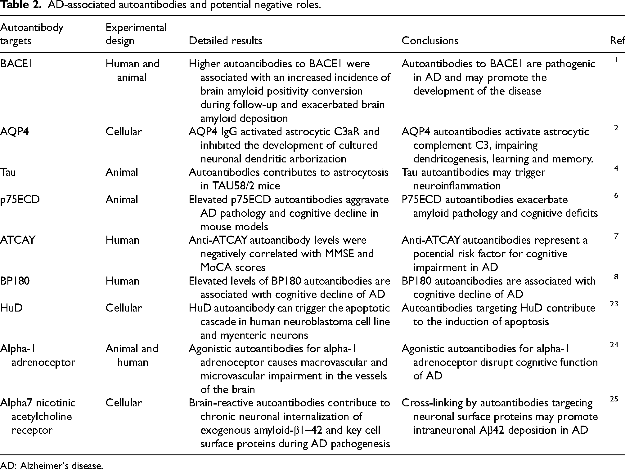
Tables 1 and 2 provide a comprehensive overview of the roles of various autoantibodies associated with AD, as identified through human, animal, and cellular studies. Table 1 enumerates six autoantibodies that potentially exhibit neuroprotective functions, including those targeting 4-hydroxynonenal adducts, tau, acrolein adducts, Presenilin-1, neuronal antigens, and Aβ. These autoantibodies may mitigate AD progression by facilitating the removal of neurotoxic substances, reducing the Aβ burden, and modulating immune pathways. Conversely, Table 2 delineates nine autoantibodies that may contribute to the pathogenic mechanisms of AD, including those against BACE1, AQP4, p75ECD, ATCAY, BP180, HuD, Alpha-1 adrenoceptor, and Alpha7 nicotinic acetylcholine receptor.16–18 These autoantibodies may exacerbate AD pathology through mechanisms such as promoting Aβ deposition, impairing neuronal development, activating neuroinflammatory processes, and inducing neuronal apoptosis. Notably, autoantibodies targeting Aβ and tau exhibit dual functions, as they can either confer protection or exacerbate pathological progression depending on specific modifications or pathological contexts.
AD-associated autoantibodies and potential positive roles.
MMSE: Mini-Mental State Examination.
AD-associated autoantibodies and potential negative roles.
AD: Alzheimer's disease.
Discussion
This review highlights the multifaceted roles of autoantibodies in AD, encompassing both pathogenic and potential neuroprotective effects. Numerous studies have identified specific autoantibodies, such as those targeting BACE1, AQP4, acrolein adducts, p75ECD, and ATCAY kinesin light chain interacting caytaxin (ATCAY), as being significantly associated with exacerbated AD pathology and cognitive decline, indicating their potential pathogenicity. Conversely, autoantibodies directed against Presenilin-1, neuronal antigens, and Aβ have been proposed to confer neuroprotective effects or to correlate with the relative preservation of cognitive function. Furthermore, discrepancies exist among study findings; for instance, some research suggests that autoantibodies related to Aβ or tau mitigate cognitive deficits, while others imply that these antibodies may facilitate pathological progression. This inconsistency indicates that the impact of autoantibodies may be contingent not only upon the specific antigen targeted but also on additional modulating factors.
Crosstalk between the immune and nervous system in AD: the role of neuroinflammation
The immune system, comprising immune organs, cells, and molecules, is integral to executing immune responses, eliminating foreign substances, and maintaining homeostasis within the internal environment. 26 As immunological research advances, accumulating evidence indicates that the immune system significantly influences neurodegenerative diseases, peripheral neurological disorders, and the aging process. 27 In AD, various components of the immune system are implicated in the disease's pathogenesis. Classical central immune elements, such as the complement system and microglia, along with peripheral immune cells, including monocytes and lymphocytes, are intricately linked to AD. 28 Research indicates that neuroinflammation is not only a significant pathological characteristic of AD but also plays a crucial role in its development and progression. 29 Microglia, as the resident immune cells within the CNS, are pivotal in AD-associated neuroinflammation Microglia have the capacity to detect and clear abnormal protein aggregates; however, their overactivation can lead to the release of inflammatory mediators, induce neurotoxicity, and exacerbate pathological progression. 28 Furthermore, peripheral monocytes and lymphocytes can traverse the blood-brain barrier, contributing to neuroinflammation and further influencing disease progression. 28 Alongside microglia, astrocytes also participate in neuroinflammatory processes related to AD. These astrocytes interact with microglial cells during the inflammatory response, collectively influencing the pathological progression of AD. 30 Furthermore, evidence suggests that dysfunction of the blood-brain barrier may permit peripheral inflammatory factors to infiltrate the CNS, thereby intensifying neuroinflammation. 31 Although the role of neuroinflammation in AD has been widely recognized, its specific mechanism still needs further study. Understanding the complex network of neuroinflammation and its specific role in AD will help to develop new therapeutic strategies to slow or prevent the progression of AD.
Production and detection techniques of autoantibodies
Autoantibodies are immunoglobulins that target intracellular, cell surface, or extracellular self-antigens within cells. 32 Natural autoantibodies, predominantly of the IgM class, are synthesized by B1 B-cells and typically exhibit low affinity for self-antigens. 33 These autoantibodies play a crucial role in the clearance of apoptotic cells and the suppression of inflammation, thereby contributing to the maintenance of immune tolerance.34,35 In contrast, autoantibodies with high affinity for self-antigens, primarily of the IgG class, are produced under conditions of inflammation or infection. Their production results from a disruption of the immune tolerance mechanisms, triggering an inflammatory cascade that can potentially lead to tissue damage. 36
In recent years, the study of AD related autoantibodies has experienced significant advancement, largely due to innovations in detection methodologies. Traditional approaches, such as immunohistochemistry, enzyme-linked immunosorbent assay (ELISA), and western blotting, are characterized by limited sensitivity and restricted target coverage, focusing primarily on autoantibodies associated with Aβ and tau proteins.19,37 In contrast, protein microarrays offer advantages including high throughput, enhanced sensitivity, and reduced sample requirements compared to conventional ELISA and western blot techniques. For instance, research conducted by Eric Nagele and colleagues utilized protein microarray technology to identify several potential autoantibody targets, including those targeting mitochondrial protein and FERM (Band 4.1–Ezrin–Radixin–Moesin) domain-containing protein 8. 20 Nevertheless, protein microarrays are typically dependent on pre-identified proteins or peptides, limiting their capacity to discover novel autoantibody markers. The phage microarray technique has further augmented the capacity to screen for previously unidentified antibodies, exemplified by the identification of autoantibodies against nuclear proteins and anthrax toxin receptor 1. 21 Nonetheless, the approach depends on preconstructed libraries that must be screened and optimized beforehand, rendering the experiment more complex. In contrast, recent advancements in immuno-mass spectrometry (Immuno-MS) techniques eliminate the requirement for predefined antigens and facilitate the detection of intact proteins, protein complexes, and post-translationally modified proteins, which more closely resemble physiological states and exhibit reduced background noise. 38 Bryant Lim et al. employed Immuno-MS methods to identify targets in cerebrospinal fluid associated with various AD pathologies, including actin-interacting proteins and metalloproteinase inhibitory factor 2. 22
These technological advancements imply that the immunodysregulatory characteristics of AD may result from the synergistic effects of multiple targets. Future research should focus on integrating high-throughput detection technologies, such as Immuno-MS and phage libraries, with longitudinal cohort studies to develop a more comprehensive autoimmune profile of AD.
Cross-disease autoantibodies association with AD
Beyond the autoantibodies identifiable in AD, those associated with other diseases have also been implicated in AD, highlighting the intricate interactions between the immune and nervous systems. In the study of CNS complications associated with COVID-19, multicenter studies have demonstrated that up to 67% of patients tested positive for autoantibodies. Furthermore, these patients exhibited a significantly improved response to treatments such as intravenous immunoglobulin compared to the control group (p < 0.01). This discovery not only unveils a novel mechanism underlying viral sequelae but also highlights the potential efficacy of immunomodulatory therapies in neurodegenerative diseases. 39 The phenomenon of immune dysregulation extends beyond acute infections, in the realm of chronic immune-mediated disorders, such as pemphigus/pemphigoid caused by autoantibodies against structural proteins, patients exhibit a 2-fold increased risk of developing AD compared to their age-matched counterparts 23 . This suggests that cross-reactivity between epidermal structural proteins and neuroproteins may constitute a shared pathological basis. In clinical practice, instances of mixed AD and dementia with Lewy bodies associated with autoantibodies against the synaptic vesicular glutamate transporter protein 2 have been reported. 24 Although based on a single case, the detection of VGlut2 autoantibodies in a patient with mixed AD and Lewy body pathology raises the hypothesis that such antibodies may contribute to synaptic dysfunction via impaired glutamate vesicle transport, thereby affecting cognitive function. However, no causal role or dynamic association with disease progression has been established to date, and further studies are needed. Additionally, there is speculation that neurochondrin autoantibodies may interact synergistically with neurodegenerative pathologies, thereby exacerbating the progression of AD. 25 The cross-disease relevance indicates that intervention strategies aimed at autoantibodies should transcend traditional disciplinary boundaries between neurodegenerative and autoimmune diseases. This approach necessitates the integration of therapeutic strategies from the field of autoimmune diseases, potentially paving the way for novel treatment avenues for AD.
Dual role of autoantibodies: Clinical perspectives
The role of autoantibodies in AD is characterized by considerable heterogeneity, encompassing both protective and pathogenic effects. Certain autoantibodies have been implicated in the clearance of neurotoxic substances, indicating a protective capacity that may contribute to delaying disease progression. For instance, autoantibodies targeting acrolein adducts have been shown to promote the removal of these substances to prevent metabolic disorders and mitigate disease advancement of AD. 40 Levels of Presenilin-1 autoantibodies may be implicated in the clearance of Aβ from the brain. Research indicates that Presenilin-1 functions as the catalytic subunit of γ-secretase, an enzyme integral to Aβ production, and its autoantibody levels are positively correlated with cognitive function. 41 Additionally, certain autoantibodies have demonstrated protective effects even in the absence of a scavenging effect on toxic substances. Autoantibodies associated with 4-hydroxynonenal adducts may constitute part of the body's defensive mechanisms, with their reduced levels potentially contributing to AD pathogenesis. 13 Additional research indicates some autoantibodies associated with paraneoplastic or autoimmune encephalitis may exhibit neuroprotective properties, particularly in the context of visual processing. 42
Nonetheless, this protective effect is not universally observed, as other autoantibodies may be implicated in mechanisms such as Aβ deposition, axonal degeneration, and abnormal neuronal differentiation. For instance, a study identified a significant correlation between elevated levels of Beta-secretase 1 autoantibodies and an increased risk of transitioning from negative to positive brain amyloid status in a follow-up cohort. 11 Furthermore, patients positive for glycine receptor autoantibodies who exhibited progressive cognitive impairment demonstrated impaired verbal memory recall, potentially linked to their axonal degeneration. 43 Additionally, although Aβ autoantibodies play a role in inhibiting Aβ aggregation and enhancing cognitive function, 44 elevated concentrations of Aβ autoantibodies can, in some cases, be not only ineffectual but also directly associated with severe complications, such as abnormal microglial activation or atypical edema observed in spontaneous amyloid-associated imaging. 45
In conclusion, autoantibodies identified in clinical studies may exert either protective effects or contribute to disease progression in the context of AD. The functions of these autoantibodies are critically dependent on their target specificity. Therefore, comprehensive investigations into their roles, as well as detailed analyses of the mechanisms of action of various autoantibodies, are essential for elucidating the pathogenesis of AD and the development of potential therapeutic strategies.
Dual role of autoantibodies: Basic research findings
Building on the intricate role of various autoantibodies in AD as revealed by clinical studies, numerous studies have further elucidated the underlying mechanisms. Generally, most autoantibodies demonstrate pathogenic effects, with the notable exception of classical Aβ and tau related autoantibodies. Among these, certain autoantibodies have been implicated in the exacerbation of AD pathology through direct mediation of neuronal damage. For instance, aquaporin-4 (AQP4) autoantibodies activate complement component C3 and inhibit neuronal dendritic development, thereby impairing learning and memory. 12 HuD ((Embryonic Lethal Abnormal Vision)-like protein 4) autoantibodies induce neuronal death by activating the Apoptotic protease-activating factor 1 dependent apoptotic pathway. 46 Additionally, agonistic autoantibodies for alpha-1 adrenoceptor can cause neuronal calcium overload, leading to neuronal dysfunction, the removal of these autoantibodies may potentially delay cognitive decline. 47 Furthermore, certain autoantibodies contribute to AD pathology by promoting the accumulation of neurotoxic substances. Autoantibodies targeting neuronal surface proteins, such as alpha7 nicotinic acetylcholine receptor, can enhance the intracellular internalization and deposition of Aβ42 via receptor cross-linking. This cross-linking effect is further intensified by the persistent peripheral or central binding of IgG, which ultimately contributes to a significant exacerbation of AD pathology. 48
In pathologies associated with Aβ and tau proteins, autoantibodies demonstrate a distinctly bidirectional influence. Within tau-related pathology, research utilizing the TAU58/2 mouse model reveals that autoantibodies targeting tau can mitigate cognitive impairments through specific immune mechanisms. However, these same autoantibodies simultaneously induce astrocyte proliferation and promote inflammatory responses. 14 The role of autoantibodies against Aβ42 is more intricate, as their function is modulated by the glycosylation state. When glycosylated, Aβ42 autoantibodies enhance the clearance mechanisms of Aβ42, thereby exerting protective effects. Conversely, following deglycosylation, these protective effects may be negated, potentially facilitating amyloid plaque formation. 49 Compounding the complexity of the situation, Aβ42 autoantibodies interact with tau proteins, α-synuclein, and other cerebral auto-proteins, thereby initiating aberrant immune responses that may exacerbate abnormal tau phosphorylation or induce trans-pathological responses. 50
In summary, diverse autoantibody types demonstrate functional heterogeneity in the pathogenesis of AD. On one hand, they exert a protective role by recognizing and eliminating misfolded protein aggregates, thereby showing potential for therapeutic application in neurodegenerative diseases. Conversely, they also contribute to neuronal damage and cognitive decline through various mechanisms, including activation of the complement system, modulation of apoptosis, influencing the aggregation of pathological proteins such as Aβ, and cross-linking cell surface receptors. Future research should focus on elucidating the specific molecular mechanisms of these autoantibodies, precisely regulating their signaling pathways, and thoroughly investigating the functional impact of autoantibody glycosylation modifications. Such efforts aim to balance the protective and pathogenic effects of autoantibodies, thereby overcoming the limitations of traditional amyloid-targeting strategies and paving the way for novel intervention approaches in AD treatment.
Heterogeneity of target antigens
Variations in the functional characteristics of target antigens are pivotal to the differing roles of autoantibodies. For instance, autoantibodies targeting neurotoxic proteins, such as Aβ, may confer a protective effect by facilitating the removal of pathogenic aggregates. In contrast, autoantibodies directed against neuroprotective proteins, such as cholinergic receptors, may trigger neuroinflammatory and demyelinating responses. Research has demonstrated that levels of protective Aβ autoantibodies are significantly diminished in patients with AD and correlate with AD pathology. This reduction may impair the clearance of pathogenic protein aggregates. 51 Moreover, mechanistic studies suggest that these autoantibodies may decelerate disease progression by promoting the depolymerization of Aβ oligomers. 52 Furthermore, certain autoantibodies have been implicated in the enhancement of cognitive function by mitigating key pathological processes associated with AD. For instance, research indicates that Bim autoantibodies can inhibit tau protein hyperphosphorylation, reduce microglial proliferation and overactivation, and decrease neuronal apoptosis, thereby exerting neuroprotective effects. 53 Additionally, a review article posits that natural IgM may contribute to tissue homeostasis, modulate inflammatory responses, and regulate immune tolerance. 54 These findings suggest that the role of specific autoantibodies in AD extends beyond the clearance of aberrant proteins, potentially offering therapeutic benefits through broader mechanisms of immune homeostasis. Autoantibodies against neuroprotective proteins are typically produced by the immune system following exposure to normal tissues and may consequently increase with neuronal apoptosis and necrosis during the progression of AD. 55 Research has indicated that degenerative neuronal lesions can result in the abnormal exposure of neuroprotective antigens, such as the alpha7 nicotinic acetylcholine receptor, which may initiate an autoimmune response targeting healthy brain tissue. This autoimmune activity can lead to the activation of microglia and the induction of neuroinflammation, thereby expediting the early pathological processes associated with AD.56,57 Furthermore, the presence of elevated IgG autoantibodies against myelin proteins in the cerebrospinal fluid of AD patients provides additional evidence supporting the hypothesis that autoantibodies may play a role in mediating demyelination. 58
Variations across disease stages
In addition to the heterogeneity of target antigens, variations in disease stages also influence autoantibody levels, a phenomenon substantiated by numerous studies. For instance, a dual correlation has been observed between aggregated Aβ25–35 autoantibodies and dementia stages, with autoantibody levels increasing during the mild to moderate stages but decreasing as the condition progresses to moderate to severe stages. 59 Furthermore, hydroxytryptamine levels of autoantibody were significantly higher in patients with mild dementia compared to healthy controls, after which they stabilized. 59 Similarly, antiphospholipid autoantibodies were elevated during the mild cognitive impairment stage but declined in the AD stage. 60 These findings suggest that autoantibodies may play roles in distinct pathological aspects of the disease, implying that their effects could vary across different stages. Concurrently, factors such as assay standardization, sample size variability, and differences in autoantibody form (free or bound state) may impact the consistency of study results.
Autoantibody subtypes and affinity
Variations in autoantibody subtypes can influence their affinity and function. For instance, in comparison to individuals with Lewy body dementia, patients with AD exhibit elevated levels of Aβ IgG and reduced levels of Aβ IgA, with an inconsistent correlation between expression levels and affinity. 61 Furthermore, another study systematically reviewed the various subtypes of Aβ autoantibodies, revealing that AD patients have increased levels of Aβ autoantibodies of the IgG class in the bloodstream and decreased levels of those in the IgM class, without significant differences in the cerebrospinal fluid. 62 It is important to consider that factors such as assay methodology and gender may influence these findings. Consequently, future research should aim to optimize experimental designs to enhance data consistency and comparability.
Glycosylation modification of autoantibodies
The glycosylation status of autoantibodies is potentially a critical determinant in the paradoxical outcomes observed in immune responses. Advances in mass spectrometry and glycoproteomics have increasingly highlighted the significance of glycosylation modifications in antibody functionality. Recent research on autoimmune diseases indicates that N-glycosylation modifications of the Fc region of IgG antibodies are crucial in determining their pro-inflammatory or anti-inflammatory functions. Specifically, IgG glycan chains that are degalactosylated and desialylated are strongly associated with heightened inflammatory responses and immune complex deposition. 63 In the context of rheumatoid arthritis, these glycoform alterations are positively correlated with increased levels of rheumatoid factor and greater disease severity. 63 Furthermore, other glycosylation changes, such as elevated core fucosylation or reduced branching of GlcNAc, influence the affinity of antibodies for immune receptors (e.g., FcγRIIIa), thereby modulating antibody-dependent cellular cytotoxicity and antibody-dependent cellular phagocytosis. 64 These dynamic glycan modifications not only regulate the effector functions of antibodies but may also serve as indicators of disease states and have been proposed as potential biomarkers for the activity of autoimmune diseases. Additionally, research indicates that N-glycosylation of the Fc region is essential for preserving the protective functions of Aβ autoantibodies. Deglycosylation of these autoantibodies may attenuate their protective effects and could potentially convert them into agents that exacerbate Aβ aggregation and toxicity. 48 While direct evidence in AD remains limited, the findings indicate that glycosylation patterns of autoantibodies may play a crucial role in determining their pathogenic or protective functions within the CNS. Consequently, targeted glycoengineering could emerge as an innovative immunomodulatory approach.
Individual immune status and genetic background
An individual's immune status, microenvironment, and genetic factors, such as the presence of the APOE4 allele, can influence the production and function of autoantibodies. Patients with autoimmune diseases frequently display elevated levels of autoantibodies. For instance, in the context of systemic lupus erythematosus (SLE), increased levels of antinuclear antibodies have been shown to mediate neurotoxicity and activate both the complement system and microglial cells, which can lead to cognitive impairment. 65 In addition, patients with SLE exhibit markedly higher autoantibody titers, and epidemiological studies indicate an approximately 2.29-fold increased risk of AD compared to matched controls. 66 Yet two-sample Mendelian-randomization analyses detect no causal association between SLE risk alleles and AD. 67 Thus, the excess dementia burden in SLE is more plausibly attributed to inflammatory cascades, vascular injury, or treatment effects than to shared susceptibility genes. Furthermore, certain members of the family Enterobacteriaceae, notably Escherichia coli and Salmonella species pluralis, secrete functional amyloid fibers termed curli, composed largely of the CsgA subunit, which promote biofilm formation and environmental persistence. 68 These proteins form complexes with extracellular DNA, resulting in heightened production of anti-double-stranded DNA autoantibodies. 69 Genetic factors also play a role in modulating autoantibody activity. For example, the levels of autoantibodies against islet amyloid polypeptide are significantly reduced in individuals carrying the APOE4 allele, and these levels are correlated with AD pathology. This suggests that APOE4 carriers may facilitate the deposition of islet amyloid polypeptide in the brain and the progression of AD by diminishing the capacity of IgA to neutralize toxic islet amyloid polypeptide through epitope masking. 70
Other factors
The outcomes of studies investigating autoantibodies associated with AD can be significantly influenced by various external factors, such as the methodologies employed in assays, the selection of antigens, and the overall study design. For instance, the use of traditional enzyme-linked ELISA techniques compared to novel protein microarrays and unbiased mass spectrometry can result in discrepancies in reported autoantibody levels across different studies, potentially impacting the conclusions drawn.22,71 Furthermore, the characteristics of Aβ antigens, including the solutions used for aggregation and the duration of aggregation, can also influence autoantibody levels. One particular study demonstrated that the duration of aggregation had a more pronounced effect on ELISA assays than the aggregation solution, with higher levels of Aβ40/42 autoantibodies detected when Aβ40/42 aggregates formed over three days were used as antigens, compared to those formed over 0.5 and 7 days. 72 Additionally, the inherent limitations of cross-sectional studies and the sample size can further impact the results. Consequently, it is crucial to thoroughly consider the effects of assay methodologies, antigenic characteristics, and study design when evaluating the levels and roles of AD autoantibodies in future research, in order to achieve more consistent and reliable conclusions.
In conclusion, the variability in autoantibody function in AD is frequently attributable to a confluence of factors, such as the specificity of the target antigen, the stage of disease progression, the modification status of the autoantibody, and the individual's immune background. These elements may account for the inconsistent findings across different studies. Consequently, when assessing the pathogenic or protective roles of autoantibodies, it is imperative to consider a multidimensional array of variables and to refine experimental designs in future research to more precisely elucidate the role of autoantibodies in AD.
Strategies for controlling modifiable influences
Paradoxical findings in AD autoantibody research, such as fluctuations in antibody levels relative to disease duration and variations in subtype functionality, have significantly impeded its clinical translational application. To mitigate these challenges, researchers have undertaken comprehensive investigations at multiple levels and have proposed several potential solutions (Figure 2).
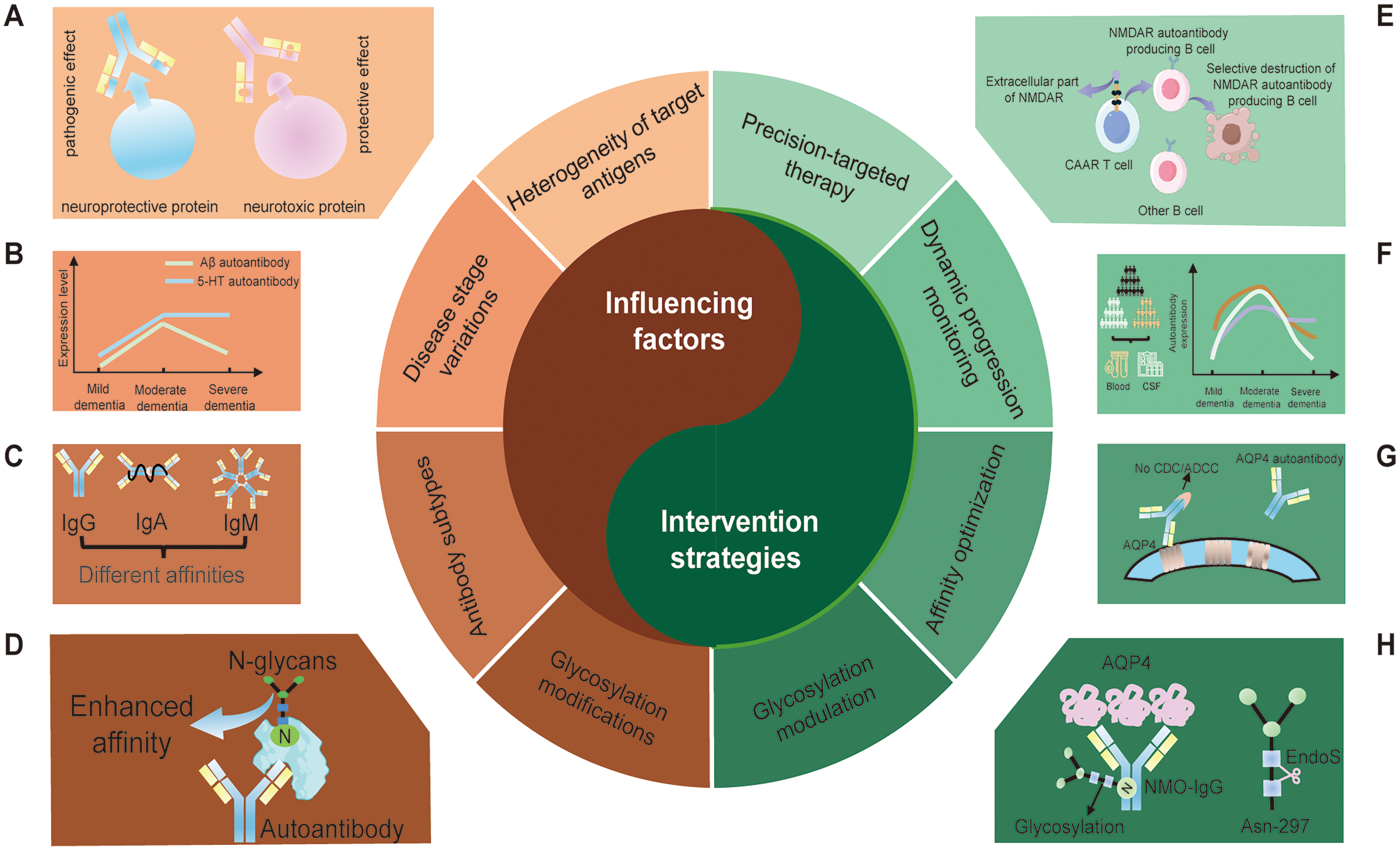
Factors influencing autoantibody function and related therapeutic strategies. (A) Variability in autoantibody responses specific to target antigens. (B) Variations in autoantibody concentrations across distinct stages of the disease. (C) Autoantibodies exhibit varying affinities for each specific subtype. (D) Modifications in glycosylation influence the affinity of autoantibodies (By figdraw). (E) A promising avenue of research involves the development of immunotherapies with high selectivity for specific antibodies (By figdraw). Chimeric autoantibody receptor (CAAR) T cells are designed to identify and eliminate B cells that are monospecific for a particular autoantibody, such as those targeting the N-methyl-D-aspartate receptor (NMDAR). This is achieved through the extracellular presentation of the target autoantigen. (F) Continuous monitoring of multiple autoantibody levels across various stages of the disease. (G) High affinity AQP4 monoclonal autoantibody competitively binds to antigen and inhibits the deleterious effects of low-affinity polyclonal IgG on host tissues. (H) Deglycosylation modification of AQP4 autoantibodies diminishes their capacity to mediate immune responses.
Precision targeted therapy
Precision-targeted therapy represents a pivotal approach in antibody intervention for AD, demonstrating substantial therapeutic efficacy by augmenting beneficial autoantibodies or neutralizing detrimental ones. Endogenously occurring Aβ autoantibodies are believed to confer protective benefits, and empirical evidence suggests that these autoantibodies exert more favorable effects compared to intravenous immunoglobulin in murine models of AD. 44 Jian et al. restored the diminished anti-Bim autoantibody using purified NAbs-Bim led to enhanced cognitive function in AD mice, alongside a reduction in cerebral Aβ and phosphorylated tau levels. 53 Additionally, antibody clinical trials targeting Aβ have thus far yielded modest cognitive benefit. Donanemab, an artificial antibody therapy, partially compensates for the deficiency of natural Aβ autoantibodies in patients with AD. However, its administration is associated with an elevated risk of treatment-related adverse events, including cerebral edema and microhemorrhages. 73 Despite the modest absolute clinical benefit observed, these findings substantiate that exogenous anti-Aβ immunotherapy represents a viable strategy for disease modification. In addition, therapies that intercept potentially harmful autoantibodies such as CAAR T cells have become a prominent area of research. This innovative approach involves the genetic engineering of T cells to selectively eradicate B cells that produce pathogenic autoantibodies, while sparing those that generate protective antibodies. 74 This methodology entails the extraction of T cells from the patient, followed by their genetic modification to express specific antigens, such as NMDAR, on their surface and to incorporate intrinsic activation domains that stimulate T cell receptors. Upon reintroduction into the patient, these CAAR T cells disseminate throughout the body. They selectively target and eliminate B cells that express autoantibodies against NMDAR, while sparing those that produce protective autoantibodies. 75 In murine models, this technique has demonstrated both efficacy and specificity in treating autoantibody-mediated neurological disorders, including NMDAR encephalitis and myasthenia gravis.75,76 Additionally, a comparable approach, which employs an anti-CD138 antibody in conjunction with ovalbumin to target and eliminate long-lived plasma cells, has been proposed as a means of achieving highly specific and durable therapeutic outcomes. 77 In summary, the evidence indicates that the supplementation of beneficial autoantibodies or the elimination of detrimental autoantibodies is feasible. However, the removal of harmful autoantibodies may inadvertently deplete normal B cells, potentially compromising immune function and elevating the risk of other autoimmune disorders. Future research should comprehensively address these adverse effects to enhance the prospects for clinical application.
Dynamic monitoring of disease stages
To address the issue of inconsistent findings in autoantibody expression levels, it is essential to implement dynamic monitoring of disease stages. Large-scale, multicenter longitudinal cohort studies can track the dynamic alterations in autoantibody profiles across various disease stages in real-time. This approach not only facilitates accurate staging of diagnoses but also enables precise interventions tailored to different disease phases. Research indicates that the interaction between central and peripheral immune systems during the progression of AD is marked by significant dynamic changes and heterogeneity, with distinct immune response patterns potentially emerging at different stages. 78 Moreover, it has been demonstrated that dynamic fluctuations in specific autoantibodies can reliably predict the onset and progression of AD, serving as potential biomarkers and targeted therapeutic tools. 10 Consequently, integrating longitudinal monitoring data to examine the relationship between autoantibody profiles and the AD process is anticipated to provide a crucial foundation for early screening, staged diagnosis, and personalized intervention strategies.
Precise regulation of glycosylation modifications
In recent years, the role of glycosylation modifications in immunomodulation has garnered significant scholarly interest. Modifications in antibody glycosylation are critical in modulating pro- and anti-inflammatory activities. Specifically, sialylated or galactosylated autoantibodies are associated with enhanced anti-inflammatory effects and reduced pathogenicity, whereas deglycosylation leads to a decrease in their immunomodulatory capabilities.79,80 Recent advancements in therapeutic strategies, including glycoengineered IVIg and monoclonal antibody, glycosylase -based methods and chemical glycosylation, have shown promise in the context of autoimmune diseases. 64 Although this approach remains in the experimental phase, its potential implications for immunomodulation in AD warrant further exploration.
Optimization of antibody affinity
The affinity of autoantibodies plays a crucial role in their functionality within AD. Enhancing the structural configuration of autoantibodies to augment their affinity and specificity can significantly mitigate their immunotoxin effects on self-tissues. For instance, a high affinity AQP4 monoclonal autoantibody was able to inhibit the detrimental effects of low affinity polyclonal IgG on self-tissues by competitively binding to the antigen. 81 Furthermore, the transformation of Aβ autoantibodies into a single-chain format, along with the multimerization of the scFv-Fc structure, modified the overall conformation of Aβ autoantibodies. This modification not only enhanced their affinity but also optimized their functional properties, thereby offering new potential for protective effects. 82
Conclusion
In conclusion, autoantibodies in AD present a highly intricate dualistic profile, with their pathogenic or protective roles influenced by a multitude of factors, including variations in target antigens, disease stage, autoantibody subtypes, glycosylation modifications, and individualized immunogenetic backgrounds. This review critically examines the existing literature to outline a range of regulatory strategies aimed at these variables. Precision targeted therapies, such as chimeric antigen receptor CAAR T-cell therapy, glycosylation modifications, and the optimization of autoantibody affinity, have been pivotal in modifying autoantibody immunity patterns. Nonetheless, the clinical applicability of these strategies requires further investigation, particularly given the lack of clarity regarding autoantibody dynamics across different disease stages and the specific impact of individual immune status and genetic background on immune regulation, which necessitates comprehensive exploration. Future research should utilize large-scale, multi-center clinical cohorts to integrate multi-omics data, enabling a dynamic analysis of the evolving patterns of autoantibodies. This approach will facilitate the development of precise treatment strategies informed by the specific characteristics of antibodyomics. Concurrently, efforts should be directed towards optimizing the specificity of targeted antigenic epitopes and incorporating glycosylation modification regulation strategies. Such advancements are expected to enhance precision immunotherapy and expedite the translation of these findings into clinical practice.
Footnotes

Author contributions
Funding
The authors disclosed receipt of the following financial support for the research, authorship, and/or publication of this article: This work was supported by the National Natural Science Foundation of China, STI2030-Major Projects (grant No.82471450, No.2021ZD0201802).
Declaration of conflicting interests
The authors declared no potential conflicts of interest with respect to the research, authorship, and/or publication of this article.