



Abstract
Background
Environmental noise pollution is increasingly recognized as a contributor to neurodegenerative processes, yet its relationship with early Alzheimer's disease biomarkers remains unclear.
Objective
This pilot study aimed to assess the feasibility of using gray-to-white matter signal intensity contrast (GWC) as a potential biomarker to explore associations between environmental noise exposure and early neurodegenerative changes.
Methods
A total of 106 participants (mean age 35.97 ± 9.21 years, range 20–55), without cognitive impairment or neurological disorders, were included. Environmental noise levels were estimated using spatial interpolation from the National Noise Information System. Based on WHO guidelines (>60 dB daytime or >55 dB nighttime), participants were categorized into high- and low-noise groups. Whole-brain and regional GWC values were derived from 3D T1-weighted MRI using FreeSurfer. Correlations between noise exposure and GWC were analyzed with Pearson's correlation.
Results
The high-noise group exhibited elevated whole-brain GWC values (20.11 ± 0.93) compared with the low-noise group (19.68 ± 0.96; p = 0.036). Regional analyses revealed higher GWC in the superior frontal gyrus, precentral gyrus, and paracentral lobules (all p < 0.05, FDR corrected). Nighttime noise exposure correlated more strongly with increased GWC (r = 0.203, p = 0.037) than daytime exposure.
Conclusions
This pilot study provides preliminary evidence of an association between environmental noise—particularly nighttime exposure—and subtle structural brain changes, as indicated by elevated GWC values. These findings suggest a potential neurobiological pathway linking noise exposure to early markers of neurodegeneration, warranting validation in larger, longitudinal studies.
Keywords
Introduction
Noise pollution is an inevitable byproduct of global urbanization and industrialization. While its auditory effects, such as hearing loss, are well established, increasing evidence suggests that noise exposure has far-reaching non-auditory consequences, including impacts on the cardiovascular, endocrine, and nervous systems. In particular, chronic noise exposure has been linked to neurodegenerative diseases, including Alzheimer's disease, through mechanisms such as chronic inflammation, increased cortisol levels, and disrupted sleep-wake cycle.1–3 Despite these insights, the biological pathways connecting environmental noise and neurodegeneration remain poorly understood.
Alzheimer's disease is the most common form of dementia, characterized by the accumulation of amyloid-β (Aβ) plaques, tau protein hyperphosphorylation, and subsequent neuronal degeneration. 4 Recent studies have highlighted the cortical gray-to-white matter signal intensity contrast (GWC) as a promising biomarker for early neurodegenerative changes.5–7 GWC, derived from conventional T1-weighted MRI, quantitatively assesses the contrast between gray and white matter, providing a non-invasive measure of structural brain alterations. Previous studies have demonstrated that GWC abnormalities are associated with Alzheimer's disease biomarkers, such as Aβ deposition, tau pathology, and cortical thinning—key pathological features of Alzheimer's disease. 5 In psychiatric research, GWC has also been interpreted as a surrogate marker of intracortical myelination. Altered GWC values have been consistently reported in specific cortical regions across a range of psychiatric disorders, including schizophrenia, bipolar disorder, and obsessive-compulsive disorder.8,9 These abnormalities have been associated with medication exposure, cognitive performance, 9 and microstructural white matter changes, 10 suggesting that intracortical myelin disruption may contribute to the pathophysiology and clinical symptoms of these conditions. Importantly, GWC has shown greater sensitivity than traditional metrics, such as cortical thickness, in detecting early neurodegeneration. 6
However, no prior research has explored the relationship between environmental noise exposure and GWC. This gap is critical, as environmental noise is a modifiable risk factor with significant public health implications. 11 Identifying its impact on neurodegeneration could pave the way for preventive interventions targeting noise pollution.
In this study, we examined the association between environmental noise exposure and GWC in a cohort of participants without cognitive impairment or neurological abnormalities. Using high-resolution MRI data and robust spatial noise exposure estimates, we investigated both whole-brain and regional GWC alterations associated with daytime and nighttime noise levels. In this study of younger adults (mean age of approximately 36 years) without cognitive impairment or neurological abnormalities, we sought to generate exploratory evidence on the association between environmental noise exposure and GWC. Using high-resolution MRI data and spatial noise exposure estimates we sought to evaluate the feasibility of methods and potential relationships to guide future larger-scale studies.
Methods
Participants and data
The datasets utilized in this study have been described previously.12,13 Specifically, we combined data from an existing regional occupational health and safety cohort study, 14 along with magnetic resonance imaging (MRI) data obtained through a research project aimed at examining associations between occupational conditions and brain structural outcomes. Between 2021 and 2023, additional volunteers, primarily healthcare workers from a large regional hospital, contributed to extra MRI assessments. Of the 137 participants, 31 were excluded from the final analysis. The reasons for exclusion are as follows: 22 participants were excluded due to insufficient demographic or noise exposure data, such as residential address information necessary for accurate spatial interpolation, and 9 participants were excluded due to low-quality MRI images that were not suitable for GWC analysis. Therefore, the final analysis included 106 participants with complete and high-quality MRI image data. Basic characteristics included in sex, educational level, dominant hand, and type of work. This study, involving human participants, was reviewed and approved by the Institutional Review Board (IRB) of Chung-Ang University (IRB number: 1041078-20231024-HR-287). All the participants provided written informed consent to participate in the study.
Noise exposure level
Environmental noise data were obtained from the National Noise Information System (NNIS; https://www.noiseinfo.or.kr/) in 2021, which provides extensive noise measurement data across various urban regions in South Korea. To estimate individual noise exposure levels, precise residential address information was collected from each participant. Due to incomplete spatial coverage of noise measurements, noise levels in unmeasured areas were estimated using Empirical Bayesian Kriging (EBK), a widely used geographic information system interpolation technique. However, the NNIS did not publicly provide validation statistics for the EBK model, limiting our ability to quantitatively evaluate interpolation accuracy. This methodological limitation should be acknowledged, and future research should incorporate model performance assessments to improve the reliability of noise exposure estimates. EBK is a spatial interpolation method that is based on an iterative process of subsets and simulations and is an effective approach for interpolating large areas of non-stationary data because it ensures stable performance even with small datasets. 15 For noise interpolation, the EBK method was used to generate a statistical noise surface from the collected noise data that covered the target area. This statistical noise surface comprised a grid that stored the estimated noise values. The noise values per administrative region were obtained by calculating the representative values of the overlapping regions on the noise surface, and the average of the noise values stored in the overlapping grid was calculated as the noise value for each region using zone-specific statistics. 16
The NNIS selected the urban land use classifications of three general sites and two roadside sites. Noise was measured during two time periods, daytime (6 am–10 pm) and nighttime (10 pm–6 am), with two nighttime measurements at 11 pm and 1 am. In this study, we used noise data from manual measurement methods, which are considered of better quality and more reliable than automated measurement data due to controlled measurement conditions and expert oversight. 17 Therefore, we converted the noise data from the measurement points into average values and used geographic coordinates.18,19 Participants were divided into two groups based on their noise exposure levels. Cutoff values of >60 dB daytime noise or >55 dB nighttime noise represented moderate noise levels and were chosen based on the WHO-recommended noise guidelines of 2009.18,20
MRI data acquisition and processing
A three-dimensional T1-weighted magnetization-prepared rapid gradient echo (T1w MPRAGE) sequence was acquired using the following parameters: repetition time, 1970 ms; echo time, 2.84 ms; inversion time, 991 ms; field of view, 256 × 256; flip angle, 9°; in-plane resolution, 0.5 × 0.5 × 1 mm3; number of slices, 192; and, scan time, 4 min 34 s.
The GWC values were derived from the acquired T1w MPRAGE images using “recon-all” in Freesurfer. 21 This process involved the exclusion of non-brain tissue, brain intensity normalization, brain segmentation, and vertex-based surface mapping to delineate the gray/white matter boundary (white matter surface) and gray matter/cerebrospinal fluid boundary (pial surface). Gray matter signal intensity (G) was measured at 30% of orthogonal distance from the white matter surface to the pial surface. White matter signal intensity (W) was measured 1.0 mm below the white matter surface, and the GWC values were calculated for each cerebral vertex using the following Eq1 22 :
GWC=100 * (W-G) / 0.5 * (W+G) (1)
To obtain the average whole-brain GWC value, the sum of the GWC values across all vertices was divided by the total number of vertices. The regional GWC values were calculated using the Desikan–Killiany atlas, which subdivides the cortex into 68 gyral-based regions of interest (ROIs). 23
Statistical analysis
We tested the normality of the GWC data using the Shapiro–Wilk test. The whole-brain GWC values were normally distributed (p = 0.405, all participants; p = 0.478, high-noise exposure group; p = 0.075, low-noise exposure group). Therefore, we used parametric analysis for further statistical tests.
Given the exploratory and pilot nature of this study, our statistical analyses primarily aimed to describe preliminary associations, estimate effect sizes, and identify potential trends rather than to conduct formal hypothesis testing. We descriptively compared GWC values between the high- and low-noise exposure groups using Student's t-tests, interpreting the results in terms of effect sizes and exploratory trends rather than emphasizing statistical significance. However, the resulting p-values from the regional GWC values were adjusted using false discovery rate (FDR) correction for multiple comparisons to guide future study design. Similarly, Pearson correlation analyses were performed to preliminarily explore potential relationships between GWC values and noise levels (daytime and nighttime), with results interpreted descriptively. Due to the limited statistical power inherent in our pilot study design, the findings are intended to generate hypotheses and guide future research rather than provide definitive statistical inference. All statistical analyses were performed using matlab 2024a (The Mathworks, Natick, MA, USA).
Results
A total of 106 participants were included in the study, with 76 participants (71.7%) in the high-noise exposure group and 30 participants (28.3%) in the low-noise exposure group. No differences were observed between groups in terms of sex, education level or occupation type. The mean age was slightly higher in the low-noise exposure group (Table 1).
Baseline characteristics of study participants according to environmental noise exposure level.
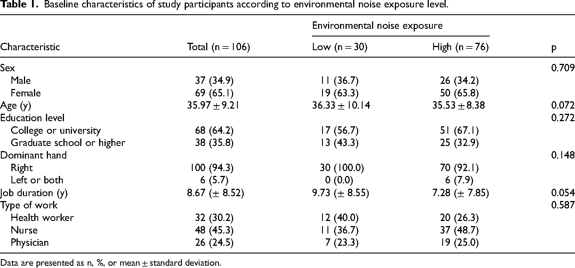
Data are presented as n, %, or mean ± standard deviation.
The whole-brain GWC was higher in the high-noise exposure group (20.11 ± 0.93) compared to the low-noise exposure group (19.68 ± 0.96; Table 2), indicating a potential 2.2% increase in the high-noise group. The GWC values in the high-noise exposure group were higher than those in the low noise exposure group in the left (5.9% higher) and right (6.0% higher) precentral gyri, left (5.9% higher) and right (6.3% higher) paracentral lobules, left caudal middle frontal gyrus (5.4% higher), and left superior frontal gyrus (7.7% higher). No regions were found where the low-noise exposure group exhibited higher GWC values than those in the high-noise exposure group. Figure 1 shows surface maps of GWC values averaged for high noise exposure group. Visual inspection revealed that the left superior frontal, caudal middle frontal, paracentral, and precentral gyri exhibit increased GWC values.

Surface maps of gray matter to white matter signal intensity contrast (GWC) values averaged across participants in the high noise exposure group (top row) and low noise exposure group (middle row), and significant comparison results (bottom row). In the bottom row, highlighted vertices indicate regions with higher GWC in the high noise exposure group compared to the low noise exposure group (Student's t-test, p < 0.05).
Group differences with statistical significance according to noise exposure level by gray matter to white matter signal intensity contrast.

p-values are corrected for false discovery rate.
Participants were categorized into high and low noise exposure based on overall noise exposure criteria defined by WHO guidelines (daytime noise >60 dB or nighttime noise >55 dB). The high-noise exposure group reflects elevated noise exposure during either daytime or nighttime periods.
Figure 1 shows surface maps of GWC values averaged for high noise exposure group. Visual inspection revealed that the left superior frontal, caudal middle frontal, paracentral, and precentral gyri exhibit increased GWC values.
Positive correlations were found between nighttime noise levels and whole-brain GWC (r = 0.203, p = 0.037; Figure 2). Regional GWC values in the precentral gyri, paracentral lobules, and frontal gyri were positively correlated with both daytime and nighttime noise levels (p < 0.05)” to “Positive correlations were found between nighttime noise levels and whole-brain GWC (r = 0.203; Figure 2). Regional GWC values in the precentral gyri, paracentral lobules, and frontal gyri were positively correlated with both daytime and nighttime noise levels. Correlation coefficients were consistently higher for nighttime noise levels compared to daytime noise levels, potentially indicating a stronger association with nighttime noise exposure (Table 3).
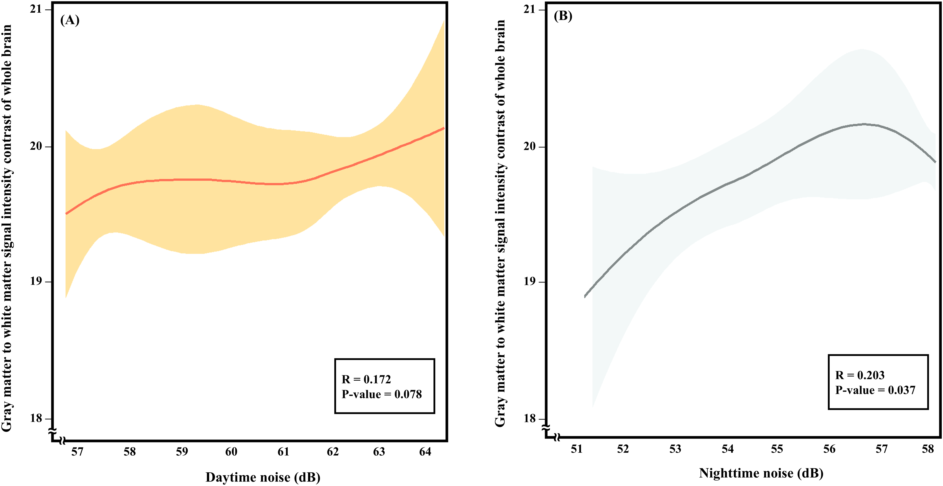
Trend plots between whole-brain gray matter to white matter signal intensity contrast values and daytime noise level (A) and night noise level (B).
Results of Pearson correlation analysis between gray matter to white matter signal intensity contrast and daytime or nighttime noise exposure level.
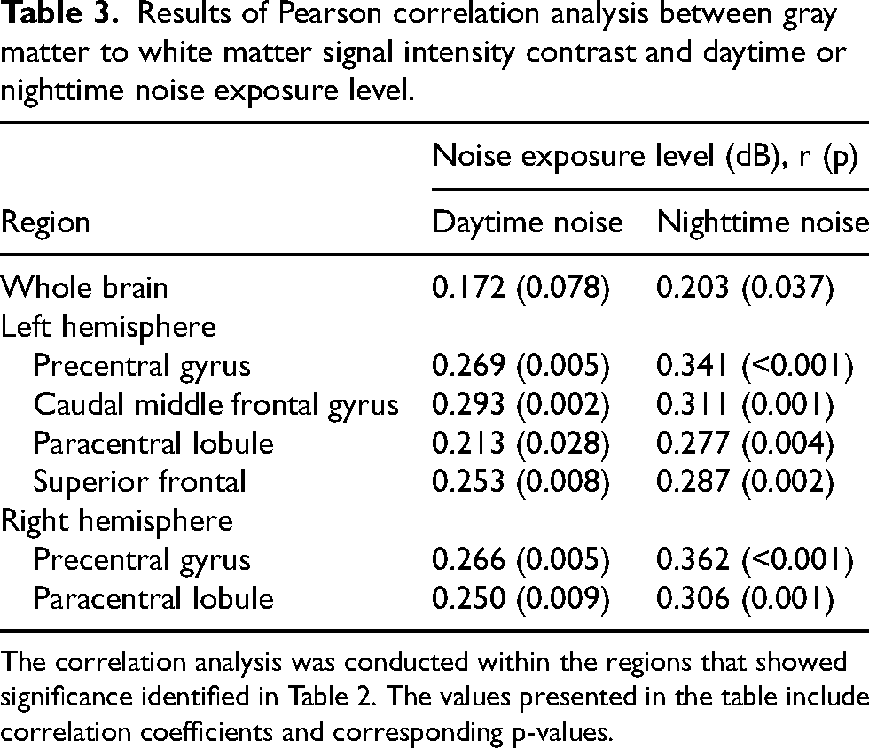
The correlation analysis was conducted within the regions that showed significance identified in Table 2. The values presented in the table include correlation coefficients and corresponding p-values.
Discussion
This pilot study provides preliminary evidence of a potential association between environmental noise exposure and alterations in GWC, a proposed biomarker of neurodegeneration. Participants residing in areas with higher estimated environmental noise levels tended to show elevated whole-brain GWC values, with specific regions such as the superior frontal gyrus, caudal middle frontal gyrus, and precentral and paracentral lobules showing more pronounced changes. Notably, nighttime noise exposure was more strongly correlated with GWC increases than daytime noise exposure, suggesting that nocturnal disturbances may play a critical role in neurodegenerative processes. These findings provide potential evidence linking environmental noise to preclinical brain changes associated with Alzheimer's disease, underscoring the importance of addressing noise pollution as a modifiable risk factor for neurodegeneration. In this relatively young group of participants (mean age approximately 36 years), our exploratory findings suggest potential associations between environmental noise exposure and alterations in GWC, emphasizing the possibility of early subclinical neurodegenerative changes detectable even decades before typical clinical onset.
Our preliminary findings suggest a potential link between environmental noise exposure and subtle alterations in brain structure, which may have implications for cognitive health, although further research is required to confirm these relationships. Previous studies on the effects of noise on adult brain structures have yielded inconsistent results. One study on military pilots demonstrated an adverse effect on the gray matter volume, 24 whereas another study indicated less local brain atrophy in the frontoparietal network. 25 Two studies reported unremarkable results for gyrification and cortical thickness.26,27 This discrepancy may arise because the study of noise and brain structure is relatively new, and the magnitude of the effect may be below the threshold of detection for the indicators employed in previous studies.
We used GWC as an indicator of changes in brain structure; it was linked to cognitive performance and cortical atrophy in regions associated with Alzheimer's disease.7,28,29 One study compared tau and amyloid PET with GWC and suggested that GWC could be used as a marker for early neurodegeneration in Alzheimer's disease. 5 Previous research has established a link between chronic environmental noise exposure and increased risk of neurodegenerative diseases, including Alzheimer's disease. 5 Mechanisms proposed to mediate these associations include chronic stress responses, inflammation, oxidative stress, sleep disruption, and dysregulation of circadian rhythms.1,4 Neuroimaging studies examining noise-induced changes in brain structures have predominantly utilized traditional measures such as cortical thickness, gray matter volume, or functional connectivity, with inconsistent or modest findings due to variability in methodological approaches and the subtlety of changes at early disease stages.24,25 The present study aims to address these limitations by utilizing the GWC, a potential MRI biomarker that captures subtle microstructural changes potentially indicative of early neurodegenerative processes. Unlike conventional measures, GWC may reflect subtle variations in tissue integrity and contrast, potentially providing increased sensitivity to early, preclinical brain changes that could precede overt structural atrophy or clinical symptoms by decades, although further research is necessary to empirically validate this hypothesis.6,22 Thus, investigating GWC could enhance early detection capabilities and clarify mechanistic links between environmental noise exposure and initial neurodegenerative processes.
Regions showing preliminary differences included the whole brain, bilateral precentral gyrus, paracentral lobule, left caudal middle frontal gyrus, and superior frontal gyrus, although these findings must be considered exploratory given the pilot nature of our study. The frontal gyri, caudal middle frontal gyrus, and superior frontal gyrus are responsible for higher cognitive and executive functions.30–32 Several studies have reported structural changes such as brain atrophy, reduced gray matter volume, and cortical thickness in these areas in neurocognitive disorders.33–36 In the middle frontal cortex, the GWC also showed a positive correlation with tau PET, but on the right side. 6 A study investigating the effects of aircraft noise exposure in military pilots revealed a reduction in resting functional connectivity in these areas, which were also observed on the right side. 24 Further studies are needed to clarify the left-right differences.
The precentral gyrus and paracentral lobule are involved in voluntary motor movement and the motor and sensory functions of the lower limbs, respectively.37,38 The relevance of these areas to Alzheimer's disease pathology and cognitive function has not been well reported. However, these areas also show a reduction in cortical thickness in patients with Alzheimer's disease.33,36 Furthermore, machine learning-based studies have classified both areas as Alzheimer's disease-related brain regions. 39 Glucose hypometabolism in the precentral frontal regions was found in a mild cognitive impairment group 40 and high perfusion changes in the paracentral lobule compensated for cognitive decline. 41
These exploratory findings suggest that nighttime noise exposure might have a stronger preliminary association with increased GWC compared to daytime exposure, highlighting a potential area for further investigation in future confirmatory studies. This heightened association may be explained by several factors. First, nighttime noise has been shown to disrupt sleep architecture, leading to fragmented sleep and reduced restorative sleep stages, such as slow-wave and rapid eye movement (REM) sleep.42,43 Sleep disturbances are closely linked to increased stress hormone levels, particularly cortisol, and elevated inflammatory cytokines, both of which are known to accelerate neurodegenerative processes. 44 Second, the human body's circadian rhythm plays a critical role in brain health, with nighttime serving as a period of essential neurophysiological repair and metabolic clearance, including the removal of Aβ via the glymphatic system. 45 Disruptions to this process caused by nocturnal noise may impair the brain's ability to clear neurotoxic waste products, contributing to structural brain changes observed in this study. Finally, nighttime noise tends to have a higher perceived annoyance factor than daytime noise, even at similar decibel levels. 46 This elevated annoyance may exacerbate stress responses, leading to sustained sympathetic nervous system activation and further dysregulation of the hypothalamic-pituitary-adrenal axis. 47 These mechanisms could amplify the impact of nighttime noise on neurodegeneration compared to daytime noise exposure. Overall, these exploratory results suggest that nighttime noise exposure might have distinct associations with brain health, which should be confirmed by future studies. Future studies should explore the physiological pathways linking nocturnal disturbances to neurodegeneration in greater detail, as well as investigate targeted interventions to mitigate the adverse effects of nighttime noise exposure.
An additional consideration regarding our findings is the relatively young average age (<40 years) of our participants, which is earlier than the typical age at clinical onset of Alzheimer's disease or related dementias. While neurodegenerative diseases like Alzheimer's disease are predominantly diagnosed later in life, current neuroscientific research highlights the importance of identifying subtle preclinical changes that occur decades before clinical symptoms manifest. Emerging evidence suggests that imaging biomarkers such as the GWC can detect early structural alterations linked to future cognitive decline and Alzheimer's disease pathology even in cognitively normal, younger adults.5,22,48 The significance of GWC as an imaging biomarker in younger populations thus lies in its potential sensitivity to subtle, preclinical neurodegenerative processes triggered by chronic exposure to environmental stressors, such as noise. Early detection of these subclinical structural brain changes offers a critical window for interventions aimed at modifying environmental risk factors well before irreversible cognitive impairment occurs.12,49 Consequently, our findings, although exploratory and preliminary, underscore the value of early and proactive public health strategies targeting modifiable risk factors like environmental noise to mitigate long-term risks for neurodegenerative diseases. Future longitudinal research will be essential to validate the clinical implications of early GWC alterations and to further elucidate their role as predictors of neurodegenerative pathology over the lifespan.
This study did not directly compare GWC with traditional neuroimaging biomarkers for Alzheimer's disease such as cortical thickness or volumetric alterations.50,51 Although not a neuroimaging approach, all participants in this pilot study undertook cognitive function assessment using the Korean version of the Mini-Mental State Examination for Dementia Screening (MMSE-DS). 52 In the present study, all participants exhibited normal cognitive scores on the MMSE-DS, limiting meaningful comparisons between cognitive function and GWC. Furthermore, direct comparison with traditional volume-based neuroimaging markers such as cortical thickness or gray matter volume was not conducted, representing an important limitation. Future studies should directly compare GWC with these traditional imaging measures to clarify whether GWC provides additional sensitivity or unique insights into early, subtle brain changes associated with neurodegeneration.
This study has several limitations that should be considered when interpreting the findings. One key limitation is related to the study population. Our participants were exclusively medical facility workers, who may differ from the general population in terms of their demographic characteristics, socioeconomic status, health behaviors, and baseline cognitive and physical health status. Consequently, the generalizability of our results to broader, more diverse populations is limited. The observed association between noise exposure and GWC changes may thus differ in magnitude or direction when studied in a general or community-based population. Future studies should incorporate broader and more heterogeneous samples to confirm the robustness and extend the applicability of our findings. Such research could better clarify the relationship between environmental noise exposure and early biomarkers of neurodegeneration across diverse populations. Second, the sample size was relatively small (n = 106), as this research was conducted as a pilot study to explore the feasibility of using the GWC as a biomarker for noise-induced neurodegeneration. Due to the small sample size and exploratory design, our pilot study results do not permit robust conclusions about causality or precise estimation of effect sizes. Future studies with larger samples are necessary to confirm these preliminary observations. Third, the uneven distribution of participants between the high-noise and low-noise exposure groups (76 versus 30) reflects the characteristics of the study region, which is heavily industrialized and includes major port and airport facilities. These environmental factors lead to higher average noise levels, resulting in a greater proportion of participants in the high-noise group. Although this context accurately represents real-world conditions in urbanized areas, it may limit the applicability of these findings to less industrialized or quieter regions. Future studies should recruit participants from a wider range of noise environments and expand on the insights from this pilot investigation to confirm and extend these findings. Fourth, the participants had normal cognitive function, which may not reflect all the factors that could affect cognitive function. Therefore, our results should be interpreted with caution. Further studies involving more correlates of cognitive function are required to confirm our results. Fifth, our estimates of location-based outdoor noise exposure may differ from the true indoor noise exposure levels. Although indoor and outdoor noise levels are well correlated, this limitation needs to be overcome through further research.53,54 Sixth, an additional limitation of our study is the lack of information on participants’ individual use of hearing protection devices. Such protective measures could substantially mitigate noise exposure, resulting in variability in actual exposure levels among participants categorized similarly based on environmental noise measurements alone. Future research should incorporate detailed assessments of personal protective equipment usage to better quantify individual exposure variability and to more accurately estimate the relationship between environmental noise and brain health outcomes. Finally, another limitation is the lack of adjustment for potential socioeconomic and environmental confounders. Although our analyses employed simple statistical comparisons without explicitly adjusting for confounders, participants in our study were homogeneous regarding key socioeconomic factors such as education, income levels, and residential environment—all resided in urban settings with similar SES characteristics. This homogeneity reduces but does not entirely eliminate the risk of confounding effects of SES on the observed associations between noise exposure and GWC changes. Future studies should utilize more detailed analytical approaches, such as propensity score matching, to explicitly adjust for socioeconomic and residential environmental variables, ensuring robust conclusions about the impact of environmental noise exposure on brain health.
This study provides potential evidence linking environmental noise exposure to early neurodegenerative changes, as measured by GWC, a promising biomarker for Alzheimer's disease. Participants exposed to higher noise levels, particularly nighttime noise, exhibited elevated GWC values both globally and in specific brain regions associated with cognitive and executive functions. These preliminary findings suggest potential associations between environmental noise exposure, particularly at nighttime, and changes indicative of neurodegeneration, demonstrating feasibility and informing the design of subsequent hypothesis-driven studies. As a pilot study, this research lays the groundwork for future investigations with larger, more diverse populations and refined methodologies to validate and expand upon these findings. Further research is needed to elucidate the causal pathways linking noise exposure to neurodegeneration and to develop evidence-based strategies for prevention and risk reduction.
Footnotes
Acknowledgements
The authors have no acknowledgments to report.
Ethical considerations
The studies involving human participants were reviewed and approved by the Chung-Ang University Ethics Center (IRB number: 1041078-20231024-HR-287).
Consent to participate
All the participants provided written informed consent to participate in the study.
Consent for publication
Not applicable.
Author contribution(s)
Funding
The authors disclosed receipt of the following financial support for the research, authorship, and/or publication of this article: This work was supported by the National Research Foundation of Korea, (grant number NRF-2021R1C1C1008871).
Declaration of conflicting interests
The authors declared no potential conflicts of interest with respect to the research, authorship, and/or publication of this article.
Data availability statement
Environmental noise data are available from the National Noise Information System (URL:  ). Other data are available in Lee et al. (2022).
14
). Other data are available in Lee et al. (2022).
14