



Abstract
Background
Recent animal studies have revealed STING (Stimulator of interferon genes) as a potential key player in Alzheimer's disease (AD). The actual impact of human STING on AD, however, is unknown. Mouse STING studies were done in WT/WT. However, TMEM173, the human gene encodes STING, has 5 common, distinct, sometimes opposite functional alleles that result in 25 TMEM173 genotypes. Only ∼50% of whites, 36% of African Americans (AA), 22% of East Asians are WT/WT. Past STING cancer immunotherapy clinic trials, which did not consider human TMEM173 heterogeneity, all failed.
Objective
(1) Discover new protective and risk AD genetic factors across populations or AA-specific. (2) Establish the physiological significance of common human TMEM173 genotypes and human diseases.
Methods
We conduct a large-scale (∼15,000 individuals) case-control analysis between TMEM173 genotypes and AD using data from The National Institute on Aging Genetics of Alzheimer's Disease Data Storage Site. The data include late-onset AD (LOAD) non-Hispanic White (NHW), early-onset AD (EOAD) NHW, and AA.
Results
A common H232/HAQ TMEM173 genotype is associated with AD protection across the populations. An AA-specific TMEM173 genotype H232/Q293 increases the risk for AA males (OR = 17.7148), especially in the APOE ε3/ε3 population.
Conclusions
The findings discovered the first AA-specific high AD risk factor and established an association between human TMEM173 and AD, paving the way for STING-targeting effective AD healthcare.
Introduction
Today, nearly 7 million Americans live with Alzheimer's disease (AD), a debilitating neurodegenerative disease. Sadly, from 2000 to 2022, deaths from AD increased by 142.4% in the United States (US). 1 In contrast, death from heart disease decreased by 1.1% during the same time period. 1 Novel AD research, especially human AD research, is urgently needed to reverse this AD death trend. Human AD research has revealed the enrichment of microglia-expressing immune genes as top genetic risk factors for late-onset AD (LOAD, ≥65 years old).2–4 In the last two years, the microglia cyclic GMP-AMP synthase (cGAS) - stimulator of Interferon Genes (STING) immune pathway has emerged as a possible key player in AD, inducing neuroinflammation, neuronal damage, and cognitive decline.5–8
cGAS senses DNA and generates cyclic GMP-AMP, which is a ligand for STING. 9 The activation of STING generates type I IFN, TNF, and lymphocyte cell death. 9 Recent studies using mouse models of AD and cGAS-STING inhibitors have shown that cGAS–STING signaling is involved in neurodegenerative diseases, including AD. 6 9–13 Using an inducible, microglia-specific cGAS knockout mouse model in the 5xFAD background, it was revealed that deleting microglial cGAS at the onset of amyloid-β (Aβ) pathology profoundly restricts plaque accumulation and protects mice from Aβ-induced cognitive impairment and neurotoxicity. 5 Consistently, deletion of STING in 5xFAD mice lowered Aβ load, improved alterations in microglial activation status, and protected against neuritic dystrophy and cognitive decline. 8 Meanwhile, pharmacological STING inhibition significantly reduced the Aβ load, tau phosphorylation, and prevented memory loss in AppNL-G-F/hTau double-knock-in mice. 14 Treating human AD macrophages (examined postmortem) with the STING inhibitor H-151 increased the uptake of Aβ after 2 h and increased the degradation of Aβ after 24 h. 15 Lastly, aging is a major risk factor for neurodegenerative diseases. cGAS-STING signaling may also be associated with aging. 13 Together, these findings suggest that the cGAS-STING pathway is a driver of detrimental immune responses associated with AD.
AD is a highly heritable disease. APOE ε4 is the strongest genetic risk factor for AD, especially early-onset AD (EOAD).16,17 In the non-Hispanic White (NHW) population, the APOE ε4/ε4 individuals have 14.9 times the odds of AD than the rest of the population. 1 18–20 Human STING is encoded by the TMEM173 gene that is highly heterogeneous in humans. 21 There are five common TMEM173 alleles (population frequency >1%): R232 (WT), H232, R71H-G230A-R293Q (HAQ), G230A-R293Q (AQ), and Q293 (Figure 1A-C).22,23 Haplotype analysis discovered that HAQ came from AQ, which was derived from Q293. 24 Molecular clock analysis showed a similar timeline with an age of H232 > Q293 > AQ > HAQ (Supplemental Figure 1A). 25 Interestingly, the HAQ allele is very young, only ∼1803.1 generations old (Supplemental Figure 1A). In Americans, < 50% are WT/WT. 22 In fact, in East Asians, WT/HAQ (34.3%), not WT/WT (22.0%), is the most common TMEM173 genotype. 23 The HAQ allele is defective in type I IFNs stimulation and STING activation-induced cell death.22,23,26 In a clinical trial (NCT02471014), we found that WT/HAQ individuals had reduced Pneumovax23®-induced antibody responses compared to WT/WT humans. 27 Anatomically modern humans outside Africa are descendants of a single Out-of-Africa migration 50,000∼70,000 years ago.28–31 The population frequency of WT/HAQ increased from 0.6% in Africans to 38% in East Asians. 24 On the contrary, the population frequency of WT/AQ decreased from 28% in Africans to 0.6% in East Asians. 24 Similarly, ∼8.14% of Africans are Q293 carriers, while ∼0.0% of East Asians have the Q293 allele (Supplemental Figure 2B). 24 The natural selection of WT/HAQ, WT/AQ, WT/Q293 indicates that HAQ, AQ, Q293 alleles are dominant over the WT allele in humans. The mouse TMEM173 gene is the equivalent of the human R232 (WT) allele. Thus, mouse STING research captures a minority of the human population (only people with WT/WT).
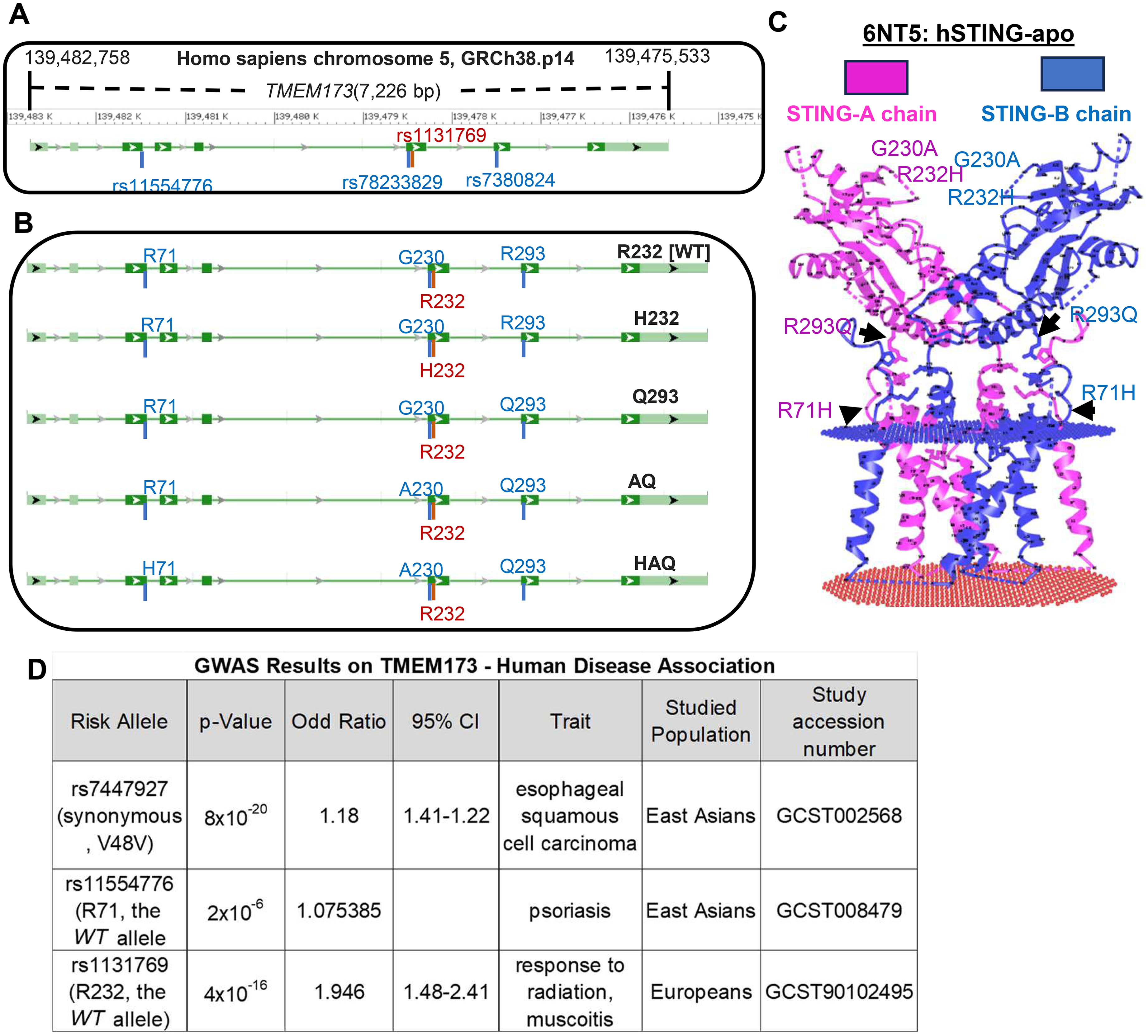
GWAS does not reveal major disease association with common human TMEM173 alleles. (A, B) An illustration of the genomic structure of the human TMEM173 gene (A), and the five common human TMEM173 alleles (B). (C) The cryo-EM structure of a full-length human STING dimer at the apo (PDB ID: 6NT5). The common human TMEM173 alleles are indicated. (D) GWAS results on the human TMEM173 gene from the NHGRI-EBI Catalog of human GWAS.
With five functionally distinct and dominant human TMEM173 alleles, there are twenty-five possible functionally distinct human TMEM173 genotypes. 21 This human TMEM173 feature is relevant. For example, in a PolSenior program that assessed the health and socioeconomic status of Polish Caucasian seniors (≥65 years, 3397 senior participants), the single nucleotide polymorphism (SNP) rs7380824 (R293Q) carriers were protected from aging-associated diseases (Odds Ratio [OR] = 0.823, p = 0.038).32,33 Subsequent analysis further showed that the rs7380824 carriers are associated with a decreased risk for obesity-associated combined aging-related diseases (OR = 0.651, p = 0.014).32,33 The rs7380824 SNP is shared by HAQ, AQ, and Q293 individuals. The AQ, Q293 alleles are African-specific. 23 Thus, in Europeans, three common TMEM173 genotypes, WT/HAQ (19.28% of Europeans), HAQ/HAQ (2.78%), HAQ/H232 (4.37%) 23 have the rs7380824 SNP. The study32,33 cannot distinguish which of the three TMEM173 genotypes (i.e., individuals) is protective in aging.
In this study, we conduct the first-ever common TMEM173 genotype association study in humans and hypothesize that functionally distinct human TMEM173 genotypes may be differentially associated with AD.
Methods
Study cohort
NG00067 – Alzheimer's Disease Sequencing Project (ADSP) Umbrella includes sequencing data and harmonized phenotypes from cohorts sequenced by ADSP and other AD and Related Dementia's studies. Samples are processed using a common workflow called VCPA (Variant Calling Pipeline and data management tool), a functionally equivalent CCDG/TOPMed pipeline. The study does not involve human subjects or animal subjects.
Procedures involving experiments on human subjects are done in accord with the ethical standards of the Committee on Human Experimentation of the institution in which the experiments were done or in accord with the Helsinki Declaration of 1975.
Sample sets analyzed
Three groups of sample sets from NG0067-ADSP Umbrella were analyzed: ADGC-AA-whole exome sequencing (WES) (snd10003), ADSP-FUS-whole genome sequencing (WGS) (snd10020, snd10031, snd10094), and EOAD-WGS (snd10032, snd10095) (Figure 2A).
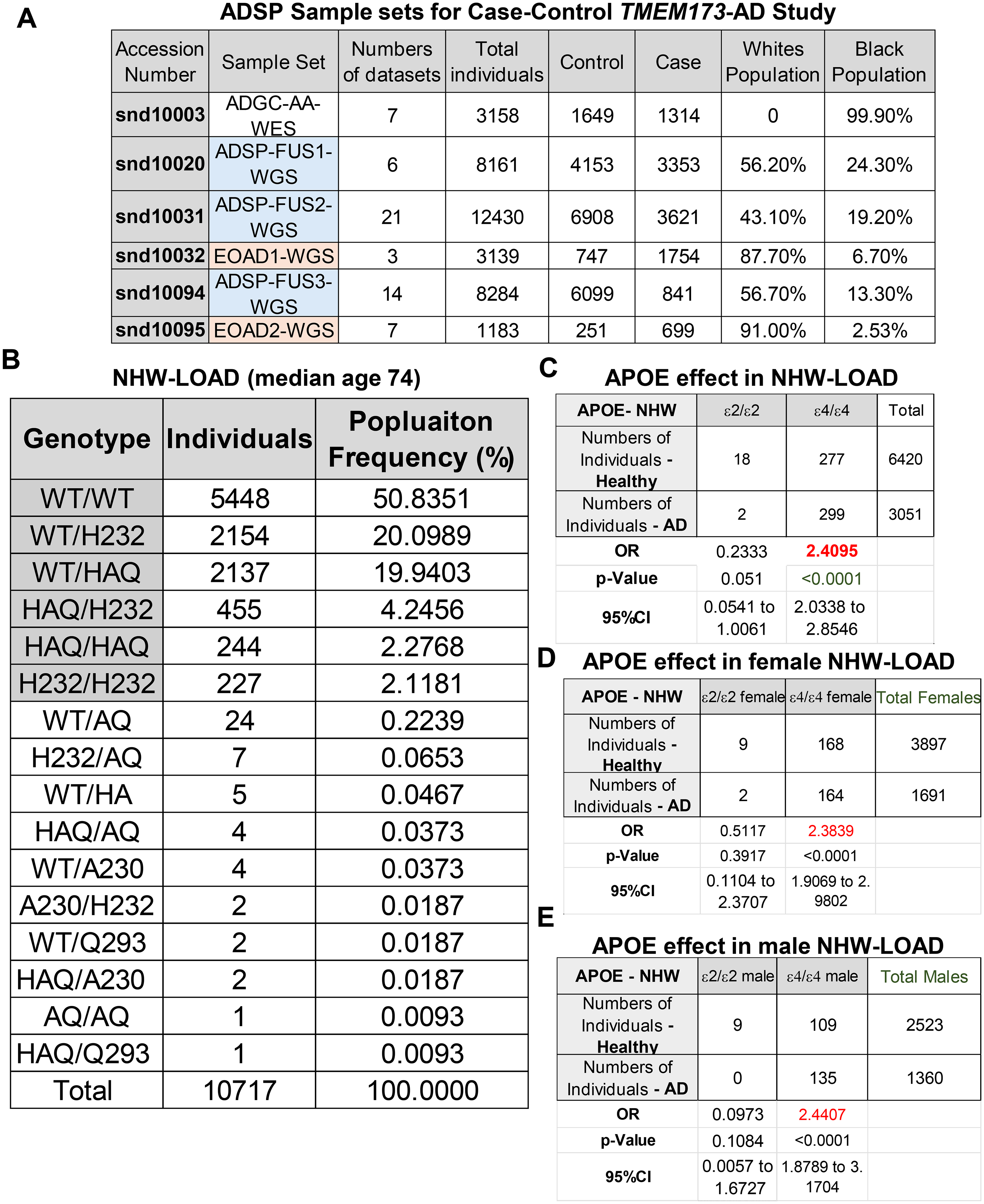
Case-control TMEM173 – AD study using ADSP sample sets. (A) A summary table of sample sets used in the case-control analysis. (B) A table of TMEM173 genotypes found in NHW LOAD from cohorts snd10020, snd10031, snd10094. (C-E) Case-control analysis of the APOE ε2/ε2, ε4/ε4, and sex impact on AD in NHW LOAD.
ADGC-AA-WES
This sample set is 99.9% African Americans. The Alzheimer's Disease Genetics Consortium (ADGC) selected subjects from the National Institute on Aging (NIA) Alzheimer‘s Disease Centers (ADCs), the University of Miami/Duke University, the Multi-Institutional Research in Alzheimer's Genetic Epidemiology Study, the Rush University Religious Orders Study and Memory and Aging Project, and the Genetic and Environmental Risk Factors for Alzheimer's Disease Among African Americans Study. All individuals self-identified as African American and had a minimum age of 60 years at onset (cases) or last exam (cognitively-normal controls). The case and control status of subjects is based on the National Institute of Neurological and Communicative Disorders and Stroke—Alzheimer's Disease and Related Disorders Association criteria. The John P. Hussman Institute for Human Genomics at the University of Miami Miller School of Medicine performed WES on 3200 samples. The Genome Center for Alzheimer's Disease (GCAD) at the University of Pennsylvania processed the data using their standardized pipeline.
ADSP-FUS
These sample sets are an NIA initiative focused on identifying genetic risk and protective variants for LOAD (onset age: >65). A concern in AD genetic studies is a lack of ethnic diversity. The ADSP-FUS sample set collects and sequences ethnically diverse and unique cohorts with clinical data from all populations. For example, ADSP-FUS1 contains 3250 AD cases, 4149 cognitively normal individuals, 194 individuals with mild cognitive impairment (MCI), and 567 with unknown or other dementia.
EOAD sample sets
Genomic studies of AD have primarily focused on NHW participants affected by the LOAD, or the study of EOAD (onset age ≤65) cases from families showing Mendelian inheritance patterns associated with mutations in the APP, PSEN1, and PSEN2 genes. However, mutations in these three genes explain ∼10% of EOAD cases. Studying EOAD in subjects without APP, PSEN1, and PSEN2 mutations is a critical gap that provides a unique opportunity for discovering novel therapeutic targets and molecular pathways. Inclusion criteria include AD cases with early onset (<65) and near-early (<70) onset, as well as cognitive controls. If APP, PSEN1, PSEN2 mutations have been typed, the individual must be negative. Samples are not excluded by race/ethnicity, so the total sample set includes non-Hispanic whites, Hispanic, and (limited) AA. The overall dataset consists of WGS derived from early-onset AD, MCI, and cognitive controls. Most “case” samples have onset 65 and under, though some up to 70 were included. All participants (AD, MCI, and cognitively intact) have standard neurocognitive/psychiatric exams and are evaluated under standard AD criteria. Primary phenotypes are AD, MCI, and cognitive controls. Phenotypes related to neurodegeneration and dementia may also be considered (neuropsychiatric phenotypes especially).
Case and control diagnostic criteria
ADSP Phenotype Harmonization Consortium (ADSP-PHC) derived inclusion and exclusion criteria for AD and control samples. Clinical AD cases were demented according to the National Alzheimer's Coordinating Center's cognitive status of dementia at the Uniform dataset (UDS) visit with a primary etiologic diagnosis of AD. Controls did not meet dementia or MCI criteria and exhibited no etiologic diagnoses. Neuropathologic definition of cases and control followed NIA-AA Alzheimer's disease neuropathologic change (ADNC) scores (ABC method), with intermediate or higher ADNC scores classified as Cases and low ADNC scores labeled Controls. When ADNC scores were not available, a similar approach was used with the BRAAK Staging System and the Consortium to Establish a Registry for Alzheimer's disease (CERAD) scores, with Cases possessing a BRAAK Stage greater than or equal to III and a CERAD score of either moderate or frequent neuritic plaques. Consistent with the ADNC definition, if a participant was lower on BRAAK or CERAD they were given a Control diagnosis (equivalent to a low score on ADNC). Individuals missing an ADNC score and either BRAAK or CERAD score were not given a neuropathologic diagnosis. Persons with Down's syndrome, neuropsychiatric, neurodegenerative, and neurologic disorders, brain structure abnormalities, non-AD tauopathies and synucleinopathies were excluded from both clinical and autopsy diagnoses of cases and controls. All autopsied controls had a clinical evaluation within two years of death. An autopsy-confirmed variable was derived from matching neuropath and clinical diagnoses when available. All cases and controls were required to be >60 years of age.
Statistics
The OR and 95% confidence interval are calculated according to Douglas G. Altman. 34 The p-value is calculated according to David J Sheskin. 35 Statistical analyses were performed using MedCalc for Windows, version 23.3.7 (MedCalc Software, Ostend, Belgium).
Results
GWAS fails to link common human TMEM173 alleles to major diseases
Animal studies have established a role for STING in infectious diseases, autoimmune diseases, cancers, lungs, liver, cardiovascular diseases, neurodegenerative diseases, obesity, and aging. 10 36–42 To establish a role for STING in human diseases, we examined the NHGRI-EBI Catalog of human genome-wide association studies (GWAS Catalog - https://www.ebi.ac.uk/gwas/genes/STING1). Surprisingly, there are only 3 significant hits for the TMEM173 gene (Figure 1D). Moreover, the OR are close to 1 (OR = 1 indicates no effect), i.e., rs7447927 (OR = 1.18), rs11554776 (OR = 1.075), and rs1131769 (OR = 1.946) (Figure 1D).
Several factors may explain the negative TMEM173 GWAS results. First, there are potentially 25 different TMEM173 genotypes, resulting from 5 common TMEM173 alleles (Figure 1B). For example, the GWAS study GCST008479 identified rs11554776 [R71H] as being associated with psoriasis in East Asians (OR = 1.075). There are three common TMEM173 genotypes in East Asians, containing the R71H allele: WT/HAQ (34.3% of East Asians), HAQ/HAQ (16.1%), and HAQ/H232 (13.1%). These different TMEM173 genotypes likely contributed to the weak rs11554776-psoriasis signal observed in GWAS. Second, common human TMEM173 alleles have distinct, sometimes opposite functions. 24 The strongest evidence came from a study showing that the HAQ allele was positively selected, while the AQ and Q293 alleles were negatively selected during the Out-of-Africa human migration. 24 (Supplemental Figure 1B). In contrast, the H232 allele was not naturally selected. 24 Lastly, some common human TMEM173 alleles are dominant. For example, during the out-of-Africa migration, the population frequency of WT/HAQ increased from 0.6% in Africans to 38% in East Asians, while the population frequency of WT/AQ decreased from 28% in Africans to 0.6% in East Asians. 24 Thus, HAQ and AQ alleles are dominant over the WT allele. In summary, due to the interplay of functionally distinct and often dominant human TMEM173 alleles, human disease association should be investigated at the TMEM173 genotype level, not SNPs or haplotypes, by GWAS. Here, we aimed to utilize case-control analysis to elucidate the physiological significance of the TMEM173 genotypes in humans.
Case-control analysis of TMEM173 genotypes and AD
We investigate the association between the TMEM173 genotypes and human AD. First, many animal studies have shown that cGAS–STING signaling is involved in neurodegenerative diseases, including AD.6,7,9,10–13,15,43–45 Second, the National Institute on Aging Genetics of Alzheimer's Disease Data Storage Site (NIAGADS) provides WGS or WES results, and AD phenotypes, including AD status, age, sex, and APOE status, from tens of thousands of individuals (Case and Control). The case and control status of individuals is based on the National Institute of Neurological and Communicative Disorders and Stroke—Alzheimer's Disease and Related Disorders Association criteria.
We focused on three population sets: the LOAD non-Hispanic White (NHW), the EOAD-NHW, and AA (Figure 2A). The LOAD-NHW were extracted from 3 sample sets: ADSP-FUS1, ADSP-FUS2, and ADSP-FUS3. The EOAD-NHW were extracted from 2 sample sets: EOAD1, and EOAD2 (Figure 2A). The EOAD-NHW cohort includes cases with early onset (≤65) and near-early (<70) onset, as well as cognitive controls. The EOAD-NHW were prescreened for negative APP, PSEN1, and PSEN2 mutations. Lastly, we added AA sample set (ADGC-AA-WES) because AA individuals have unique TMEM173 genotypes not found in non-Africans. 23
We first extracted NHW from ADSP-FUS1, ADSP-FUS2, ADSP-FUS3 (total 10,717 NHW) (Figure 2B). Next, we extracted TMEM173 genotypes from NHW in the ADSP-FUS sample sets, resulting in a total of 16 TMEM173 genotypes (Figure 2B). Six TMEM173 genotypes are common with a population frequency greater than 2% (Figure 2B). APOE ε4 is the #1 AD risk allele in humans.16,17 Third, we conducted case-control analysis. The OR, p-value, and 95% confidence interval are calculated according to Douglas G. Altman 34 and David J Sheskin 35 using MedCalc Software. The analysis confirmed that APOE ε4/ε4 is associated with increased AD risk (OR = 2.4095, p < 0.0001) in the NHW LOAD sample sets (Figure 2C). Furthermore, females (OR = 2.3839, p < 0.0001) and males (OR = 2.4407, p < 0.0001) have similar AD risks in the NHW LOAD (Figure 2D and E). Finally, we examined the association between the TMEM173 genotypes and AD in NHW LOAD.
The common H232/HAQ TMEM173 genotype is associated with AD protection in NHW LOAD
Among all 10,717 NHW-LOAD individuals, 9661 have a known AD status, including the Case (3204 individuals, median age 74) and Control (6457 individuals, median age 74) (Figure 3A and B). The H232/HAQ genotype is associated with AD protection (OR = 0.7556, p = 0.0128) (Figure 3C). The remaining 5 common TMEM173 genotypes do not show significant AD association (Figure 3C). Males and females had similar H232/HAQ - AD association (male: OR = 0.7484, p = 0.0782) (female: OR = 0.7489, p = 0.062) (Figure 3D). The presence of APOE ε4 does not affect H232/HAQ association with AD (Figure 3E). Next, we examined TMEM173 genotype association with EOAD.
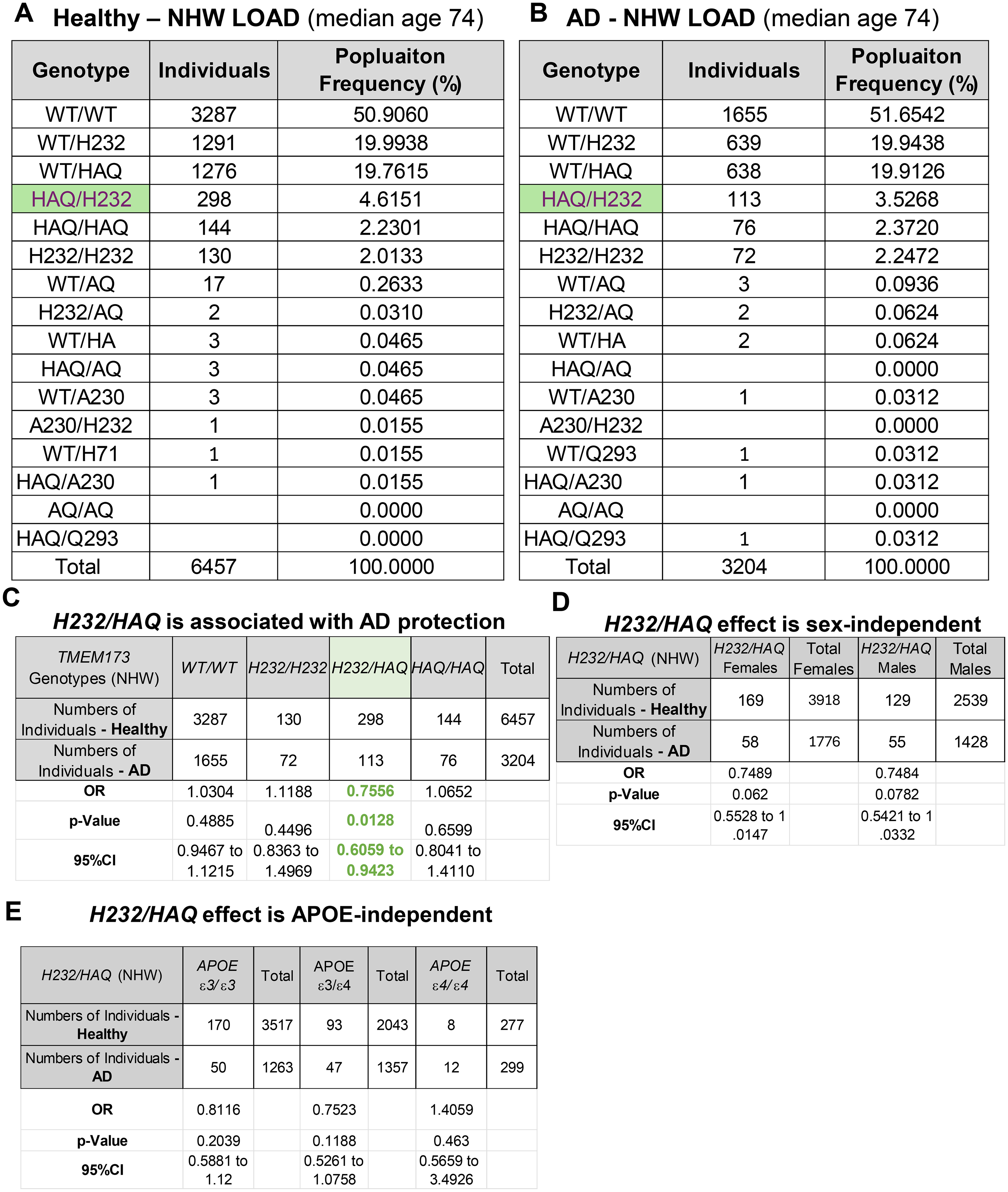
The H232/HAQ TMEM173 genotype is associated with AD protection in NHW LOAD. (A, B) TMEM173 genotypes in the case control samples from NHW LOAD. (C) Case–control analysis of H232/HAQ genotype and AD association. (D, E) Case-control analysis of the sex and APOE ε2/ε2, ε4/ε4 impact on AD–H232/HAQ association in NHW LOAD.
The common H232/HAQ TMEM173 genotype is associated with AD protection in NHW EOAD
Unlike LOAD NHW patients, EOAD NHW patients have a median age of 62 (Figure 4A and B). Furthermore, the Case (1722 individuals)-Control (776 individuals) (Figure 4A and B) study showed that APOE ε4/ε4 risk in EOAD NHW is over 10 times higher (OR = 32.1385) than LOAD NHW in both males (OR = 20.5772) and females (OR = 49.5584) (Figure 4C and D). Nevertheless, the study in EOAD NHW identified H232/HAQ genotype as associated with AD protection with a similar OR (OR = 0.5941, p = 0.0221) to LOAD (Figure 4E). Unlike the drastic OR difference by APOE ε4/ε4 between EOAD and LOAD, the OR of H232/HAQ is similar between EOAD and LOAD (Figure 3C and 4E). Lastly, similar to the LOAD NHW, the H232/HAQ association in EOAD is comparable in males and females (Supplemental Figure 2A), and APOE does not influence the H232/HAQ - EOAD association (Supplemental Figure 2B).
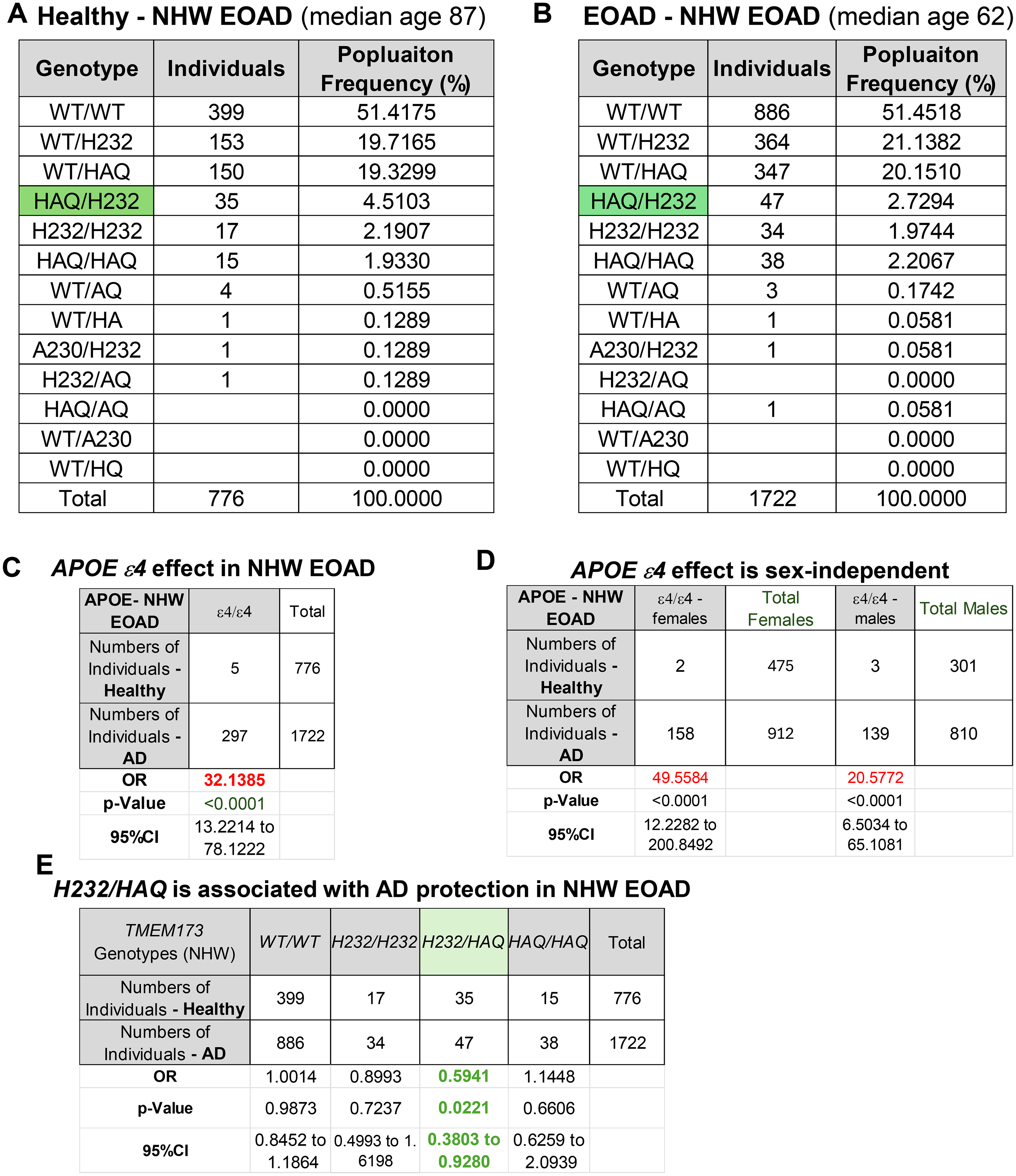
The H232/HAQ TMEM173 genotype is associated with AD protection in NHW EOAD. (A, B) TMEM173 genotypes in the case control samples from NHW EOAD. (C, D) Case-control analysis of the sex and APOE ε4/ε4 impact on AD association in NHW EOAD. (E) Case–control analysis of H232/HAQ genotype and AD association in NHW EOAD.
The common H232/HAQ genotype is associated with AD protection in AA
Lastly, we investigated AA for TMEM173 genotype and AD association. First, the median age of AA is 73 (Control: 72, Case: 75), similar to the LOAD NHW (Figure 5A and 6A). Second, APOE ε4/ε4 has a higher risk in AA (OR = 5.3853, p < 0.0001) than in NHW LOAD (Figure 5B–D). Notably, APOE ε2/ε2 is associated with AD protection in AA (OR = 0.242, p = 0.0101) (Figure 5B–D). Third, the common TMEM173 genotypes are vastly different from NHW. 7 out of 12 common TMEM173 genotypes in AA are not found in NHW (Figure 5A). Fourth, the Case (1280 individuals, media age 75) - Control (1482 individuals, media age 72) (Figure 6A and B) study revealed that the H232/HAQ genotype is also associated with AD protection in AA with a similar OR (OR = 0.402, p < 0.0387) to NHW LOAD, EOAD (Figure 6C and D). Same as in NHW LOAD and EOAD, the association of H232/HAQ and AD is independent of sex and APOE ε4 (Figure 7A and B).
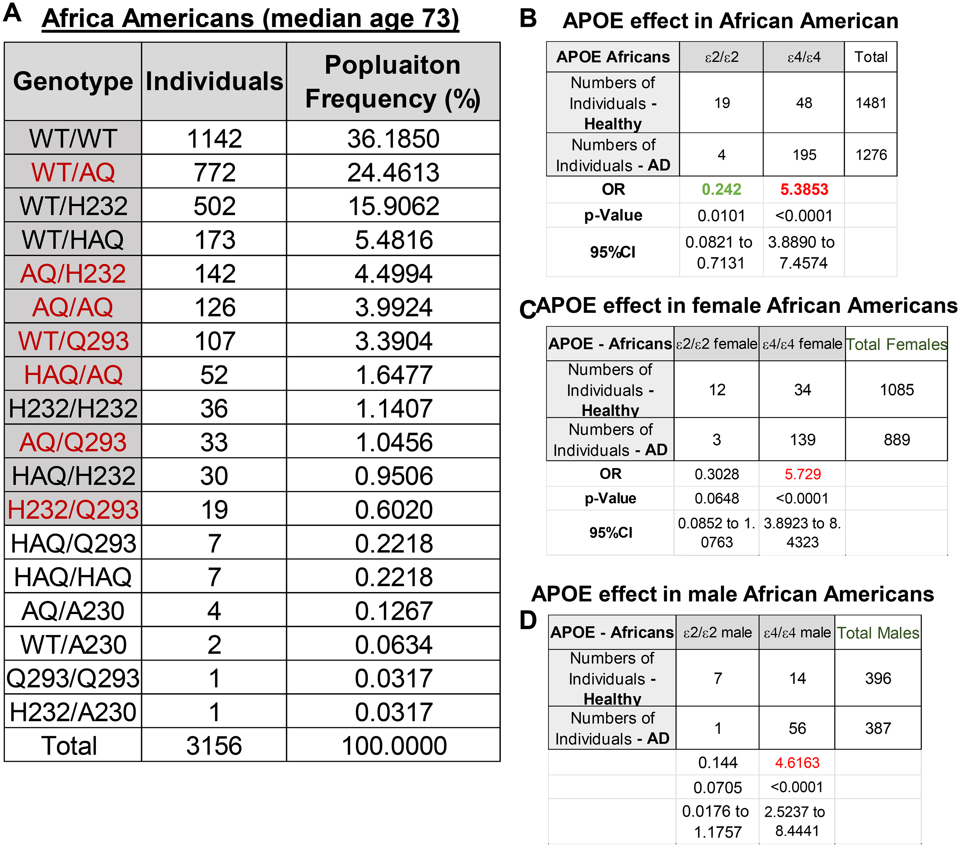
The APOE ε4 allele is associated with increased AD risk in African cohorts in both sexes. (A) A table of TMEM173 genotypes found in AA from cohorts snd10003. (B-D) Case-control analysis of the APOE ε2/ε2, ε4/ε4, and sex impact on AD association in AA.
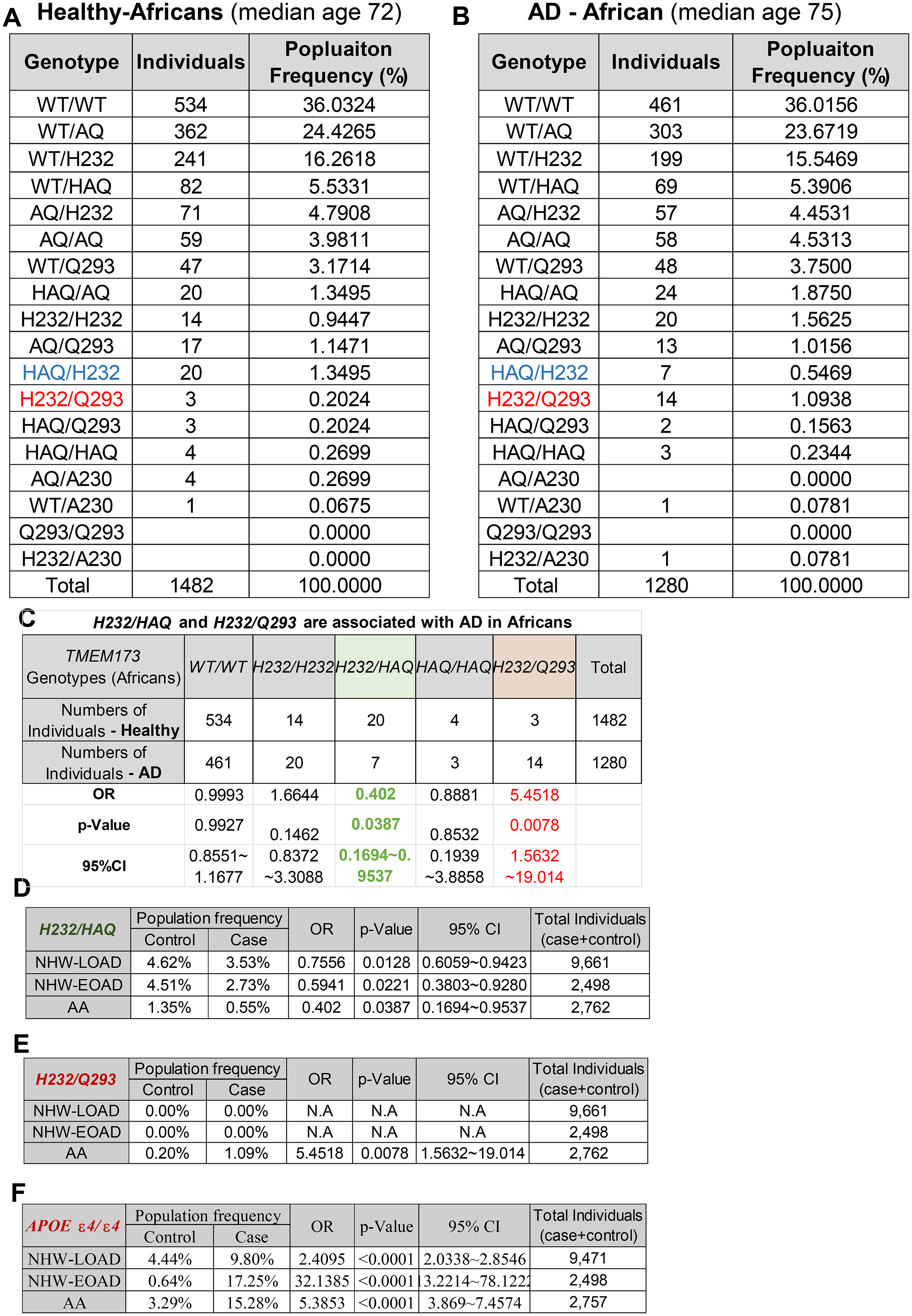
The H232/HAQ TMEM173 genotype is associated with decreased, while the H232/Q293 is associated with increased AD risk in AA. (A, B) TMEM173 genotypes in the case control samples from AA. (C) Case–control analysis of H232/HAQ, H232/Q293 genotypes and AD association in AA. (D) Comparing the H232/HAQ-AD association in NHW-LOAD, NHW-EOAD, and AA. (E) Comparing the H232/Q293-AD association in NHW-LOAD, NHW-EOAD, and AA. (F) Comparing the APOE ε4/ε4-AD association in NHW-LOAD, NHW-EOAD, and AA.
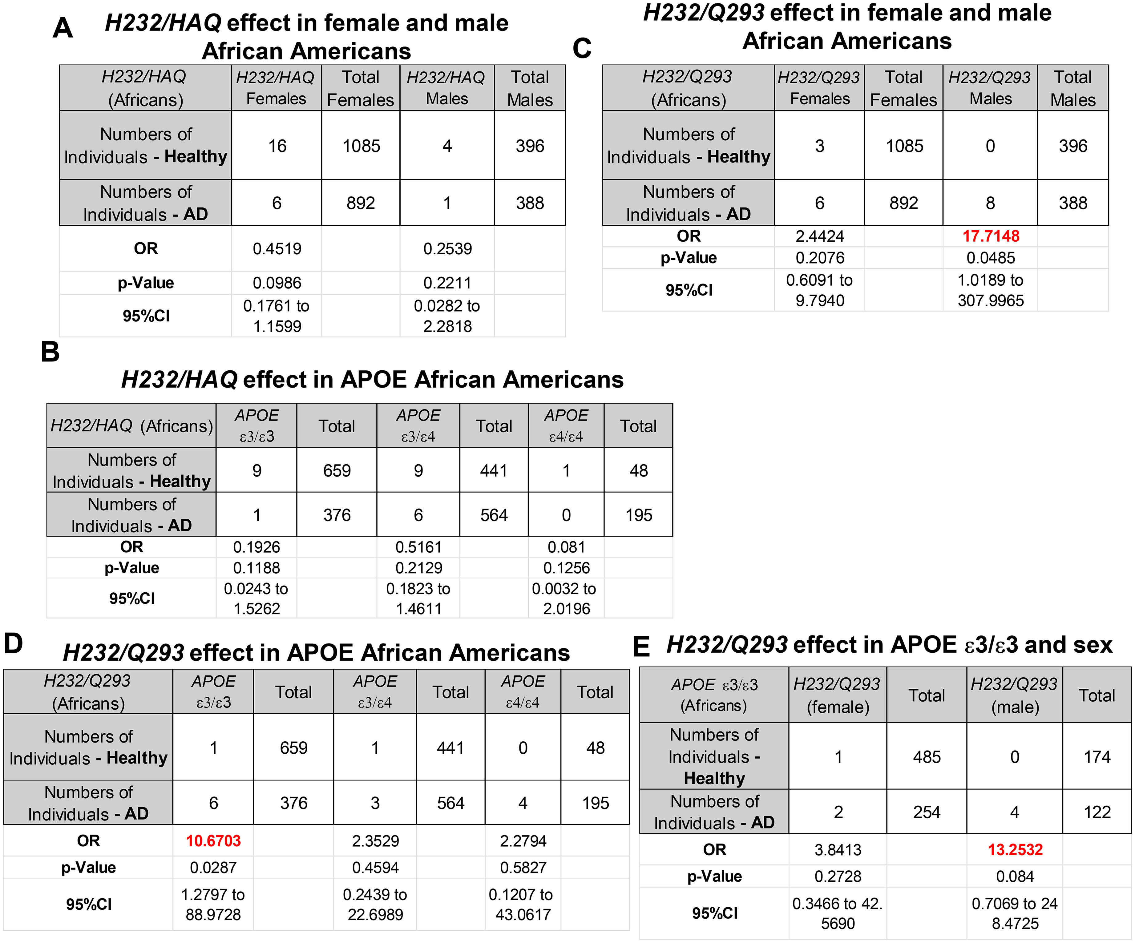
The H232/Q293 TMEM173 genotype selectively increases AD risk in males and APOE ε3/ε3 AA. (A, B) Case-control analysis of the sex and APOE impact on H232/HAQ -AD association in AA. (C-E) Case-control analysis of the sex and APOE impact on H232/Q293 -AD association in AA.
The African-specific H232/Q293 TMEM173 genotype is associated with increased AD risk in male African Americans with APOE ε3/ε3
Surprisingly, the Case-Control analysis in AA revealed that a H232/Q293 genotype is associated with an increased AD risk, with an OR of 5.4518 (p < 0.0078) (Figure 6C). The H232/Q293 genotype is unique to AA (Figure 6E). The OR for H232/Q293 is similar to the APOE ε4/ε4 risk in AA (Figure 6F). Unlike the APOE ε4/ε4 risk, the H232/Q293-associated AD risk is male-specific with an OR of 17.7148 (p = 0.0485), much higher than APOE ε4/ε4 in male Africans (OR = 4.6163) (Figure 5C and 7C). Intriguingly, the H232/Q293-associated AD risk is found in APOE ε3/ε3 individuals (OR = 10.6703, p = 0.0287), not the APOE ε4/ε4 risk group (Figure 7D and E). Thus, the H232/Q293 TMEM173 genotype is an AA-specific, APOE ε4-independent, high AD risk factor.
Discussion
STING plays a crucial role in AD pathogenesis in mice.6,7,910–13,15 Here, we identified two common TMEM173 genotypes associated with AD in humans. Surprisingly, while the H232/HAQ is associated with AD protection, the H232/Q293 genotype is associated with increased AD risk, suggesting TMEM173 genotype-specific influence on AD. Notably, ∼4% of NHW are H232/HAQ, while ∼1% of AA are H232/Q293, impacting hundreds of millions of people worldwide. It is worth emphasizing that our study did not conclude that these TMEM173 genotypes are protective or detrimental to AD. To advance STING-targeting AD therapy, the next immediate step is to establish a causal relationship between the H232/HAQ, H232/Q293 genotypes, and AD.
This study cannot detect an association between AD and low-frequency TMEM173 genotypes due to sample size. Thus, negative results here do not mean that these low-frequency TMEM173 genotypes do not impact AD. For example, the WT/AQ genotype has the highest population frequency among the low-frequency TMEM173 genotypes in NHW (0.26%) (Figure 3A). The WT/AQ – AD association has an OR = 0.3550, p = 0.0984 in NHW-LOAD, Power analysis (α = 0.05, β = 0.2, power = 1-β = 0.8, 0.26% in control and 0.09% in case) revealed that the population size has to be 18,976 to yield an OR = 0.3994, p = 0.0085. The actual population size is 9661. In the future, when more AD data are available, we will revisit the AD association in low-frequency TMEM173 genotypes.
The STING inflammation pathway has been linked to neurodegenerative diseases such as traumatic brain injury, spinal cord injury, subarachnoid hemorrhage, Parkinson's, and AD in mice.6,9–11,13,46 However, there is no report on the impact of common human TMEM173 alleles on AD or neurodegenerative diseases. The discovery here, if confirmed with causality, may reveal a new paradigm for the mode of action of STING in neurodegenerative diseases. The present paradigm is that STING promotes neuroinflammation that drives neurodegenerative diseases. Using mouse models of the common HAQ, AQ, and Q293 TMEM173 allele, we showed that AQ completely suppressed the constitutively active STING SAVI mutation-induced organ inflammation. 26 In comparison, the HAQ allele partially inhibited organ inflammation. 26 Nevertheless, here it is the HAQ/H232, not the common AQ/H232, genotype that is associated with AD protection (Figure 6). Furthermore, we found that the Q293 allele also suppresses STING-SAVI-induced organ inflammation in mice (data not shown). 26 However, the H232/Q293 genotype is associated with an increased risk of AD (Figure 6). Together, we propose a STING-inflammation-independent paradigm for AD and neurodegenerative disease.
The H232/HAQ association with AD protection seems to be universal, across populations, sex, LOAD, EOAD, and APOE status, with a similar OR (Figure 3). Most studies suggested that STING promotes AD.6,7,910–13,15 How does the H232/HAQ STING protect the elderly from AD? Using immobilized B cells from H232/HAQ Europeans, we showed that H232/HAQ STING had defective CDNs-STING-Type I IFNs responses. 23 However, HAQ/HAQ from Europeans also had reduced STING-Type I IFNs responses. 23 but the common HAQ/HAQ genotype is not associated with AD protection (Figure 3C, 4E and 6C). STING activation also induced cell death in human CD4 T cells and monocytes that is independent of type I IFNs.47,48 Neuron cell death is a hallmark of AD. We discovered that HAQ is resistant to STING activation-induced cell death. 26 Nevertheless, HAQ/H232, not HAQ/HAQ, is associated with AD protection. Further research is required to understand how HAQ/H232 STING provides AD protection in LOAD, EOAD, and AA.
The H232/Q293 is associated with significantly increased AD risk with an OR > 10. Unexpectedly, this risk association is restricted to male Africans and in APOE ε3/ε3 population. APOE ε3 is the most common APOE allele. Unlike the APOE ε2 or ε4, APOE ε3 does not affect AD risk. Thus, the H232/Q293 likely represents a non-APOE ε4-mediated risk factor. Second, sexual dimorphism in STING function is underappreciated. A recent report found that activating STING in microglia through intrathecal injections of ADU-S100 and DMXAA STING agonists, significantly alleviated both allodynia and hyperalgesia in male mice, but not female mice. 49 Another study found that the STING activation in infiltrating immune cells was substantially higher in males compared with their age-matched female type 2 diabetic nephropathy (T2DN) rats, with these differences becoming more pronounced with aging. 50 In addition, STING expression increased more than 3-fold in the old male rats than in the old female rats with aging (∼50 weeks old). 50 H232/Q293 associated AD risk is specific in male AA. There is no report on H232/Q293 STING. Further research is needed to reveal sexual dimorphism by H232/Q293 STING in aging cohorts.
H232/Q293 exists only in AA with a large AD risk OR. AA has the highest risk of dementia among US ethnic groups. 51 Among AA aged 70 and older, approximately 21.3% are living with AD, which is significantly higher compared to NHW.52,53 The higher prevalence is partially attributed to AA-specific genetic factors.54–56 For example, the cumulative risk of dementia among first-degree relatives of AA who have AD is 43.7%. 52 In comparison, for spouses (who share environmental but not genetic backgrounds), the cumulative risk was 18.4%. 52 However, the AA-specific genetic risk factors for the high prevalence of AD are unknown.55,57 To our knowledge, the common H232/Q293 TMEM173 is the first AA-specific high AD genetic risk factor. More research on H232/Q293 STING should be done to lower AD mortality in AA.
The current mode of action of STING cannot explain why H232/HAQ and H232/Q293 have the opposing AD effects. Given that they share the H232 allele, the difference lies in the HAQ and Q293 alleles. Both HAQ22,23,26,58,59 and Q293 22,26 are defective in type I IFNs responses. Furthermore, HAQ and Q293 STING are resistant to STING-activation-induced cell death, 26 which has been linked to the proton channel function of STING.60,61 Notably, the HAQ is positively selected while the Q293 allele was negatively selected in non-Africans (Supplemental Figure 1B). 24 However, the underlying cause of the natural selection of human TMEM173 alleles remains unknown. 24 Further research is needed to dissect the opposite function of Q293 and HAQ STING.
Finally, the major barrier to current STING research is the poor translation from animals to humans because most of the STING knowledge was from WT/WT in mice. Consequently, despite the promising results of STING agonists for multiple cancers in WT/WT mice,39,62–64 STING cancer clinical trials are marred by failures.65–67 Learning from these failures, future STING-targeting therapy clinical trials should be personalized. For example, our results indicate that the H232/Q293 individuals are over-represented in the AD patient cohorts. But there is no data at all on H232/Q293 STING responses, whether it is the classic type I IFNs, TNF stimulation, or the nonclassical lymphocyte cell death, energy metabolism function of STING. Blindly applying STING regents to H232/Q293 AD patients is neither data-driven nor ethical.
In summary, the H232/HAQ, H232/Q293 TMEM173 genotypes are associated with the opposite impact on AD risk. Non-WT/WT TMEM173 genotypes are prevalent in humans. Animal studies in WT/WT mice, thus, do not reflect the functionally different human TMEM173 genotypes. In addition, AA has the highest risk of AD among US ethnic groups. Identification of H232/Q293 as the first African American-specific AD genetic factor opens the door for personalized AD care to reduce disease burden in AA.
Supplemental Material
sj-docx-1-alz-10.1177_13872877251396973 - Supplemental material for Identification of common human TMEM173 genotypes associated with Alzheimer's disease
Supplemental material, sj-docx-1-alz-10.1177_13872877251396973 for Identification of common human TMEM173 genotypes associated with Alzheimer's disease by Mollie R Usher, Alexandra A Aybar-Torres and Lei Jin in Journal of Alzheimer's Disease
Footnotes
Acknowledgements
The authors have no acknowledgments to report.

Author contribution(s)
Funding
The authors disclosed receipt of the following financial support for the research, authorship, and/or publication of this article: The study is supported by the UF Gatorade Fund and NIH-HL152163 (to L.J).
Data for this study were prepared, archived, and distributed by the National Institute on Aging Alzheimer's Disease Data Storage Site (NIAGADS) at the University of Pennsylvania (U24-UFAG041689), funded by the National Institute on Aging.
The Alzheimer's Disease Sequencing Project (ADSP) is comprised of two Alzheimer's Disease (AD) genetics consortia and three National Human Genome Research Institute (NHGRI) funded Large Scale Sequencing and Analysis Centers (LSAC). The two AD genetics consortia are the Alzheimer's Disease Genetics Consortium (ADGC) funded by NIA (U01 AG032984), and the Cohorts for Heart and Aging Research in Genomic Epidemiology (CHARGE) funded by NIA (R01 AG033193), the National Heart, Lung, and Blood Institute (NHLBI), other National Institute of Health (NIH) institutes and other foreign governmental and non-governmental organizations. The Discovery Phase analysis of sequence data is supported through UF1AG047133 (to Drs. Schellenberg, Farrer, Pericak-Vance, Mayeux, and Haines); U01AG049505 to Dr Seshadri; U01AG049506 to Dr Boerwinkle; U01AG049507 to Dr Wijsman; and U01AG049508 to Dr Goate and the Discovery Extension Phase analysis is supported through U01AG052411 to Dr Goate, U01AG052410 to Dr Pericak-Vance and U01 AG052409 to Drs. Seshadri and Fornage.
•
Sequencing for the Follow Up Study (FUS) is supported through U01AG057659 (to Drs. PericakVance, Mayeux, and Vardarajan) and U01AG062943 (to Drs. Pericak-Vance and Mayeux). Data generation and harmonization in the Follow-up Phase is supported by U54AG052427 (to Drs. Schellenberg and Wang). The FUS Phase analysis of sequence data is supported through U01AG058589 (to Drs. Destefano, Boerwinkle, De Jager, Fornage, Seshadri, and Wijsman), U01AG058654 (to Drs. Haines, Bush, Farrer, Martin, and Pericak-Vance), U01AG058635 (to Dr Goate), RF1AG058066 (to Drs. Haines, Pericak-Vance, and Scott), RF1AG057519 (to Drs. Farrer and Jun), R01AG048927 (to Dr Farrer), and RF1AG054074 (to Drs. Pericak-Vance and Beecham).
•
The National Institutes of Health, National Institute on Aging (NIH-NIA) supported this work through the following grants: ADGC, U01 AG032984, RC2 AG036528; samples from the National Centralized Repository for Alzheimer's Disease and Related Dementias (NCRAD), which receives government support under a cooperative agreement grant (U24 AG21886) awarded by the National Institute on Aging (NIA), were used in this study. Sequencing data generation and harmonization is supported by the Genome Center for Alzheimer's Disease, U54AG052427, and data sharing is supported by NIAGADS, U24AG041689. We thank contributors who collected samples used in this study, as well as patients and their families, whose help and participation made this work possible.
NIH grants supported enrollment and data collection for the individual studies including the Alzheimer's Disease Centers (ADC, P30 AG062429 (PI James Brewer, MD, PhD), P30 AG066468 (PI Oscar Lopez, MD), P30 AG062421 (PI Bradley Hyman, MD, PhD), P30 AG066509 (PI Thomas Grabowski, MD), P30 AG066514 (PI Mary Sano, PhD), P30 AG066530 (PI Helena Chui, MD), P30 AG066507 (PI Marilyn Albert, PhD), P30 AG066444 (PI John Morris, MD), P30 AG066518 (PI Jeffrey Kaye, MD), P30 AG066512 (PI Thomas Wisniewski, MD), P30 AG066462 (PI Scott Small, MD), P30 AG072979 (PI David Wolk, MD), P30 AG072972 (PI Charles DeCarli, MD), P30 AG072976 (PI Andrew Saykin, PsyD), P30 AG072975 (PI David Bennett, MD), P30 AG072978 (PI Neil Kowall, MD), P30 AG072977 (PI Robert Vassar, PhD), P30 AG066519 (PI Frank LaFerla, PhD), P30 AG062677 (PI Ronald Petersen, MD, PhD), P30 AG079280 (PI Eric Reiman, MD), P30 AG062422 (PI Gil Rabinovici, MD), P30 AG066511 (PI Allan Levey, MD, PhD), P30 AG072946 (PI Linda Van Eldik, PhD), P30 AG062715 (PI Sanjay Asthana, MD, FRCP), P30 AG072973 (PI Russell Swerdlow, MD), P30 AG066506 (PI Todd Golde, MD, PhD), P30 AG066508 (PI Stephen Strittmatter, MD, PhD), P30 AG066515 (PI Victor Henderson, MD, MS), P30 AG072947 (PI Suzanne Craft, PhD), P30 AG072931 (PI Henry Paulson, MD, PhD), P30 AG066546 (PI Sudha Seshadri, MD), P20 AG068024 (PI Erik Roberson, MD, PhD), P20 AG068053 (PI Justin Miller, PhD), P20 AG068077 (PI Gary Rosenberg, MD), P20 AG068082 (PI Angela Jefferson, PhD), P30 AG072958 (PI Heather Whitson, MD), P30 AG072959 (PI James Leverenz, MD). The Miami ascertainment and research were supported in part through: RF1AG054080, R01AG027944, R01AG019085, R01AG028786-02, RC2AG036528. The Columbia ascertainment and research were supported in part through: R37AG015473 and U24AG056270. The University of Washington ascertainment and research were supported in part through R01AG044546, RF1AG053303, RF1AG058501, U01AG058922 and R01AG064877.
•
NIH grants supported enrollment and data collection for the individual studies including: GenerAAtions R01AG20688 (PI M. Daniele Fallin, PhD); Miami/Duke R01 AG027944, R01 AG028786 (PI Margaret A. Pericak-Vance, PhD); NC A&T P20 MD000546, R01 AG28786-01A1 (PI Goldie S. Byrd, PhD); Case Western (PI Jonathan L. Haines, PhD); MIRAGE R01 AG009029 (PI Lindsay A. Farrer, PhD); ROS P30AG10161, R01AG15819, R01AG30146, TGen (PI David A. Bennett, MD); MAP R01AG17917, R01AG15819, TGen (PI David A. Bennett, MD); MARS R01AG022018 (PI Lisa L. Barnes).[CL1] [KA2] The NACC database is funded by NIA/NIH Grant U24 AG072122. NACC data are contributed by the NIA-funded ADCs: P30 AG019610 (PI Eric Reiman, MD), P30 AG013846 (PI Neil Kowall, MD), P30 AG062428-01 (PI James Leverenz, MD) P50 AG008702 (PI Scott Small, MD), P50 AG025688 (PI Allan Levey, MD, PhD), P50 AG047266 (PI Todd Golde, MD, PhD), P30 AG010133 (PI Andrew Saykin, PsyD), P50 AG005146 (PI Marilyn Albert, PhD), P30 AG062421-01 (PI Bradley Hyman, MD, PhD), P30 AG062422-01 (PI Ronald Petersen, MD, PhD), P50 AG005138 (PI Mary Sano, PhD), P30 AG008051 (PI Thomas Wisniewski, MD), P30 AG013854 (PI Robert Vassar, PhD), P30 AG008017 (PI Jeffrey Kaye, MD), P30 AG010161 (PI David Bennett, MD), P50 AG047366 (PI Victor Henderson, MD, MS), P30 AG010129 (PI Charles DeCarli, MD), P50 AG016573 (PI Frank LaFerla, PhD), P30 AG062429-01(PI James Brewer, MD, PhD), P50 AG023501 (PI Bruce Miller, MD), P30 AG035982 (PI Russell Swerdlow, MD), P30 AG028383 (PI Linda Van Eldik, PhD), P30 AG053760 (PI Henry Paulson, MD, PhD), P30 AG010124 (PI John Trojanowski, MD, PhD), P50 AG005133 (PI Oscar Lopez, MD), P50 AG005142 (PI Helena Chui, MD), P30 AG012300 (PI Roger Rosenberg, MD), P30 AG049638 (PI Suzanne Craft, PhD), P50 AG005136 (PI Thomas Grabowski, MD), P30 AG062715-01 (PI Sanjay Asthana, MD, FRCP), P50 AG005681 (PI John Morris, MD), P50 AG047270 (PI Stephen Strittmatter, MD, PhD).
Declaration of conflicting interests
The authors declared no potential conflicts of interest with respect to the research, authorship, and/or publication of this article.
Data availability statement
The data supporting the findings of this study are available within the article and/or its supplemental material.
Supplemental material
Supplemental material for this article is available online.
References
Supplementary Material
Please find the following supplemental material available below.
For Open Access articles published under a Creative Commons License, all supplemental material carries the same license as the article it is associated with.
For non-Open Access articles published, all supplemental material carries a non-exclusive license, and permission requests for re-use of supplemental material or any part of supplemental material shall be sent directly to the copyright owner as specified in the copyright notice associated with the article.
