



Abstract
Background
With the aging population, the number of Alzheimer's disease (AD) patients has been increasing annually, creating an urgent need for AD therapeutic drugs. Donepezil combined with nimodipine (DN) has demonstrated therapeutic potential in the treatment of AD, but its mechanism of action remains unclear.
Objective
To reveal the mechanism of DN in the treatment of AD rats.
Methods
The AD rat model was established and evaluated by behavioral experiments and pathology. The therapeutic mechanism of DN in AD treatment was investigated through lipidomics and hippocampal metabolomics analyses based on ultra-high-performance liquid chromatography-quadrupole/time-of-flight mass spectrometry (UPLC-Q-TOF/MS).
Results
The learning and memory ability of AD rats can be improved after DN treatment. Significant changes were observed in 40 serum lipid metabolites and 19 hippocampal metabolites. These metabolites are mainly involved in glycerophospholipid metabolism, sphingolipid metabolism, amino acid metabolism, unsaturated fatty acid metabolism and other processes in AD rats.
Conclusions
DN could improve cognitive function and nerve damage in AD rats. It may plays a therapeutic role in AD rats by regulating cholinergic damage, Ca2+ overload, oxidative stress, neuroinflammation, and energy deficiency caused by metabolic disorders, which has practical significance for further research and clinical application of DN.
Introduction
Alzheimer's disease (AD) is the most prevalent neurodegenerative disorder and the leading cause of dementia. 1 Its clinical manifestations mainly involve functional impairments in multiple aspects such as cognition, memory, language and movement. 2 Affected by population aging, the number of AD cases globally continues to rise and is projected to reach 19.117 million cases by 2036. 3 The pathological hallmarks of AD are deposition of amyloid-β protein (Aβ) and neurofibrillary tangles (NFTs) caused by the hyperphosphorylation of tau protein. 4 The pathophysiology of AD remains obscure. Currently recognized hypotheses include the cholinergic nerve damage hypothesis, the amyloid hypothesis, and the excitatory amino acid neurotoxicity hypothesis. 5 The Food and Drug Administration (FDA) has approved two types of medicines for the treatment of AD. The first category is cholinesterase inhibitors, which include donepezil, rivastigmine, and galanthamine. The other is N-methyl-D-aspartate receptor antagonists, represented by memantine. 6 Current medications can only alleviate the symptoms of AD, they cannot cure or prevent the disease. Therefore, finding a new treatment regimen is one of the most pressing issues that must be resolved. 7
Given the complex pathogenesis of AD, traditional single-target therapies often fail to acquire the desired effect. Therefore, multi-target therapies are increasingly viewed as viable therapeutic alternatives for AD. At present, many studies have demonstrated that DN is more effective and safer than donepezil alone in the treatment of AD. 8 Donepezil is the first choice for mild-to-moderate AD. It is an acetylcholinesterase (AChE) inhibitor that reversibly binds and inactivates AChE to increase the concentration of acetylcholine (ACh) at cholinergic synapses, hence enhancing cognitive performance. 9 Nimodipine is a highly lipid-soluble calcium (Ca2+) channel blocker that can block Ca2+ channels, prevent their aberrant inflow, and selectively dilate blood vessels to protect brain cells. 10 Previous studies have revealed that abnormal Ca2+ accumulation in neurons can induce the production and deposition of Aβ and hyperphosphorylated tau protein, leading to the aggravation of AD patients. 11 Moreover, nimodipine can alleviate AD symptoms by decreasing Aβ-dependent stimulation of nuclear factor kappa-B (NF-κB) and NLRP3 inflammasome and preventing Aβ-mediated mitochondrial damage. 12
As the most recent omics technology, metabolomics is the study of the end products of biological activities. It focuses primarily on the qualitative and quantitative analysis of polar small molecule metabolites, such as carbohydrates, amino acids, and fatty acids, which is a crucial technique for investigating the pathophysiology of diseases and identifying prospective treatment targets. 13 Recently, ultra-high-performance liquid chromatography-quadrupole/time-of-flight mass spectrometry (UPLC-Q-TOF/MS) metabolomics technology has remained popular due to having advantages like rapid analysis speed, high mass accuracy, good repeatability, high sensitivity and resolution. 14 This technology offers a fresh vantage point for investigating complex disease causes and assessing treatment efficacy. The majority of current AD metabolomics investigations rely on the examination of cerebrospinal fluid (CSF), urine, blood, and other biological materials. 15 But the hippocampus is the first brain region affected by AD, and accumulating data suggests that metabolic abnormalities in the hippocampus play a crucial role in the pathogenesis of AD.16–18 Lipids play a crucial role in various physiological processes, including cellular development, differentiation, apoptosis, and inflammation. Changes in the levels of lipids are correlated with the development of disease. The identification of metabolites enables the rapid development of biomarkers and provides crucial data for elucidating the illness mechanism. 19 Based on UPLC-Q-TOF/MS technology, this study investigated serum and hippocampus metabolism that may be altered by DN therapy in rats with AD. It offered a theoretical and experimental foundation for the clinical therapy of AD.
Methods
Animal modeling and administration
Adult male Sprague Dawley (SD) rats (Peng Yue Experimental Animal Breeding, Jinan, China) weighing 180∼200 g were employed in this study. All animal experiments were approved by the Ethics Committee of Shandong First Medical University (No. SYXK (Lu) 20190022). After one week of adaptive feeding (humidity 50–60%, temperature 24 ± 1°C, 12-h light-dark cycle), 72 rats were randomly assigned to six groups: the AD model (MOD) group, the Sham-operated (SHA) group, the donepezil (DNP) group, the nimodipine (NMD) group, the low-dose donepezil and nimodipine (DNP + NMD L) group, the high-dose donepezil and nimodipine (DNP + NMD H) group with 12 rats in each group. The AD rat model was established by injecting 5 μL of Aβ25–35 protein (Sigma Aldrich, St Louis, MO, USA) into the CA1 region of the bilateral hippocampus of rats in the MOD group, DNP group, NMD group, DNP + NMD L group and DNP + NMD H group. 20 The SHA group received the same volume of normal saline (Chen Xin Pharmaceutical, Jining, China). Two weeks later, they had eight consecutive weeks of intragastric donepezil, nimodipine, donepezil and nimodipine administration (Aladdin Biochemical Technology, Shanghai, China). The dose supplied to rats was derived from the maximum daily dose per person based on body surface area. Finally, the DNP group was administered donepezil (0.91 mg/kg), the NMD group was administered nimodipine (0.25 mg/kg), the DNP + NMD L group was administered donepezil (0.91 mg/kg) and nimodipine (0.25 mg/kg), and the DNP + NMD H group was given donepezil (1.82 mg/kg) and nimodipine (0.5 mg/kg), all groups were administered once daily. Both the MOD and SHA groups received the same amount of normal saline.
Y-maze test
The Y-maze apparatus (CSI, Long Island, NY, USA) consisted of three arms of equal length with an angle of 120° each. Each rat was placed in the Y-maze test apparatus to explore freely for 10 min. The total number of times the rat entered the arm and the order in which it entered the arm were recorded. Consecutive entry into three different arms was defined as a alternation behavior. The alternation ratio of each rat was calculated to reflect the learning ability of different groups of rats. After each rat was tested, the apparatus was wiped with 75% alcohol to eliminate odors and reduce interference. Alternation ratio = the number of alternations / (total number of entries −2) × 100%.
Active avoidance test
The experimental apparatus (CSI, Long Island, NY, USA) is composed of two connected chambers, one light and one dark, with conductive metal strips placed on the bottom. Each rat was placed from the light chamber and allowed to freely explore for 90 s before formal test. When the rat was completely in the dark chamber, it was given two shocks (0.6 mA, 3 s) with a 10 s interval between each shock. The formal test was performed 1 h later. The escape latency (the time when the rat first entered the dark chamber) and the time of errors (the number of times the rat entered the dark chamber) were recorded for five consecutive days of training. If the rats did not enter the dark chamber, the escape latency was recorded as 90 s. After each rat was tested, the apparatus was wiped with 75% alcohol to eliminate odors and reduce interference.
Sample collection
Serum samples. After completing the behavioral tests, rats were euthanized for sample collection. Blood samples were centrifuged at 3000 rpm for 10 min at 4°C, and the supernatant was taken to obtain serum samples. The prepared serum samples were stored at −80°C. Serum samples were processed as follows: after thawing at 4°C, next, 200 µL serum was mixed by vortex for 5 min followed by incubation for 10 min at room temperature, which was added to 1.5 mL of MeOH/MTBE/H2O (1:5:2, v:v:v). The sample was then frozen at −20°C to promote protein precipitation. After that, the samples were centrifuged at 14000 rpm for 20 min. Under nitrogen, the supernatant was collected and blow-dried. Finally, the samples were subsequently redissolved in 1 mL of isopropanol/acetonitrile (1:1, v:v) and filtered through 0.22 µm organic filter membrane for UPLC-Q-TOF/MS analysis.
Hippocampus samples. The hippocampus was separated and frozen at −80°C. At the start of the experiment, frozen hippocampus thawed at 4°C was combined with 1 g:10 mL of precooled pure MeOH, one 3 mm steel ball, and two 1 mm steel balls. The homogenate obtained by three times homogenization at 60 Hz for 120 s was vortexed for 5 min, then centrifuged at 13000 rpm for 10 min at 4°C, and the supernatant was kept at −20°C overnight. The next day, the precipitated protein was centrifuged at 13000 rpm for 15 min. Finally, the supernatant was vortexed and then filtered via a 0.22 µm organic filter membrane for UPLC-Q-TOF/MS analysis.
QC samples of serum was obtained by mixing 100 µL of each processed sample. The Hippocampal QC sample was collected in the same manner as the serum. In the process of metabolomics analysis, a QC sample is injected after the analysis of every six individual samples to assess the system's stability and reproducibility.
Hematoxylin and eosin (HE) staining and immunohistochemical staining of the hippocampus. Following fixation with fixative solution, dehydration, wax immersion, embedding, and sectioning, the rat hippocampus was removed and cut into paraffin sections. Afterward, the slices were stained with HE solution and sealed with neutral gum. The cell structure and morphology of hippocampus tissue were finally studied using a high-powered microscope and processed with an image acquisition system.
The same paraffin sections were subjected to antigen repair, endooxygenase inactivation, serum blocking, incubation with primary and secondary antibodies, color development, counterstained and dehydrated and air-dried before sealing and being observed under a microscope. The Average Optical Density (AOD) of Iba-1, NeuN and GFAP was quantified using ImageJ software (National Institutes of Health, USA).
Metabolomic UPLC-Q-TOF/MS conditions
Chromatographic conditions. The samples were analyzed by the Waters Acquity UPLC system (Waters, Milford, Massachusetts, United States). The chromatographic column used was the BEH C18 column (1.7 µm, 2.1 mm × 100 mm, Waters), with a column temperature was 40°C, injection volume of 5 µL, and a flow rate was 0.2 mL/min.
For serum samples, mobile phase A was a 0.1% formic acid-water solution containing 10 mM ammonium formate, and mobile phase B was an isopropanol/acetonitrile (1:1, v/v), solution containing 10 mM ammonium formate. The gradient elution procedure in positive ion mode was: 0–7.5 min 50%-95% B; 7.5–12.5 min 95%-100% B; 12.5–17 min 100% B; 17–18 min 100%-50% B. And in negative ion mode was: 0–7 min 50%-95% B; 7–9 min 95%-100% B; 9–10 min 100%-50% B; 10–15 min 50% B.
For hippocampus samples, mobile phase A consisted of a 0.1% formic acid-water solution, and mobile phase B was acetonitrile. The gradient elution procedure in positive ion mode was: 0–11 min 5%-80.5% B; 11–13 min 80.5%-100% B; 13–15 min 100%-5% B; 15–17 min 5% B. And in negative ion mode was: 0–12 min 5%-80.5% B; 12–15 min 80.5%-100% B; 15–16 min 100%-5% B; 16–20 min 5% B.
MS conditions. The positive and negative ion modes of the Xevo-G2-XS Q-TOF mass spectrometer (Waters, Milford, Massachusetts, United States) and the ESI ion source were utilized to acquire data. Sodium formate was employed to correct the mass spectrometer, and leucine enkephalin was used to calibrate the instrument in real-time ([M + H]+ = 556.2766, [M + H]− = 554.2620). The temperatures of the source and desolvation were 100°C and 450°C, respectively. The cone gas and desolvation gas were nitrogen, and the flow rate was 50 L/h and 800 L/h, respectively. The voltage of the cone was 40 V. In both positive and negative ion modes, the capillary voltage of serum samples was 1.5 kV, whereas that of hippocampus samples was 2.5 kV in positive ion mode and 2.0 kV in negative ion mode.
Data analysis
UPLC-Q-TOF/MS data were processed using Progenesis QI and EZinfo 2.0 software. For preliminary screening of potential biomarkers, principal component analysis (PCA) and orthogonal partial least squares discriminant analysis (OPLS-DA) was utilized. By using Variable important in projection (VIP) VIP > 1 and p < 0.05 as screening conditions, biomarkers with significant inter-group differences were searched. HMDB (http://www.hmdb.ca/) database was employed to discover potential biomarkers for initially screened differential metabolites. The potential biomarkers were then imported into the website MetaboAnalyst 5.0 (https://www.metaboanalyst.ca) for pathway analysis and heat map generation.
Results
Y-maze test
Y-maze test was used to evaluate the short-term learning and memory ability of rats. As shown in Figure 1(A), compared with the SHA group, the altern ratio of the MOD group was significantly decreased (p < 0.01), indicating that the learning and memory abilities of the ADM group was significantly reduced. Compared with ADM group, the alternating ratios of the DNP, NMD, DNP + NMD H and DNP + NMD L groups were significantly increased (p < 0.01). The results showed that DN could improve the learning and memory abilities of AD rats

Alternating ratio of rats in six groups (compared with SHA group, *p < 0.05, **p < 0.01; compared with MOD group, #p < 0.05, ##p < 0.01) (A); the escape latency (B) and times of errors (C) of rats in six groups (color figure available online).
Active avoidance test
The Active avoidance test was used to reflect the ability of rats to remember punitive learning. Figure 1(B, C) showed that, compared with the SHA group, the escape latency of the MOD group was significantly decreased, and compared with the MOD group, the escape latency of the DNP, NMD, DNP + NMD L and DNP + NMD H groups were significantly increased. The results indicated that DN could significantly improve the memory impairment of AD rats in response to punitive learning.
HE staining and immunohistochemical staining of the hippocampus
Studies have shown that the brain of AD patients is accompanied by the necrosis of nerve cells and the activation of microglia and astrocytes. As depicted in Figure 2(A), the SHA group's hippocampus contained a great number of cells that were packed tightly together. The majority of the nuclei were big and spherical with distinct nucleoli. They were also dyed uniformly with a mild bluish-purple hue (black arrow). In the MOD group, the number of cells reduced dramatically, and cells with abnormal morphology were placed in a disordered and disorganized manner. The cells deteriorated and shrunk, the nucleoli vanished, and the nucleus turned a dark purple color (red arrow). Comparatively to the MOD group, the size, number, and shape of hippocampus cells in the DNP, NMD, DNP + NMD L and DNP + NMD H group were similar to those in the SHA group. Significant pathological alterations were observed in the hippocampus of AD rats, and DN was able to ameliorate hippocampal damage in AD rats.

He staining of histopathological changes of rat hippocampus (scale = 50 µm) (A) representative images (magnification, × 200) and corresponding mean optical densities of GFAP (B, E), Iba-1 (C, F), and NeuN (D, G) immunohistochemistry in rat hippocampal (compared with SHA group, **p < 0.01; compared with MOD group, #p < 0.05, ##p < 0.01) (color figure available online).
GFAP is a specific marker of astrocyte activation, and Iba-1 is a marker of microglial activation. These markers are associated with Aβ deposition in the brain and may contribute to neuroinflammatory processes. NeuN is a marker of mature neurons. As shown in Figure 2(B,C), compared with the SHA group, the GFAP and Iba-1 positive areas in the ADM group were significantly increased (p < 0.01), and compared with the ADM group, the areas in the DN group were significantly decreased (p < 0.01), indicating that astrocytes and microglia were heavily activated in AD rats. DN can inhibit the expression of GFAP and Iba-1, reduce hippocampal tissue damage caused by inflammation, and improve the condition of AD.
As shown in Figure 2(D), compared with the SHA group, the MOD group had a large loss of neurons, loose arrangement of cells, and irregular morphology, indicating that AD rats had fewer neurons and reduced cognitive function. Compared with MOD group, the number of neurons in the hippocampus of SHA group, NMD, DNP, DNP + NMD L, and DNP + NMD H group was significantly increased, the arrangement of cells was more compact. The results showed that DN could upregulate the expression of NeuN in the hippocampus, alleviate nerve damage, and alleviate the symptoms of AD.
Metabolic profile analysis of serum and hippocampus
The pharmacodynamic results demonstrated that the DNP + NMD L group exhibited the best effect. Therefore, this group was chosen for lipidomic and hippocampal metabolomic analyses. Figure 3 shows the Base peak intensity (BPI) chromatogram of QC samples of hippocampus (Figure 3(A, B)) and serum (Figure 3(C, D)) in positive and negative ion modes, which shows that the samples have good separation under these chromatographic conditions. Figure 3(E) revealed the serum lipid biomarker PC[18:2(2E,4E)] m/z 520.3416 identified under the positive ion mode, the 6 fragment ions primarily produced are 542.3222, 520.3405, 502.3301, 483.2492, 443.2571, and 337.2747. The fragment ions they denote are [M + Na]+, [M + H]+, [M + H-H2O]+, [M + Na-C3H9N]+, [M + H-C3H12NO]+, and [M + H-C5H14NO4P]+, respectively. The UPLC-Q-TOF/MS data were imported into the Progenesis QI program, and new assays for serum and hippocampal samples under positive and negative ion modes, respectively, were developed. Then, the new tests were imported into EZinfo to screen potential biomarkers using PCA and OPLS-DA. The PCA plot depicted in Figure 4 demonstrated that each sample group had excellent within-group repeatability and inter-group difference. The clustering of the QC samples indicates the instrument's good stability and repeatability. As depicted in Figure 4, OPLS-DA demonstrated clear separation between every two groups in both positive and negative ion modes, and the respective model parameters R2Y and Q2 were greater than 95%, suggesting high applicability and predictability of OPLS-DA. Compounds with VIP > 1 are denoted in red on the S-plot by red-colored metabolites.

BPI chromatograms of QC samples. Serum positive ion (A) and negative ion (B) mode; hippocampal positive ion (C) and negative ion (D) mode, mass spectrogram PC[18:2(2E,4E)/0:0] in positive ion mode (E).
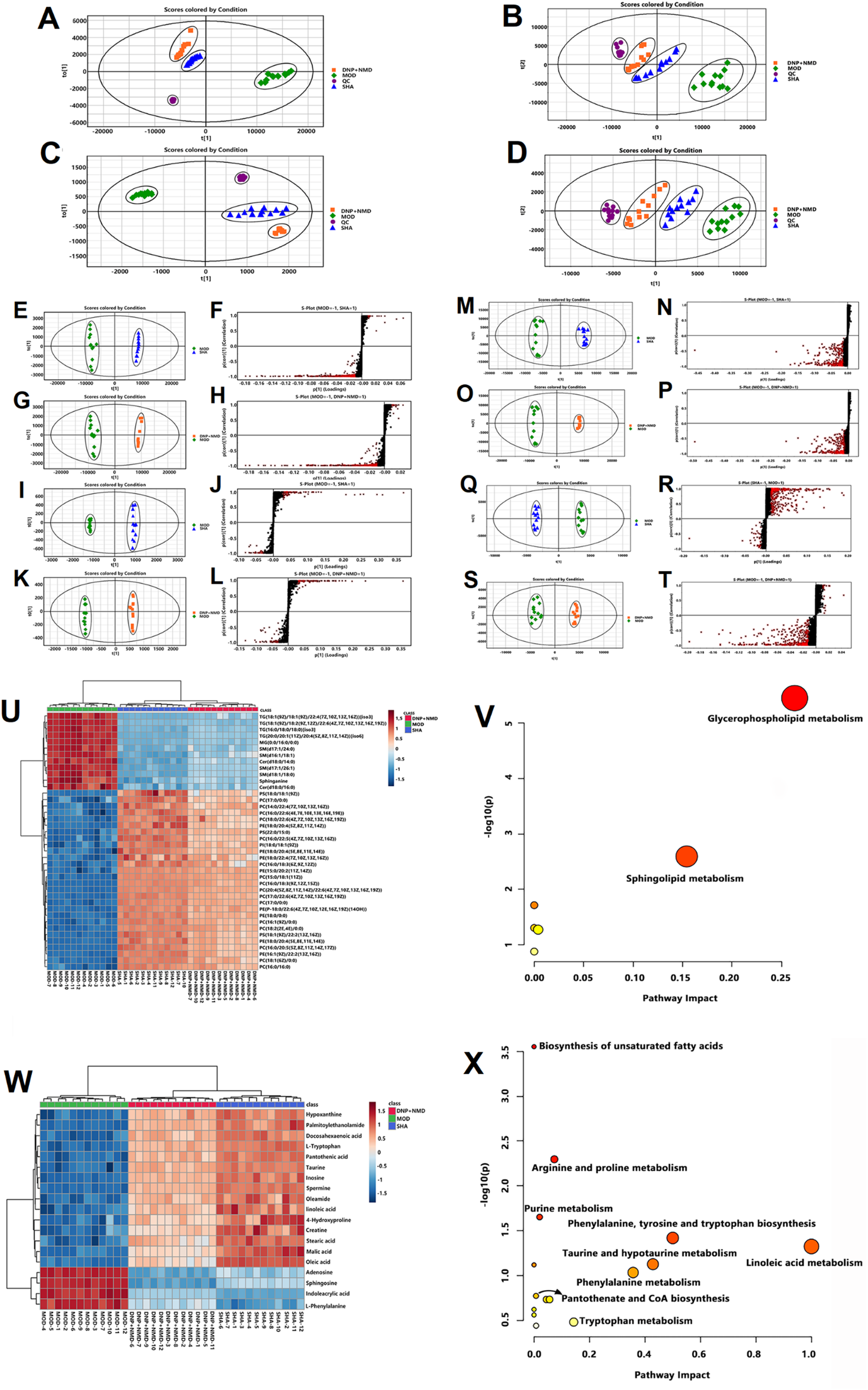
PCA plots in positive and negative ion mode. (A) (C) serum sample; (B) (D) hippocampus sample. OPLS-DA and S-plot plots in positive and negative ion mode, (E-L) serum sample; (M-T) hippocampus sample. Heat map of differential metabolite content in serum (U) and hippocampus (W). Red represents increased content and blue represents decreased content (color figure available online).
Identification of potential biomarkers
The potential biomarkers screened by VIP > 1 and p < 0.05 were imported into the Progenesis QI software, and then identified by HMDB database and MS/MS secondary fragment ion. Finally, 40 serum lipid metabolites and 19 hippocampus metabolites that may serve as potential biomarkers were discovered.
Table 1 provides information on potential biomarkers identified by serum and hippocampus metabolomics. Considering the normalized abundance of all potential biomarkers, MetaboAnalyst 5.0 (https://www.metaboanalyst.ca/) was applied to create a heat map to visually illustrate the variation of biomarker content in distinct groups. The heat map is depicted in Figure 4.
Information of potential biomarkers.

Metabolic pathway analysis
For metabolic pathway analysis, the identified potential biomarkers were imported into MetaboAnalyst 5.0 (https://www.metaboanalyst.ca/). These metabolites are mainly involved in sphingolipid metabolism, glycerophospholipid metabolism, phenylalanine metabolism, taurine and hypotaurine metabolism, Arginine and proline metabolism, Tryptophan metabolism, Purine metabolism, tricarboxylic acid cycle and Biosynthesis of unsaturated fatty acids. The results are depicted in Figure 4.
Discussion
In this study, the results of the Y-maze test and active escape test indicated that DN could enhance the learning and memory capabilities of AD rats, thereby alleviating their cognitive impairments. Histopathological analysis via HE staining revealed that DN treatment reduced inflammatory cell infiltration and mitigated hippocampal inflammation in AD rats. Through metabolomics analysis, a total of 59 metabolites were identified as potential biomarkers, including 40 serum lipid biomarkers and 19 hippocampal biomarkers. They are predominantly involved in glycerophospholipid metabolism, sphingolipid metabolism, linoleic acid metabolism, taurine and hypotaurine metabolism, phenylalanine metabolism, arginine and proline metabolism, biosynthesis of unsaturated fatty acids, pantothenate and CoA biosynthesis, purine metabolism and tryptophan metabolism.
Glycerophospholipid (GP) is an essential component of cell membrane lipids and regulates ion channel function, lipoprotein secretion, neuronal proliferation, and membrane function. The loss of GP may not only result in the production of NFTs and the pathological accumulation of Aβ, but it may also impact the permeability, fluidity, and ion homeostasis of the membrane, leading to an increase in oxidative stress in AD patients. 21 GP is composed of phosphatidyl acid and a substituent group attached to phosphate. Due to the various substituents, GP can be subdivided into phosphatidylcholine (PC), phosphatidyl-inositol (PI), phosphatidyl-ethanolamine (PE), and phosphatidyl-serine (PS), among others.
Phospholipase D catalyzes the hydrolysis of PC into phosphatidic acid and choline. Choline is the precursor of ACh, a crucial neurotransmitter in the cholinergic system that plays a crucial role in memory, cognition, and emotion regulation of the nervous system. 22 Depolarization of cholinergic neurons will decrease the level of PC, the amount of free choline released by PC, and the synthesis of ACh, 23 leading to AD induced by cholinergic nerve damage. Moreover, phospholipase A2 (PLA2) can hydrolyze PC to generate lyso-phosphatidylcholine (LPC) and free fatty acids. These metabolites stimulate the release of inflammatory cytokines and are implicated in the direct activation of microglia and astrocytes. In addition, they can produce neuroinflammation and oxidative stress, resulting in neuronal damage induced by Aβ aggregation. 24
Similar to PC, PE can be hydrolyzed by PLA2 to create LPC and free fatty acids, resulting in an increase in the synthesis of free radicals and pro-inflammatory cytokines and neurotoxic consequences. The distinction is that PE is essential for the start of autophagy. 25 Autophagy is the primary breakdown route for removing Aβ and hyperphosphorylated tau protein, which helps neurons maintain homeostasis. 26 In addition, PE can restore mitochondrial function and reduce the aberrant increase in intracellular Ca2+ concentration and Aβ deposition resulting from mitochondrial dysfunction, so as to repair, thereby repairing neuronal damage and enhancing cognitive performance. 27 Ca2+ signaling control is crucially dependent on PI. And Ca2+ signaling dysregulation is associated with AD pathogenic characteristics such as Aβ deposition, NFTs, and synaptic dysfunction. 28 Moreover, the metabolic equilibrium of PI promotes functional autophagy. 29 PI can be hydrolyzed to produce diacylglycerol and Phosphoinositide (PIP) under the catalysis of phospholipase C. PIP is an important regulator of neuroinflammation mediated by microglia. And the aforementioned process is considered as a potential mechanism for the onset or progression of neurodegenerative diseases. 30
PS can boost the release of dopamine and the creation of ACh, which protects cells against oxidative stress and Aβ deposits, hence promoting nervous system function and memory and cognition. In 2006, the Korea Food and Drug Administration (KFDA) approved that dietary PS supplementation can alleviate negative mood, boost cognition, and lower the risk of AD. 31 PS also stimulates signaling pathways, including AKT, PKC, and Raf-1, to modify the nature and function of certain membrane-bound receptors, which are essential for neuronal survival, neurite development, and synaptogenesis. 32 With aging and the onset of the degenerative condition of AD, the amount of PS in the body will gradually diminish, the synthesis of ACh will be inhibited, and the neuroprotective effect will be lost, leading to a reduction in memory and cognitive function. Exogenous PS supplementation is capable of alleviating symptoms. 33
In this study, the levels of PC, PE, PI, and PS in the MOD group were considerably lower than in the SHA group. After the administration of DN, their levels progressively reverted to normal. It is hypothesized that DN may improve memory and cognitive function by controlling cholinergic damage, autophagy, and mitochondrial dysfunction caused by glycerophospholipid metabolism abnormalities, having a significant effect on the treatment of AD.
Sphingolipids (SM) are a class of complex lipids having the Sphinganine (Sph) skeleton, they are essential components of biofilm and can help maintain the homeostasis of neuronal membranes. SM metabolites, meantime, are involved in the control of cell growth, differentiation, senescence, and apoptosis and play a crucial role in neuronal survival, signaling, synaptic transmission, and stability. 34
Bioactive molecules such as Ceramide (Cer) and Sph are the principal metabolites of sphingolipid metabolism. Cer is the core molecule among them. As a second messenger, it affects development, apoptosis, and stress responses, contributing to neuronal signaling and function. Cer is predominantly produced via the hydrolysis of SM by acid sphingomyelinase (ASM). 35 Cer and Aβ intracellular levels are discovered to be in equilibrium to sustain neuronal homeostasis under physiological conditions. However, in the pathological condition of AD, ASM activity is increased, leading to an increase in Cer synthesis. Cer can stabilize and extend the half-life of β-secretase, hence promoting Aβ production. In turn, the produced Aβ drives a further increase in Cer via boosting ASM activity, generating a positive feedback regulatory mechanism. 36 Some ASM inhibitors have been used as treatment targets for AD due to their ability to decrease Cer levels, ameliorate autophagy abnormalities, and remove Aβ and tau protein deposition. 37 In addition, Cer is linked to decreased mitochondrial respiratory chain activity, decreased mitochondrial membrane potential, elevated reactive oxygen species (ROS), and oxidative stress. 38 The central nervous system is susceptible to mitochondrial injury, resulting in a loss of cognitive function and the eventual onset of AD. Consequently, Cer biosynthesis pathways and enzymes may be viable targets for regulating Cer levels and preventing the development of AD. 39
Sph is a product of Cer hydrolysis catalyzed by ceramidase. It can engage in cell arrest and apoptosis as a cell growth inhibitor. 40 Therefore, an increase in Sph concentration might promote the apoptosis of nerve cells and it can induce the development of AD in extreme situations in severe cases. Sphingosine-1-phosphate (S1P), the product of Sph phosphorylated by Sphingosine kinase (SphK), is also notable in SM metabolism. In contrast to the apoptosis-promoting effects of Cer and Sph, S1P suppresses apoptosis, enhances cell survival, stimulates proliferation and differentiation, and protects neurons from Aβ toxicity. 41 In addition, it enhances glutamate secretion and promotes memory consolidation. 42 Due to their opposing effects, restoring the metabolic balance among Cer, Sph, and S1P provides a new therapy option for AD. 43
In this study, the MOD group had considerably higher levels of SM, Cer, and Sph than the SHA group. After treatment with DN, the levels of biomarkers were regulated, suggesting that DN may be used to treat AD by preventing neuronal apoptosis, anti-inflammation, and anti-oxidation.
As an essential amino acid in the human body, phenylalanine is a crucial component in the creation of neurotransmitters. 44 L-amino acid oxidase (IL-4I1) catalyzes the transformation of phenylalanine into phenylpyruvate. Liu et al. 45 conducted untargeted and targeted metabolomics analyses on postmortem brain tissues of AD patients and discovered that phenylalanine, phenylpyruvate, and IL-4I1 were upregulated. These findings suggested that phenylalanine dysmetabolism may play a key role in the etiology of AD. In the meantime, poor phenylalanine conversion may influence the production of the neurotransmitters dopamine, norepinephrine, and epinephrine, 46 resulting in neuronal dysfunction and AD-related dementia. In this study, phenylalanine was upregulated in the MOD group compared to the SHA group and downregulated after treatment with DN. Therefore, DN may play a therapeutic effect by boosting the neurotransmitter content in the brain and normalizing phenylalanine levels.
Taurine, one of the most abundant amino acids in the brain, is synthesized and released in astrocytes. It can function intracellularly and extracellularly in neurons as a glial transmitter. Taurine's intracellular function is its participation in the Ca2+ homeostasis pathway, which prevents excitotoxicity, apoptosis, and mitochondrial stress by lowering Ca2+ deposition. The extracellular effect is that taurine binds to γ-aminobutyric acid (GABA) and glycine receptors in neurons, resulting in cell hyperpolarization and glutamate excitotoxicity mediated by N-methyl-aspartate (NMDA). 47 AD is intimately associated with neuronal degeneration, mitochondrial stress, and excitatory glutamate toxicity. Therefore, it is anticipated that taurine and hypotaurine metabolism would give novel treatment targets for AD patients. In this study, the taurine content in the MOD group was significantly reduced compared to the SHA group, and it was restored after treatment with DN, indicating that DN may play a therapeutic role by inhibiting excitatory cytotoxicity, oxidative stress and Ca2+ overload caused by taurine and hypotaurine metabolic disorders.
L-tryptophan (Trp) affects protein homeostasis and plays a function in neurotransmitter-mediated physiological activity. Moreover, Trp can work as a precursor of 5-hydroxytryptamine (5-HT) and is intimately associated with cognitive function. Widner et al. 48 discovered that systemic chronic immunological activation in AD patients was associated with substantial Trp degradation. Specifically, immunological activation can activate tryptophan 2,3-dioxygenase (TDO), leading to an increase in tryptophan breakdown. Indoleacrylic acid is a Trp metabolite that inhibits the generation of pro-inflammatory cytokines by lymphocytes and diminishes the inflammatory response of immune cells. 49 To the best of our knowledge, however, there are no reports linking it to AD. In this study, the MOD group had lower levels of Trp and higher levels of indoleacrylic acid than the SHA group. And their levels were restored following DN therapy. By reducing the inflammatory response, DN may therefore exert a neuroprotective impact.
The contents of creatine (Cr), spermine, and hydroxyproline considerably changed in the MOD group relative to the SHA group during the arginine and proline metabolism. Cr is produced by combining arginine, glycine, and methionine. Cr protects rat hippocampus neurons against glutamate toxicity, as demonstrated. 50 Cr supplementation can eliminate ROS and improve adenosine triphosphate (ATP) generation to reduce ATP use resulting from Aβ toxicity. 51 Spermine is an anti-inflammatory polyamine with antioxidant properties. Studies have revealed that spermine can diminish AChE overactivation, hyperphosphorylated tau protein, deposition of Aβ, and neural apoptosis by activating the NF-κB pathway. 52 These mechanisms suggest that spermine exerts a substantial neuroprotective impact. Regulating spermine levels is important for preventing and treating neurological disorders associated with aging. Hydroxyproline is a structurally and physiologically significant sub-amino acid in animals; it scavenges oxidants, regulates the intracellular environment, and stimulates the development of antioxidant enzymes in cells. 53 In this study, the MOD group had lower amounts of Cr, spermine, and hydroxyproline than the SHA group. After treatment with DN, the levels of these metabolites increased, indicating that DN may regulate the metabolic abnormalities produced by arginine and proline by decreasing glutamate toxicity, anti-inflammation, and anti-oxidation.
Purines are essential precursors for the synthesis of DNA and RNA. It can also operate as metabolic signals, brain function regulators, and energy converters throughout the body. In this investigation, the purine metabolic pathway found three purine metabolites, including hypoxanthine, inosine, and adenosine. The intermediate product of purine metabolism is hypoxanthine. Studies indicate that aberrant purine metabolism can lead to varying degrees of neurodevelopmental delay and neurological dysfunction. 54 Kaddurah-Daouk et al. 55 analyzed postmortem ventricular CSF samples from dementia patients and non-dementia patients using an electrochemical-based metabolomics technique and found that the downregulation of many metabolites in purine pathways, such as hypoxanthine and xanthine was significantly in accordance with the formation of NFTs, highlighting the importance of purine metabolism in the formation of AD. Inosine is an endogenous nucleoside derived from the interaction of hypoxanthine and ribose. It is involved in the activation of the pyruvate oxidase system, increases coenzyme A (CoA) activity, and has a neuroprotective impact on neurons and astrocytes injured by hypoxia and oxidative stress. Previous research has demonstrated that the prevention and improvement of AD memory impairment by inosine may be strongly related to the control of multiple brain pathways, including ion pump activity, oxidative stress, and cholinergic signaling. 56 Additionally, inosine ameliorates memory problems, controls the levels of inflammatory and neurotrophin, adenosine receptors, synaptic proteins, and cholesterol, 57 and is anticipated to be employed as a multi-target medication to prevent and treat AD. As a neuromodulator and neuroprotectant, adenosine regulates glutamate release. 58 It has been shown that the overexpression and translocation of adenosine receptors in the frontal cortex of AD patients are more active in neurons damaged by Aβ deposition or hyperphosphorylated tau protein. Although a high level of adenosine in the brain is associated with natural aging, experimental data indicate that AD patients create an additional rise. 59 In this study, the levels of hypoxanosine and inosine were significantly lower in the MOD group compared to the SHA group, whereas the amount of adenosine was elevated. The progressive recovery to normal levels of the three metabolites following treatment with DN suggests that DN has a regulatory influence on purine metabolism disorders.
The TCA cycle is the primary route for the oxidative breakdown of acetyl-CoA into water and carbon dioxide to generate energy. 60 PA often known as vitamin B5, is an essential member of the vitamin B family and precursor of acetyl-CoA. PA shortage can lead to decreased acetyl-CoA synthesis and TCA cycle blockage, resulting in brain energy imbalance and eventually causing neurodegeneration and dementia in AD. 61 Malic acid is an essential molecule that links the metabolism of sugar, protein, and fat and provides energy for numerous bodily processes. Lack of bioenergy is a significant cause of neurodegenerative diseases. 62 Compared to the SHA group, the levels of PA and malic were downregulated in the MOD group, and their levels increased after treatment with DN, demonstrating that DN can effectively ameliorate the energy shortage caused by AD.
The levels of oleic acid, linoleic acid (LA), stearic acid (SDA), and docosahexaenoic acid (DHA) in the MOD group were significantly different from those in the SHA group during the biosynthesis of unsaturated fatty acids. Oleic acid is a member of the omega-9 fatty acid family, which is a vital energy ingredient for the human body and is essential for maintaining cerebrovascular health. Kandel et al. 63 found that oleic acid can influence hippocampus neurogenesis by acting as a ligand for the orphan nuclear receptor TLX/NR2E1, consequently increasing neural stem cell proliferation and neurogenesis. Omega-3 polyunsaturated fatty acids include LA, SDA, and DHA (PUFAs). Several epidemiological studies have linked a reduced intake of omega-3 PUFAs to cognitive decline and an increased risk of dementia, particularly AD. 64 Numerous pathways contribute to the neuroprotective effects of omega-3 PUFAs, including neuroprotective DHA metabolites, reduced arachidonic acid metabolites, and increased nutritional factors. DHA has a particularly protective effect against AD via multiple mechanisms: it limits the development of AD and the deposition of Aβ, and it inhibits two key kinases that cause the creation of hyperphosphorylated tau protein and NFTs. 65 Moreover, the majority of investigations on LA and its derivatives 66 indicate that LA has neuroprotective and anti-inflammatory effects on neurodegenerative diseases. 67 In this study, the levels of oleic acid, LA, SDA, and DHA in the MOD group were lower than in the SHA group. And the levels of the four metabolites were recovered after treatment with DN, suggesting that DN may have a neuroprotective and anti-inflammatory role in the treatment of AD.
In this study, the successful establishment of AD rat model and the improvement effect of DN on AD rats were proved by pharmacodynamic tests. Based on UPLC-Q-TOF/MS technology, this study established the research approach of serum and hippocampus metabolomics in AD rats treated with DN, and finally 40 serum lipid and 19 hippocampal potential biomarkers were identified. Through biological analysis of potential biomarkers in serum and hippocampus, it is speculated that DN primarily plays a therapeutic role in AD rats by regulating cholinergic damage, Ca2+ overload, oxidative stress, neuroinflammation, and energy deficiency caused by metabolic disorders, which has practical significance for further research and clinical application of DN.
Footnotes
Acknowledgements
This experiment was made possible by the joint efforts of all the authors and was supported by the funding mentioned above.
Ethical considerations
This study was carried out according to the Ethics Committee of Shandong First Medical University (Shandong Academy of Medical Sciences).
Consent to participate
Not applicable
Consent for publication
Not applicable
Author contribution(s)
Funding
The authors disclosed receipt of the following financial support for the research, authorship, and/or publication of this article: This work was supported by the National Natural Science Foundation of China, (grant number 82204749).
Declaration of conflicting interests
The authors declared no potential conflicts of interest with respect to the research, authorship, and/or publication of this article.
Data availability statement
The data supporting the findings of this study are available on request from the corresponding author.