



Abstract
Background
Alzheimer's disease (AD) is marked by cognitive decline, depressive symptoms, and gut microbial dysbiosis. Yoga may support cognitive and emotional health while modulating gut microbiota, but integrative clinical evidence is limited
Objective
To evaluate the effects of a 12-week yoga intervention on cognition, depressive symptoms, and gut microbial diversity, composition, and function in Indian patients with mild AD.
Methods
In this hospital-based case-control study, 16 AD patients and 17 cognitively healthy controls (HCs) were recruited at AIIMS, New Delhi. AD diagnosis followed NIA-AA criteria, supported by Montreal Cognitive Assessment (MoCA) and Patient Health Questionnaire-9 (PHQ-9) assessments. AD participants underwent 60-min supervised yoga sessions daily for 12 weeks. Cognitive performance, depressive symptoms, and stool microbiota were assessed pre- and post-intervention. Metagenomic sequencing enabled taxonomic and functional profiling, with alpha diversity, beta diversity (Bray-Curtis distance), and differential abundance analyses performed using standard bioinformatics tools.
Results
Yoga was associated with improved cognition (MoCA: 22.33 ± 2.34 → 25.44 ± 2.01; p = 0.001) and reduced depressive symptoms (PHQ-9: 5.78 ± 3.11 → 2.22 ± 1.71; p = 0.007). Alpha diversity remained stable, while beta diversity shifted post-yoga AD samples toward the HC cluster. Beneficial taxa (Faecalibacterium prausnitzii, Roseburia intestinalis, Bifidobacterium, Akkermansia) increased, whereas pro-inflammatory taxa (Collinsella aerofaciens, Klebsiella spp.) decreased. Functional analysis showed partial recovery of metabolic and short-chain fatty acid pathways.
Conclusions
A 12-week yoga intervention was associated with cognitive and mood improvements and partial normalization of gut microbial function in mild AD. Larger randomized trials with lifestyle monitoring and multi-omics integration are warranted to confirm causal mechanisms.
Keywords
Introduction
Alzheimer's disease (AD) is a complex, multifactorial neurodegenerative disorder characterized by progressive cognitive decline, synaptic dysfunction, and chronic neuroinflammation. While amyloid-β plaques and hyperphosphorylated tau tangles remain central to its pathological diagnosis and therapeutic targeting, emerging evidence highlights the contributory role of peripheral systems particularly the gut microbiota in disease onset and progression.1,2 Disruption of the gut–brain axis through dysbiosis can impair short-chain fatty acid (SCFA) signaling, increase intestinal permeability, promote systemic inflammation, and compromise blood-brain barrier integrity, collectively accelerating neurodegenerative cascades.3–5
High-throughput metagenomic and 16S rRNA sequencing studies have consistently revealed altered gut microbial diversity and composition in individuals with AD compared to cognitively healthy controls. Reductions in SCFA-producing commensals such as Faecalibacterium prausnitzii and Roseburia intestinalis, alongside depletion of Bifidobacterium spp., have been observed, whereas pro-inflammatory taxa including Escherichia/Shigella, Klebsiella, and members of Proteobacteria are enriched.1,2,6,7 Despite their potential as predictive biomarkers, most existing studies are cross-sectional, lack longitudinal follow-up, and rarely investigate whether lifestyle-based interventions can restore microbial homeostasis in AD.
Yoga, a holistic mind–body practice integrating physical postures (asanas), breath regulation (pranayama), and meditative awareness (dhyana), has demonstrated neuroprotective, cognitive, and psychosocial benefits in aging populations and individuals with cognitive impairment. It modulates hypothalamic-pituitary-adrenal (HPA) axis activity, enhances vagal tone, reduces circulating pro-inflammatory cytokines, and improves autonomic balance-mechanisms linked to better executive function, emotional regulation, and brain structural integrity.8–11 Clinical and interventional studies have reported improvements in cognition, mood, and caregiver burden following structured yoga programs in patients with mild AD.8–11 Similar benefits have been observed in older adults with mild cognitive impairment (MCI) and subjective cognitive decline (SCD), where yoga and meditation produced outcomes comparable to structured memory training.12–15 Emerging evidence also suggests that yoga may beneficially modulate gut microbial composition and reduce dysbiosis-related inflammation, thereby influencing the gut–brain axis. 9 However, its direct impact on the gut microbiome in AD remains largely unexplored. However, its direct impact on the gut microbiome in AD remains largely unexplored.
To address this gap, we conducted an exploratory metagenomic study in clinically diagnosed AD participants undergoing a standardized 12-week yoga intervention, compared with age-matched healthy controls in an Indian cohort. Cognitive and mood assessments (Montreal Cognitive Assessment (MoCA) and Patient Health Questionnaire-9 (PHQ-9)) were combined with gut microbial analyses, including diversity metrics, beta-diversity profiling, and comprehensive taxonomic and functional annotations, to capture intervention-associated changes. Our results demonstrated improvements in cognition and mood, alongside increased microbial evenness, enrichment of SCFA-producing taxa, and partial restoration of key metabolic pathways. These findings provide novel evidence that yoga can beneficially modulate the gut-brain axis in AD, supporting its potential as an adjunctive lifestyle-based strategy for neurodegenerative disease management.
Methods
Study design and participant recruitment
This hospital-based case–control study was conducted at the Outpatient Department of Neurology, All India Institute of Medical Sciences (AIIMS), New Delhi. Thirty-three participants were enrolled, including 16-patients with clinically diagnosed mild AD and 17-cognitively healthy controls (HCs). The study received approval from the Institutional Ethics Committee (IEC No. AIIMSA00394/12-01-2024; RP-13/2024), and written informed consent was obtained from all participants in accordance with the Declaration of Helsinki.
Inclusion criteria for the AD group were: age 45–75 years, diagnosis of mild AD according to the National Institute on Aging–Alzheimer's Association (NIA-AA) criteria, 14 supported by neuropsychological impairment assessed using MoCA, and corroborated by neuroimaging (MRI and FDG-PET). HCs were age- and sex-matched individuals with normal MoCA scores and no cognitive impairment. Exclusion criteria for all participants included major psychiatric disorders, neurological diseases other than AD, systemic vascular or malignant conditions, chronic renal/hepatic/pulmonary dysfunction, recent major illness or gastrointestinal surgery (within the last year), antibiotic use in the past three months, and unwillingness to provide consent (Supplemental Figure 1).
Cognitive and clinical assessments
Baseline demographic and clinical data including age, sex, education, disease onset, lifestyle habits, and family history of dementia were recorded. Cognitive performance was assessed at baseline (T0) and after intervention (T12) using MoCA, while depression severity was measured using the PHQ-9. Improvements in cognitive and mood scores were observed following the yoga intervention.
Yoga intervention
AD participants underwent a 12-week standardized, supervised yoga program comprising daily 60-min sessions. Each session began with Sukshma Vyayam (neck, shoulder, hand, and ankle rotations) to enhance circulation and mobility, followed by gentle supported asanas Tadasana, Vrikshasana, Bhujangasana, Marjariasana, Shashankasana, and Shavasana to improve posture, flexibility, balance, and relaxation. Pranayama techniques (Anulom Vilom, Bhramari, Kapalabhati, and Deep Diaphragmatic Breathing) were practiced to reduce stress and enhance mental focus. Sessions concluded with Mindfulness Meditation, OM Chanting, and Yoga Nidra to promote emotional balance and better sleep. All sessions were conducted by certified yoga instructors, with adherence tracked through attendance logs and instructor feedback. Stool samples were collected at baseline (T0, n = 16) and post-intervention (T12, n = 9, due to dropout).
Stool sample collection and DNA extraction
Fresh stool samples were collected in sterile containers Zymo Research DNA/RNA Shield Fecal Collection Tubes (Zymo Research, USA) and transported on dry ice for storage at −80°C. Microbial DNA was extracted using the QIAamp Fast DNA Stool Mini Kit (Qiagen, Germany) with bead-beating for efficient lysis of Gram-positive bacteria. DNA quality and concentration were measured using NanoDrop spectrophotometry (Thermo Fisher Scientific) and Qubit fluorometry (Invitrogen).
Metagenomic sequencing
Whole-genome shotgun sequencing was performed on the Illumina NovaSeq 6000 platform (paired-end 2 × 150 bp), achieving an average depth of ∼10 million reads per sample. Libraries were prepared using the Nextera XT DNA Library Preparation Kit (Illumina, USA). Raw reads were quality-filtered with Trimmomatic v0.39 (sliding window 4 bp, Q ≥ 20) and adapter sequences removed. Host-derived reads were excluded by aligning to the human reference genome (GRCh38) using Bowtie2. High-quality non-host reads were retained for downstream analyses.
Taxonomic and diversity analysis
Raw sequencing reads were first quality-checked using FastQC and trimmed with Trimmomatic to remove low-quality bases and adapters. High-quality reads were classified using the Kraken2 tool with the Kraken NT and NCBI RefSeq databases for comprehensive taxonomic identification. Abundance estimation at genus and species levels was refined using Bracken. Alpha diversity indices, including Observed species, Chao1, ACE, Shannon, Simpson, Inverse Simpson, and Fisher, were calculated using QIIME2. Group-wise comparisons were performed using Kruskal–Wallis tests followed by FDR-corrected pairwise Wilcoxon tests. Beta diversity was assessed using Bray-Curtis dissimilarity and visualized via Principal Coordinates Analysis (PCoA). Statistical differences between groups were tested using PERMANOVA with 999 permutations.
Differential abundance and functional profiling
Differential abundance of taxa across groups was determined using DESeq2, applying the Benjamini–Hochberg correction (FDR < 0.05), and taxa with a log2 fold change ≥ 1.5 were considered significant. Functional annotation was conducted using HUMAnN3, mapping reads to the UniRef90 and MetaCyc databases to determine pathway abundance and coverage. Additional functional classification was carried out using eggNOG-mapper with the eggNOG database, providing annotations for KEGG Orthology (KO) and Clusters of Orthologous Groups (COGs). Pathways related to short-chain fatty acid (SCFA) metabolism, carbohydrate utilization, amino acid biosynthesis, and microbial defense mechanisms were specifically examined and compared between groups. Heatmaps and hierarchical clustering analyses were generated using the ComplexHeatmap package in R.
Statistical analysis
All statistical analyses were performed using SPSS Statistics (version XX, IBM Corp., USA), and graphical representations were generated using GraphPad Prism (version XX, GraphPad Software, USA). Data were tested for normality using the Shapiro–Wilk test. Continuous variables were expressed as mean ± standard deviation (SD), and categorical variables as frequencies and percentages. Between-group comparisons (Pre-Yoga AD versus Healthy Controls) were conducted using independent samples t-tests or Mann–Whitney U tests, as appropriate. Within-group comparisons (Pre- versus Post-Yoga AD) were analyzed using paired t-tests or Wilcoxon signed-rank tests. Categorical variables were compared using Chi-square or Fisher's exact tests. Alpha diversity indices (Observed species, Chao1, se.Chao1, ACE, se.ACE, Shannon, Simpson, Inverse Simpson and Fisher) were analyzed using independent or paired t-tests, depending on the comparison. Beta diversity differences were evaluated using PERMANOVA based on Bray–Curtis dissimilarity. A p value < 0.05 was considered statistically significant.
Results
Demographic and clinical characteristics
The demographic and clinical characteristics of pre-Yoga AD participants (n = 16) and HCs (n = 17) are summarized in Table 1. The groups were comparable in age (Pre-Yoga AD: 65.87 ± 3.14 years; HC: 64.29 ± 5.40 years; p = 0.32) and sex distribution (Pre-Yoga AD: 7 males/9 females; HC: 10 males/7 females; p = 0.49). Educational attainment did not differ significantly (Pre-Yoga AD: 12.37 ± 2.25 years; HC: 13.29 ± 4.79 years; p = 0.49). The mean age at AD onset was 62.81 ± 2.79 years. Family history of AD or dementia was reported in 2/16 participants (12.5%), while smoking and alcohol use were reported in 3/16 (18.75%) and 4/16 (25%) participants, respectively. Baseline cognitive performance, assessed using the MoCA, was significantly lower in pre-Yoga AD participants than in HCs (21.56 ± 2.55 versus 27.59 ± 1.42; p < 0.001; mean difference: −6.02, 95% CI: −7.48 to −4.57). Depressive symptoms, measured with the PHQ-9, were higher in pre-Yoga participants (5.56 ± 2.85 versus 0.29 ± 0.59; p < 0.001; mean difference: 5.27, 95% CI: 3.83–6.71) (Table 1).
Demographic and clinical characteristics outcomes between Alzheimer's disease (AD) participants with yoga intervention and healthy controls.
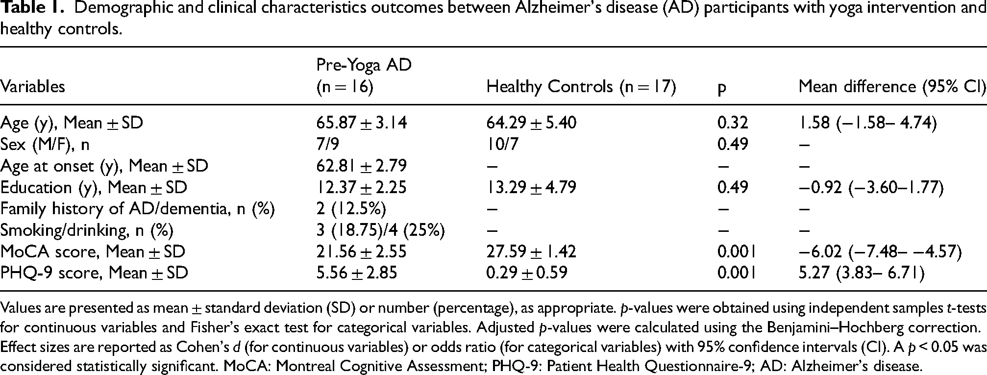
Values are presented as mean ± standard deviation (SD) or number (percentage), as appropriate. p-values were obtained using independent samples t-tests for continuous variables and Fisher's exact test for categorical variables. Adjusted p-values were calculated using the Benjamini–Hochberg correction. Effect sizes are reported as Cohen's d (for continuous variables) or odds ratio (for categorical variables) with 95% confidence intervals (CI). A p < 0.05 was considered statistically significant. MoCA: Montreal Cognitive Assessment; PHQ-9: Patient Health Questionnaire-9; AD: Alzheimer's disease.
Attrition and baseline comparisons
Of the 16 pre-Yoga AD participants, 9 completed the 12-week intervention (∼44% attrition). Baseline comparisons between completers (n = 9) and non-completers (n = 7) revealed no significant differences in age, education, disease duration, MoCA or PHQ-9 scores, or gut microbial alpha diversity indices (Observed species, Chao1, ACE, Shannon, Simpson, Inverse Simpson, Fisher; all p > 0.05) (Supplemental Table 1), indicating comparable baseline profiles and minimal attrition bias.
Cognitive and psychological outcomes post-yoga intervention
The 12-week yoga intervention resulted in significant improvements in cognitive and psychological measures. Consistent trends were observed both when comparing the full baseline cohort (n = 16) to completers (n = 9) and within the completer group from baseline to post-intervention (n = 9) (Table 2, Supplemental Table 2). MoCA scores increased from 22.33 ± 2.34 at baseline to 25.44 ± 2.01 post-intervention (p = 0.001; mean difference: −3.11, 95% CI: −4.47 to −1.75), indicating improved global cognitive function. PHQ-9 scores decreased from 5.78 ± 3.11 pre-intervention to 2.22 ± 1.71 post-intervention (p = 0.007; mean difference: 3.55, 95% CI: 2.33–4.78) These findings suggest that yoga participation was associated with improved cognitive scores and lower depressive symptoms, though causal inference is limited by the non-randomized design.
Cognitive and psychological outcomes in Alzheimer's disease (AD) participants before and after yoga intervention.

Values are presented as mean ± standard deviation (SD). p-values were calculated using paired t-tests comparing pre- and post-yoga intervention scores. Mean differences are shown with 95% confidence intervals (CI). A p < 0.05 was considered statistically significant. MoCA: Montreal Cognitive Assessment; PHQ-9: Patient Health Questionnaire-9; AD: Alzheimer's disease.
Alpha diversity of gut microbiota in AD participants and effects of Yoga intervention
At baseline, no significant differences were observed between AD and HC groups. Observed species counts were slightly lower in AD participants (5137.5 ± 1277.9) than in controls (5712.82 ± 1165.74; p = 0.19). Richness indices-Chao1 (8991.46 ± 881.62 versus 9221.28 ± 740.97; p = 0.42), ACE (10215.2 ± 686.35 versus 10170.16 ± 405.07; p = 0.82), and Fisher (1003.64 ± 287.77 versus 1117.28 ± 252.17; p = 0.24) were comparable. Diversity indices, including Shannon (5.21 ± 0.29 versus 5.01 ± 0.21; p = 0.07) and Simpson (0.97 ± 0.01 versus 0.98 ± 0.01; p = 0.10), showed modest but non-significant differences.
Post-intervention comparisons between pre-Yoga (n = 16) and post-Yoga (n = 9) AD participants revealed no statistically significant shifts in most alpha diversity indices. However, a notable reduction in se.ACE (66.35 ± 4.53 to 56.94 ± 12.31; p = 0.01) indicated reduced variability in microbial richness after yoga. Other indices, including Observed species (5137.5 ± 1277.9 versus 5082 ± 2749.7; p = 0.94), Chao1 (8991.46 ± 881.62 versus 8232.58 ± 2901.98; p = 0.34), Shannon (4.85 ± 0.29 versus 4.83 ± 0.58; p = 0.88), and Simpson (0.97 ± 0.01 versus 0.97 ± 0.03; p = 0.78), remained stable. Similar patterns were observed in completers (n = 9) (Figure 1, Table 3).

Alpha diversity indices of gut microbiota across Pre-Yoga AD, Post-Yoga AD, and Healthy Control (HC) groups. Boxplots illustrate comparisons of microbial alpha diversity based on (A) Observed species, (B) Chao1, (C) se.Chao1, (D) ACE, (E) se.ACE, (F) Shannon, (G) Simpson, (H) Inverse Simpson, and (I) Fisher indices. Each point represents an individual sample, with boxes indicating interquartile range (IQR) and whiskers representing 1.5 × IQR. Triangles denote group means. No significant differences were observed between Pre-Yoga AD and HC groups across most indices (p > 0.05). Following the 12-week yoga intervention, alpha diversity indices remained largely stable, except for a significant reduction in se.ACE (p = 0.01), indicating decreased variability in microbial richness and enhanced community stability post-intervention.
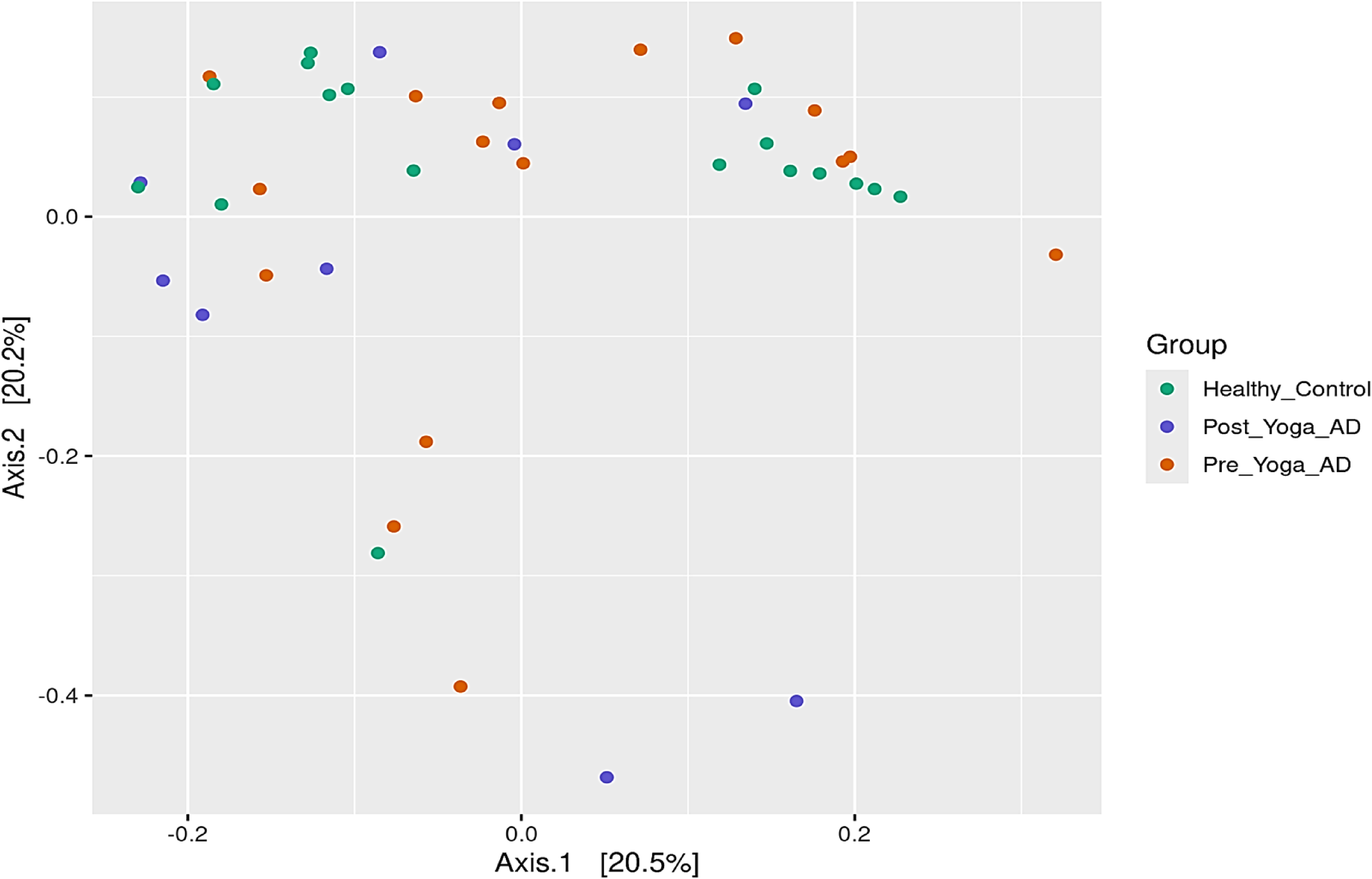
Principal Coordinate Analysis (PCoA) of gut microbiota reveals Yoga-induced compositional shifts in Pre-Yoga AD, Post-Yoga AD, and Healthy Control (HC) groups, Principal Coordinate Analysis (PCoA) plot illustrating the compositional differences in gut microbiota across three groups: Healthy Control (green), Pre-Yoga AD (orange), and Post-Yoga AD (purple). The axes represent the first two principal coordinates—Axis.1 (20.5% variance explained) and Axis.2 (20.2%) derived from [Bray–Curtis dissimilarity]. Healthy Control samples cluster distinctly, while Pre-Yoga AD samples show broader dispersion. Post-Yoga AD samples exhibit a directional shift toward the Healthy Control cluster, suggesting partial restoration of microbial community structure following yoga intervention. Each point represents an individual sample. Ellipses or clustering trends (if applicable) reflect group-level compositional similarity.
Alpha diversity indices of gut microbiota between pre- and post-yoga intervention in Alzheimer's disease (AD) participants and with healthy controls.

The table summarizes alpha diversity measures of gut microbiota across three groups: pre-yoga AD (n = 16), post-yoga AD (n = 9), and healthy controls (n = 17). Comparisons between pre-yoga AD and healthy controls were analyzed using independent samples t-tests, whereas pre- versus post-yoga AD comparisons (n = 9 matched participants) were analyzed using paired t-tests. Diversity indices include Observed species richness, Chao1 and ACE estimators (with their standard errors), and diversity metrics such as Shannon index, Simpson index, Inverse Simpson index, and Fisher's alpha, which collectively reflect microbial richness, evenness, and overall community diversity. Values are presented as mean ± standard deviation (SD). Mean differences are shown with 95% confidence intervals (CI). A p < 0.05 was considered statistically significant. AD: Alzheimer's disease.
Collectively, these results indicate that while overall alpha diversity remained comparable, yoga participation was associated with reduced inter-individual variability in richness, suggesting a possible stabilizing shift in gut microbial structure rather than a marked compositional change. A similar trend was observed in the completer subgroup (n = 9), from baseline to post-intervention, where alpha diversity indices remained stable, with minor shifts in richness and diversity and a significant reduction in se.ACE (Supplemental Table 2). These results indicate that while yoga did not significantly alter overall gut microbiota richness or diversity, it may contribute to stabilization of microbial community structure in AD participants.
Beta diversity indicates microbial shifts following yoga intervention
Principal Coordinates Analysis (PCoA) based on Bray–Curtis dissimilarity metrics revealed distinct microbial community structures across HC, Pre-Yoga AD, and Post-Yoga AD groups. The first two principal coordinates explained 20.5% and 20.2% of the total variance, respectively Figure 2. HC samples formed a tight and cohesive cluster, whereas Pre-Yoga AD samples were more dispersed, reflecting greater inter-individual variability. Following the yoga intervention, AD samples shifted closer to the HC cluster, suggesting a partial normalization of the gut microbial composition. Statistical analysis using PERMANOVA confirmed significant group-level differences, supporting the presence of yoga-associated microbial shifts in AD participants.
Hierarchical clustering reveals yoga-associated shifts in gut microbial composition
Hierarchical clustering of normalized taxonomic abundance profiles revealed distinct group-specific microbial signatures across Pre-Yoga AD, Post-Yoga AD, and HC groups (Supplemental Figure 2A–C). In the Post-Yoga AD versus HC comparison (Supplemental Figure 2A), Post-Yoga AD samples formed a separate but closely aligned cluster with HCs, suggesting partial convergence of microbial profiles toward healthy patterns. Post-Yoga AD participants displayed increased relative abundance of Bacteroides, Clostridium sensu stricto, Desulfovibrio, and Collinsella, along with partial recovery of beneficial taxa such as Faecalibacterium and Bifidobacterium. In contrast, HCs exhibited higher proportions of SCFA-producing and anti-inflammatory genera, including Faecalibacterium, Bifidobacterium, Akkermansia, and Roseburia, which are associated with gut barrier integrity and neuroprotection.
The Pre-Yoga AD versus HC heatmap (Supplemental Figure 2B) displayed a clear separation between groups, reflecting pronounced gut dysbiosis in AD. Pre-Yoga AD samples showed enrichment of potentially pro-inflammatory or pathogenic taxa, such as Escherichia/Shigella, Veillonella, Prevotella copri, and Clostridium sensu stricto, accompanied by marked depletion of beneficial SCFA-producing bacteria, including Faecalibacterium prausnitzii, Bifidobacterium catenulatum, Lachnospira eligens, and Akkermansia muciniphila. These patterns indicate a disrupted microbial ecosystem favoring inflammation and metabolic stress.
Comparative analysis between Pre- and Post-Yoga AD groups (Supplemental Figure 2C) revealed yoga-associated modulation of gut microbiota composition. Post-intervention samples showed enrichment of beneficial taxa such as Faecalibacterium prausnitzii, Roseburia intestinalis, Akkermansia muciniphila, and Bifidobacterium adolescentis, along with reductions in potentially pathogenic genera including Klebsiella variicola, Collinsella aerofaciens, Sutterella spp., and Escherichia/Shigella. These compositional shifts suggest partial restoration of microbial balance and improved gut homeostasis following yoga intervention.
Differential abundance analysis across 1000 OTUs further supported these findings (Table 4; Supplemental Table 3A–C). Twenty OTUs showed significant alterations between Pre- and Post-Yoga AD groups. Post-intervention increases were noted in SCFA-producing and anti-inflammatory species, including Roseburia intestinalis, Schaalia meyeri, and several Lactobacillus and Clostridia taxa, while reductions were observed in potentially pathogenic taxa such as Collinsella aerofaciens, Klebsiella variicola, Sutterella spp., and Duodenibacillus massiliensis (Supplemental Table 3A). Pre-Yoga AD samples were characterized by depletion of Faecalibacterium spp., Lachnospira eligens, and Bifidobacterium catenulatum, and enrichment of Prevotella copri and Veillonella spp. relative to HCs (Supplemental Table 3B). Post-Yoga AD samples exhibited recovery of Faecalibacterium, Roseburia, and Phocaeicola, with concurrent reduction of Collinsella aerofaciens, Veillonella parvula, Klebsiella spp., and Haemophilus parainfluenzae (Supplemental Table 3C).
Differential analysis of gut microbial operational taxonomic units (OTUs) among study groups.
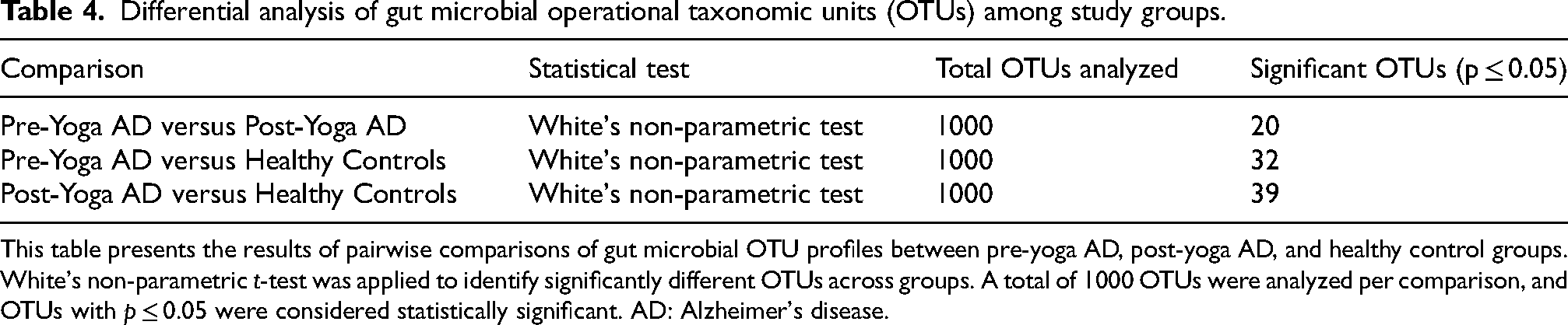
This table presents the results of pairwise comparisons of gut microbial OTU profiles between pre-yoga AD, post-yoga AD, and healthy control groups. White's non-parametric t-test was applied to identify significantly different OTUs across groups. A total of 1000 OTUs were analyzed per comparison, and OTUs with p ≤ 0.05 were considered statistically significant. AD: Alzheimer's disease.
Consistent taxonomic profiling across multiple levels (Figure 3A–C) reinforced these trends. At the phylum level, Pre-Yoga AD samples showed dominance of Bacillota and Bacteroidota, along with elevated Candidatus Marinamimicrobia and Fusobacteriota, whereas beneficial Actinomycetota and Verrucomicrobiota were depleted. Post-yoga intervention led to rebalancing of Bacillota and Bacteroidota and increased representation of Actinomycetota and Verrucomicrobiota, indicating improved microbial diversity and stability.

Heatmaps showing the top 20 most abundant gut microbial taxa across Pre-Yoga AD, Post-Yoga AD, and Healthy Control (HC) groups at three taxonomic levels: (a) phylum, (b) genus, and (c) species. Each heatmap includes hierarchical clustering dendrograms to visualize sample-level similarity and taxonomic co-occurrence patterns. (A) Phylum-level profiles. Colors indicate relative abundance (blue = low, red = high). Bacteroidota and Bacillota are enriched in Pre-Yoga and Post-Yoga AD groups, whereas Healthy Controls show higher relative abundance of Verrucomicrobiota, Mycoplasmatota, and Actinomycetota. (B) Genus-level profiles. Healthy Controls exhibit higher abundance of Faecalibacterium, Prevotella, Collinsella, Candidatus Anaerobutyricum, and Mediterraneibacter, while Methanobrevibacter and Coprococcus are more abundant in AD groups. (C) Species-level profiles. Post-Yoga AD samples show increased abundance of Faecalibacterium prausnitzii, Blautia wexlerae, Bifidobacterium adolescentis, Ruminococcus torques, and Bacteroides eggerthii, indicating a restorative shift following the yoga intervention.
At the genus level, Pre-Yoga AD individuals exhibited overrepresentation of pro-inflammatory genera (Prevotella, Veillonella, Collinsella) and reduced levels of SCFA-producing genera (Faecalibacterium, Roseburia, Bifidobacterium). Post-Yoga AD samples demonstrated a compositional shift toward increased Faecalibacterium and Bifidobacterium and reduced Prevotella and Collinsella, aligning more closely with the HC profile.
At the species level, Pre-Yoga AD participants showed depletion of Faecalibacterium prausnitzii, Bifidobacterium adolescentis, Roseburia intestinalis, and Lachnospira eligens, alongside enrichment of Prevotella copri and Collinsella aerofaciens. Post-Yoga AD samples displayed partial restoration of the beneficial butyrate producers Faecalibacterium prausnitzii, Bifidobacterium adolescentis, and Roseburia intestinalis, with reduced abundance of inflammatory taxa such as Prevotella copri and Collinsella aerofaciens. Overall, these results demonstrate that yoga participation was associated with stabilization of gut microbial community structure, partial restoration of SCFA-producing and anti-inflammatory taxa, and rebalancing of functional pathways related to metabolism and cellular homeostasis. These microbial and functional improvements were accompanied by enhanced cognitive performance and reduced depressive symptoms, suggesting beneficial modulation of the gut–brain axis in AD participants
Functional and taxonomic profiling of gut microbiome across study groups
Functional annotation of metagenomic sequences revealed distinct variations in microbial gene distribution among the Healthy Control, Pre-Yoga AD, and Post-Yoga AD groups (Figure 4A–D). As shown in Figure 4A, Clusters of Orthologous Groups (COG) analysis demonstrated that genes involved in amino acid transport and metabolism, carbohydrate metabolism, energy production and conversion, and signal transduction mechanisms were predominant across all groups. The Post-Yoga AD group exhibited a noticeable increase in genes related to coenzyme transport and metabolism, defense mechanisms, and cell wall/membrane/envelope biogenesis, indicating enhanced metabolic adaptability and improved cellular function following yoga intervention.
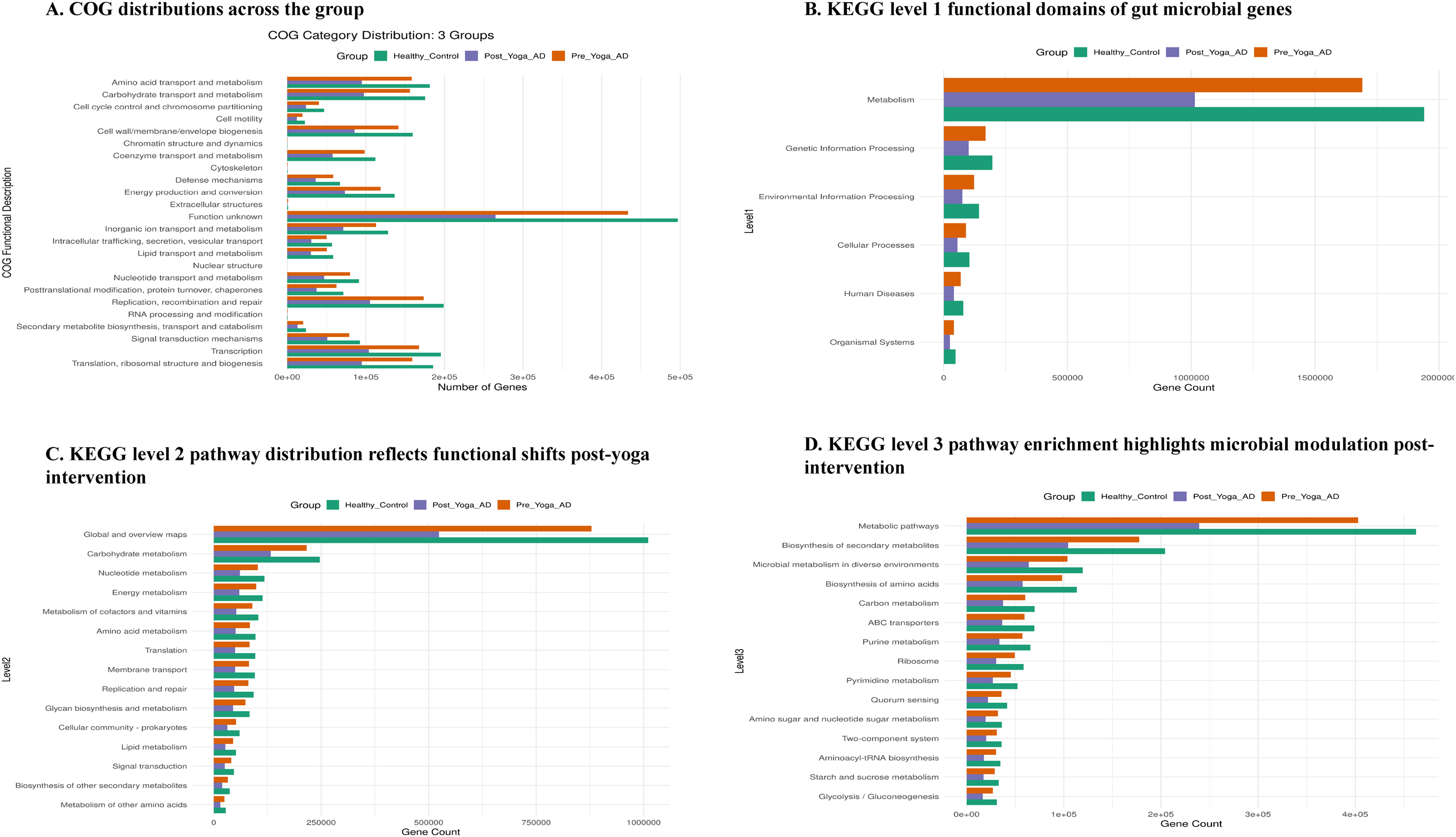
(A) COG-based functional classification of gut microbiota across study groups. Bar chart showing the distribution of annotated genes across 20 COG (Clusters of Orthologous Groups) functional categories in Healthy Controls (green), Pre-Yoga AD (orange), and Post-Yoga AD (purple) groups. Categories include amino acid transport and metabolism, energy production and conversion, signal transduction mechanisms, and replication/recombination/repair. Healthy Controls samples exhibit elevated gene counts in biosynthetic and metabolic pathways, while Post-Yoga AD samples show partial restoration compared to Pre-Yoga-AD, suggesting functional recovery following yoga intervention. (B) KEGG level 1 functional domains of gut microbial genes. Comparative analysis of gene counts across six KEGG Level 1 functional domains Metabolism, Genetic Information Processing, Cellular Processes, Environmental Information Processing, Organismal Systems, and Poorly Characterized among Healthy Controls (green), Pre-Yoga AD (orange), and Post-Yoga AD (purple) groups. Metabolic functions dominate in Healthy Controls, with Post-Yoga AD samples showing intermediate recovery, indicating a shift toward a healthier functional profile post-intervention. (C) KEGG level 2 pathway distribution reflects functional shifts post-yoga intervention. Bar chart depicting gene counts across KEGG Level 2 pathways including carbohydrate metabolism, lipid metabolism, membrane transport, and environmental adaptation. Pre- Yoga AD samples show reduced representation across most pathways, while Post-Yoga AD samples demonstrate increased gene counts in key metabolic and adaptive functions, suggesting yoga-associated enhancement of microbial functional potential. (D) KEGG level 3 pathway enrichment highlights microbial modulation post-intervention. Detailed functional annotation of gut microbial genes across KEGG Level 3 pathways such as biosynthesis of secondary metabolites, microbial metabolism in diverse environments, amino acid biosynthesis, quorum sensing, and ABC transporters. Post-Yoga AD samples show increased abundance in catabolic and amino acid biosynthetic pathways compared to Pre-Yoga AD, indicating functional modulation and partial restoration of microbial activity following yoga intervention.
At the KEGG Level 1 classification (Figure 4B), metabolism was identified as the most abundant functional category, followed by genetic information processing and environmental information processing. The Healthy Control group displayed the highest gene counts for metabolic functions, which were markedly reduced in the Pre-Yoga AD group but showed partial restoration in the Post-Yoga AD group after yoga intervention. At KEGG Level 2 (Figure 4C), the predominant pathway categories included global and overview maps, carbohydrate metabolism, nucleotide metabolism, energy metabolism, and amino acid metabolism. The Post-Yoga AD group showed increased representation in membrane transport, glycan biosynthesis and metabolism, and cellular community prokaryotes, suggesting a recovery of microbial functional capacity toward a healthy metabolic profile.
Further analysis at KEGG Level 3 (Figure 4D) highlighted metabolic pathways, biosynthesis of secondary metabolites, and microbial metabolism in diverse environments as dominant across all groups. The Healthy Control group exhibited the highest diversity and abundance of these pathways, whereas Pre-Yoga AD showed reduced functional richness. The enhanced representation of metabolic pathways, biosynthesis of secondary metabolites, amino acid biosynthesis, carbon metabolism, and ABC transporter pathways in the Post-Yoga AD group supports the restorative effect of yoga on gut microbial metabolic capacity and diversity, promoting a shift toward a healthier functional state.
Taxonomic profiling based on metagenomic sequencing revealed the hierarchical distribution of microbial taxa across multiple taxonomic levels, depicting the overall gut microbiome composition in the study cohort (Supplemental Figure 3). The microbiome was dominated by members of the domain Bacteria, with Bacteroidota and Bacillota emerging as the predominant phyla, followed by minor contributions from Actinomycetota and Euryarchaeota. Within Bacteroidota, the class Bacteroidia and order Bacteroidales were highly represented, primarily comprising the genera Bacteroides, Prevotella, Alistipes, and Parabacteroides. The Bacillota phylum was chiefly represented by the class Clostridia, with abundant genera including Blautia, Faecalibacterium, Roseburia, and Ruminococcus, known producers of butyrate that support gut homeostasis. A smaller fraction of reads corresponded to Viruses, predominantly from the families Caudoviricetes (order Caudovirales), Demerecviridae, and Gilesvirus, suggesting the presence of bacteriophages potentially involved in regulating bacterial population dynamics. Overall, the taxonomic composition underscores the dominance of commensal anaerobes within the gut ecosystem, alongside subtle contributions from archaeal and viral taxa, reflecting a complex and interactive microbial environment associated with AD.
Discussion
This study demonstrates that a 12-week yoga intervention in individuals with AD led to significant improvements in cognitive performance, reduced depressive symptoms, and measurable modulation of gut microbial composition and functional capacity. These findings support growing evidence that mind-body interventions can beneficially influence gut microbial ecology and host physiology through stress reduction, neuroendocrine regulation, and vagal activation. Similar improvements reported in populations with MCI and SCD further underscore yoga's potential as an adjunct strategy to restore gut–brain axis balance and mitigate cognitive deterioration in AD.11,16–19
Consistent with the baseline comparisons, alpha diversity analyses revealed that overall microbial richness and diversity in AD participants were comparable to healthy controls, with no major differences across indices such as Observed species, Chao1, ACE, Shannon, and Simpson. Following yoga, alpha diversity remained largely stable; however, a significant reduction in se.ACE indicated reduced inter-individual variability in microbial richness, suggesting a stabilization of the gut ecosystem. This stabilization without dramatic compositional change supports ecological models in which stress reduction and autonomic balance promote microbial evenness and community resilience rather than expansion of diversity.9,20,21
Beta diversity analyses further supported these findings. Principal Coordinates Analysis revealed that while pre-yoga AD samples were widely dispersed, reflecting dysbiotic heterogeneity, post-yoga samples clustered more closely to healthy controls, suggesting partial normalization of the gut microbial community structure. PERMANOVA confirmed significant between-group differences, indicating that yoga was associated with distinct microbial shifts toward a healthier configuration. These results are in line with previous reports showing that AD is associated with increased inter-individual microbial variability and that interventions reducing systemic inflammation can promote microbial reorganization toward a balanced state.2,22–24
Hierarchical clustering and differential abundance analyses revealed specific taxonomic changes driving these compositional shifts. Pre-yoga AD participants exhibited enrichment of pro-inflammatory or opportunistic taxa such as Prevotella copri, Veillonella, Collinsella aerofaciens, and Escherichia/Shigella, accompanied by depletion of SCFA-producing and anti-inflammatory taxa such as Faecalibacterium prausnitzii, Bifidobacterium adolescentis, Roseburia intestinalis, and Akkermansia muciniphila. Following yoga intervention, partial restoration of these beneficial taxa was observed, alongside reductions in the pro-inflammatory genera. These yoga-associated improvements in SCFA-producing bacteria are mechanistically significant, as butyrate supports intestinal barrier integrity, modulates neuroinflammation, and regulates neurotransmission key pathways implicated in AD pathology.25–29 The observed partial recovery (approximately 20–45% normalization toward control levels) suggests that while yoga adopts a favorable microbial environment, complete restoration may require longer interventions or complementary microbiome-targeted strategies such as dietary fiber or probiotics.30–32
Functional annotation of metagenomic sequences provided additional evidence for microbial functional restoration. COG and KEGG pathway analyses revealed that metabolic and biosynthetic functions predominated across all groups, with the Post-Yoga AD group showing increased representation of genes involved in coenzyme transport, defense mechanisms, cell wall biogenesis, amino acid metabolism, and energy production. These shifts reflect enhanced microbial metabolic adaptability and suggest recovery of microbial biosynthetic capacity following yoga intervention. In contrast, pre-yoga AD samples were enriched in stress-response pathways such as quorum sensing and two-component systems, reflecting adaptive responses to host inflammation. The observed upregulation of pathways related to translation, ribosomal structure, and secondary metabolite biosynthesis in the Post-Yoga AD group indicates functional reorganization of the gut microbiome toward a more efficient and metabolically balanced state. These functional signatures align with prior studies linking improved microbial metabolism with better neurocognitive outcomes and reduced systemic inflammation.2,6,33
The taxonomic profiling across hierarchical levels corroborated these trends, showing dominance of Bacteroidota and Bacillota phyla, with beneficial Clostridia (e.g., Faecalibacterium, Roseburia) and Actinomycetota members enriched post-intervention. The detection of bacteriophages, predominantly Caudoviricetes and Demerecviridae, suggests a possible role in shaping bacterial community dynamics through phage-bacteria interactions. The overall taxonomic structure reflects a complex, yet adaptive microbial ecosystem in AD, with yoga-associated stabilization of commensal taxa contributing to homeostasis and gut-brain axis integrity.
Taken together, these results support a mechanistic model in which yoga exerts beneficial effects on cognition and mood through multi-level modulation of the gut–brain axis. Stress reduction likely attenuates HPA axis overactivation, enhances vagal tone, and reduces gut permeability, collectively creating a microenvironment favorable to beneficial anaerobes and SCFA producers.3,33 Enrichment of these taxa promotes anti-inflammatory signaling and neuroprotective metabolite production, potentially contributing to the observed cognitive and psychological improvements. Although causality cannot be fully established in this non-randomized design, the consistent convergence of microbial, functional, and behavioral outcomes suggests yoga's potential as a non-pharmacological intervention to restore gut microbial and neurocognitive health in AD. 34
Despite the promising findings, several limitations must be acknowledged. The modest sample size and post-intervention attrition reduced statistical power. Possible confounders, including dietary variability, physical activity, and medication use, were not fully controlled. Moreover, while functional inference from metagenomic data provides valuable insights, integration with metatranscriptomic, metabolomic, and immune profiling would offer a more comprehensive understanding of host–microbe interactions. Future studies with larger, randomized cohorts, longer follow-up, and multimodal integration are warranted to delineate causal pathways linking yoga, microbial functional recovery, and cognitive resilience in AD.
Limitations and future directions
This exploratory study has several limitations. First, the sample size was relatively small (16 AD patients and 17 healthy controls), and post-intervention samples from AD participants were further reduced to nine due to dropouts, limiting statistical power. The observed partial restoration of microbial diversity and composition may be more pronounced in a larger cohort. Notably, attrition in the AD group was 44%, primarily due to challenges in adhering to the yoga intervention, reflecting cognitive impairments and difficulties in performing the modules. This highlights the practical challenge of implementing structured mind-body interventions in older, cognitively impaired populations. Second, moderate sequencing depth may have affected estimates of microbial richness and diversity. Functional predictions were based on taxonomic data, and the absence of direct metabolomic, inflammatory, and gut–brain axis measurements limits confirmation of predicted pathway alterations. Third, several uncontrolled variables may have influenced outcomes. Dietary intake, sleep patterns, and unrecorded physical activity were not strictly monitored, each of which can significantly impact gut microbiome composition. Additionally, data on AD medications were limited, preventing a comprehensive analysis of pharmacological exposures during the study. Medication use may confound both cognitive outcomes and microbial composition and should be considered when interpreting the results. Fourth, cognitive assessments were repeated over a short interval of 12 weeks, introducing the possibility of practice or learning effects that could contribute to observed improvements. Longer-term interventions with follow-up assessments are necessary to evaluate sustained cognitive effects while minimizing potential test–retest biases. Finally, the absence of an attention-matched or active control group limits causal inference regarding the specific effects of yoga. Methodological aspects, including small sample size, partial data reporting, and limited intervention monitoring, reduce the overall strength of the conclusions.
Future studies should address these limitations by conducting larger, multi-center randomized controlled trials with adequate sample sizes to enhance statistical power and generalizability. Standardized monitoring of dietary intake, sleep, physical activity, and medication use is essential to control for potential confounders. Inclusion of attention-matched control groups and longer follow-up periods will help isolate the specific contributions of yoga to cognitive and microbiome outcomes. Mechanistic investigations integrating metabolomic, cytokine, and autonomic measures (e.g., heart rate variability) will be critical to link yoga-induced microbial changes with physiological and cognitive outcomes. Combinatorial interventions, such as yoga combined with prebiotic or probiotic supplementation, may further enhance microbiome restoration and clarify causal pathways. Finally, future research should explore dose–response relationships, adherence patterns, and the relative contributions of specific yoga components (e.g., meditation versus physical postures) to identify which elements most strongly mediate cognitive and microbiome benefits in AD.20,34
Conclusion
This study demonstrates that a 12-week yoga intervention in individuals with AD was associated with significant improvements in cognitive function and mood, accompanied by measurable shifts in gut microbial composition and function. While overall microbial richness and diversity remained stable, reduced inter-individual variability (decrease in se.ACE) suggested enhanced community stability following yoga. Beta-diversity analyses further indicated a directional shift in gut microbial structure, with post-intervention AD samples clustering closer to healthy controls, reflecting partial restoration of microbial balance.
Taxonomic and functional analyses revealed enrichment of SCFA-producing and anti-inflammatory taxa such as Faecalibacterium prausnitzii, Roseburia intestinalis, Bifidobacterium, and Akkermansia alongside reductions in pro-inflammatory and potentially pathogenic genera including Collinsella aerofaciens, Klebsiella spp., and Escherichia/Shigella. These compositional shifts were paralleled by functional recovery, with post-yoga samples showing upregulation of genes involved in translation, energy metabolism, and amino acid biosynthesis, and downregulation of stress-related pathways such as cell wall biogenesis and quorum sensing.
Collectively, these findings suggest that yoga exerts beneficial effects on the gut–brain axis by stabilizing microbial community structure, enriching neuroprotective and metabolically beneficial taxa, and promoting partial restoration of microbial metabolic potential. Through mechanisms involving stress reduction, improved autonomic regulation, and modulation of gut microbiota-derived metabolites such as SCFAs, yoga may contribute to cognitive and emotional resilience in AD. Although the small sample size and lack of a control arm limit causal inference, the observed convergence of clinical and microbial improvements highlights yoga as a promising, accessible, and non-pharmacological adjunct to support cognitive function and mitigate gut dysbiosis in AD. Future large-scale, controlled, multi-omics studies are warranted to confirm these findings and elucidate mechanistic pathways underlying yoga-mediated gut-brain modulation.
Supplemental Material
sj-docx-1-alz-10.1177_13872877261415612 - Supplemental material for Association of yoga with cognitive and gut microbiome changes in Alzheimer's disease: An exploratory case-control study
Supplemental material, sj-docx-1-alz-10.1177_13872877261415612 for Association of yoga with cognitive and gut microbiome changes in Alzheimer's disease: An exploratory case-control study by Prabhakar Tiwari, Anu Gupta, Meenakshi Kaushik, Rekha Dwivedi, Manjari Tripathi and Rima Dada in Journal of Alzheimer's Disease
Footnotes
Acknowledgements
We acknowledge the All India Institute of Medical Sciences (AIIMS), New Delhi, for providing intramural grants (F.8.55/AC-55/2023/RS; F.8.13/AT-13/2024/RS) that supported the conduct of this study. We thank Dr Richa Mishra (Yoga Instructor) for facilitating the yoga sessions and diligently maintaining the yoga activity diary. We are also thankful to Dr Ashish Datt Upadhyay, Clinical Research Unit, AIIMS, New Delhi, for his valuable assistance with statistical analysis. We acknowledge the support provided by N2 Genomics lab, New Delhi, India for metagenomic sequencing, data analysis, and figures generation. Also, we thanks to Mr Dinesh Tomar for his technical support throughout the study.

Ethical considerations
The study was approved by the Institutional Ethics Committee (IEC) of AIIMS, New Delhi (IEC approval no. AIIMSA00394/12-01-2024, RP-13/2024). The research was conducted in accordance with the relevant institutional IEC guidelines.
Consent to participate
All human subjects involved in this study provided informed consent prior to participation.
Consent for publication
Not applicable
Author contribution(s)
Funding
The authors disclosed receipt of the following financial support for the research, authorship, and/or publication of this article: This study was funded by the AIIMS, New Delhi, intramural grant (F.8.55/AC-55/2023/RS; F.8.13/AT 13/2023/RS).
Declaration of conflicting interests
The authors declared no potential conflicts of interest with respect to the research, authorship, and/or publication of this article.
Data availability statement
Supplemental material
Supplemental material for this article is available online.
References
Supplementary Material
Please find the following supplemental material available below.
For Open Access articles published under a Creative Commons License, all supplemental material carries the same license as the article it is associated with.
For non-Open Access articles published, all supplemental material carries a non-exclusive license, and permission requests for re-use of supplemental material or any part of supplemental material shall be sent directly to the copyright owner as specified in the copyright notice associated with the article.
