



Abstract
Alzheimer’s disease (AD) is pathologically characterized by the accumulation of amyloid-β (Aβ) plaques and neurofibrillary tangles composed of hyperphosphorylated tau protein. In recent years, two cellular processes have emerged as pivotal drivers of neurodegeneration in AD: mitophagy, the selective autophagic clearance of damaged mitochondria, and ferroptosis, an iron-dependent form of regulated cell death. This review outlines the molecular mechanisms of mitophagy and ferroptosis, with a focus on their interplay in AD. We propose that impaired mitophagy disrupts intracellular redox and iron homeostasis, thereby increasing neuronal susceptibility to ferroptosis. Conversely, ferroptosis-executing events, such as lethal lipid peroxidation, can further exacerbate mitochondrial dysfunction. This establishes a self-amplifying vicious cycle that accelerates disease progression. Furthermore, we summarize potential therapeutic strategies targeting this interactive network (e.g., Urolithin A, ferroptosis inhibitors) and highlight promising directions for future research. In contrast to previous reviews that have focused on each process in isolation, this work synthesizes evidence for a self-amplifying feedback loop between impaired mitophagy and exacerbated ferroptosis in AD. We posit that targeting this self-amplifying loop between mitophagy and ferroptosis may offer a novel and effective therapeutic paradigm for halting Alzheimer’s disease progression.
Introduction
Alzheimer’s disease (AD) is a progressive neurodegenerative disorder that poses an immense and growing global health burden, with prevalence projected to rise dramatically in the coming decades. 1 The classical hallmarks of AD—amyloid-β (Aβ) plaques and neurofibrillary tangles composed of hyperphosphorylated tau protein—have long guided research.2,3 However, the repeated failure of therapies targeting Aβ and tau underscores the complexity of AD pathogenesis and highlights the urgent need to explore alternative pathogenic mechanisms. 4
Mitochondrial dysfunction is increasingly recognized as a critical early event in AD, contributing to bioenergetic deficits and oxidative stress. 5 This dysfunction implicates two closely related yet distinct cellular processes: mitophagy, the selective autophagy of damaged mitochondria, and ferroptosis, an iron-dependent form of regulated cell death characterized by lethal lipid peroxidation. Dysregulation of both processes has been independently linked to neuronal loss in AD models. 6
Crucially, emerging evidence suggests that mitophagy and ferroptosis are not independent pathways but engage in a complex interplay. 7 Impaired clearance of damaged mitochondria via mitophagy may promote iron overload and oxidative stress, both of which are key drivers of ferroptosis. 8 Conversely, damage executed during ferroptosis can exacerbate mitochondrial dysfunction. This self-amplifying cycle is hypothesized to be a significant contributor to neurodegeneration in AD. Therefore, this review aims to: (1) outline the fundamental mechanisms of mitophagy and ferroptosis; (2) synthesize evidence for their dysregulation in AD; and (3) focus specifically on their mechanistic interplay and resulting therapeutic implications. By integrating these two fields, we provide a novel perspective on AD pathogenesis and highlight promising avenues for therapeutic intervention.
Mitophagy in Alzheimer’s disease
Mechanism of mitophagy
Mitophagy is a critical, evolutionarily conserved quality control mechanism that selectively targets damaged or superfluous mitochondria for autophagic degradation, thereby maintaining cellular health and bioenergetic efficiency. 9 The dysregulation of this precise process is implicated in numerous age-related diseases, including AD. 10 In mammals, mitophagy is primarily orchestrated through two distinct yet sometimes complementary molecular pathways: the ubiquitin (Ub)-dependent pathway, which relies on specific protein tagging for recognition, and the Ub-independent pathway, which utilizes dedicated receptor proteins on the mitochondrial surface (Figure 1).

Overview of the mitophagy mechanisms. The mechanisms of mitophagy are generally categorized into two main pathways: Ub-dependent pathway and Ub-independent pathway. Black arrows indicate activating or promoting steps. ROS: reactive oxygen species; PINK1: PTEN-induced putative kinase 1; Ub: ubiquitin; TBK1: TANK-binding kinase 1; ULK1: UNC51-like kinase-1; OPTN: optineurin; NDP52: nuclear dot protein 52; SMURF1: smad ubiquitination regulation factor-1; GP78: glycoprotein 78; MUL1: mitochondrial E3 ubiquitin ligase 1; NIX: Nip3like protein X; BNIP3: BCL2/adenovirus E1B 19 kDa interacting protein-3; FUNDC1: FUN14 domain containing 1; OMM: outer mitochondrial membrane; LC3: microtubule-associated protein 1A/1B-light chain 3.
Ub-dependent pathway. The ubiquitin-dependent pathway is predominantly governed by the PINK1/Parkin signaling axis, a system essential for responding to acute mitochondrial damage. 11 Under normal conditions with healthy membrane potential, PINK1 is constitutively imported into mitochondria and degraded. 12 However, upon mitochondrial depolarization—a hallmark of dysfunction—PINK1 import is halted, leading to its stabilization and accumulation on the outer mitochondrial membrane (OMM). 13 Here, PINK1 undergoes autophosphorylation, activating its kinase activity. Active PINK1 then phosphorylates both ubiquitin molecules already present on the OMM and the cytosolic E3 ubiquitin ligase, Parkin. 14 This phosphorylation event serves as a powerful recruitment and activation signal for Parkin, which translocates to the damaged mitochondrion. Once activated, Parkin catalyzes the extensive ubiquitination of numerous OMM proteins, constructing polyubiquitin chains. These chains function as universal “eat-me” signals, which are specifically recognized by autophagy adapter proteins such as OPTN (optineurin) and NDP52 (nuclear dot protein 52). 15 These adapters, in turn, bind to LC3-II proteins embedded in the expanding phagophore membrane, thereby physically tethering the damaged mitochondrion to the nascent autophagosome for subsequent engulfment and lysosomal destruction. 16 Importantly, the efficiency of PINK1/Parkin-mediated mitophagy is often enhanced by prior mitochondrial fission, a process where Parkin can modulate the activity of the fission GTPase Drp1 via ubiquitination, facilitating the segregation of damaged mitochondrial segments for more efficient clearance. 17
Ub-independent pathway. The ubiquitin-independent pathway provides an alternative and often more stimulus-specific route for mitochondrial clearance, crucial under conditions like hypoxia or developmental remodeling. 18 This pathway bypasses the need for extensive ubiquitination by employing a set of OMM-resident receptor proteins that possess LC3-interacting regions (LIRs), enabling them to directly engage the autophagic machinery. 19 A key hypoxia-induced receptor is FUN14 domain-containing 1 (FUNDC1), whose interaction with LC3 is tightly regulated by phosphorylation; dephosphorylation under low oxygen enhances binding, while phosphorylation by kinases like SRC inhibits it. 20 ULK1, a key initiator kinase of autophagy, can also phosphorylate FUNDC1 to positively regulate mitophagy under certain stresses. 21 Additionally, the Bcl-2 family homologs BCL2/adenovirus E1B 19 kDa-interacting protein 3 (BNIP3) and its close relative NIX/BNIP3L are critical receptors. 22 They are upregulated in response to various stresses and directly recruit autophagosomes via their LIR motifs. 23 The activity of these receptors can be further fine-tuned; for example, phosphorylation of serine residues adjacent to the LIR in NIX strengthens its affinity for LC3, promoting efficient mitochondrial turnover. 24 These pathways ensure cellular survival, particularly when the canonical PINK1/Parkin system is overwhelmed or impaired, as often observed in chronic neurodegenerative settings. 25
Dysregulation of mitophagy in Alzheimer’s disease: evidence and pathogenic consequences
A compelling body of evidence from diverse experimental systems—including post-mortem analyses of human AD brain tissue, neurons derived from AD patient-induced pluripotent stem cells (iPSCs), and transgenic animal models—consistently demonstrates a profound impairment in mitophagic flux. This defect is not merely a bystander effect but correlates with key pathological markers and cognitive decline. 26 Biochemically, this impairment manifests as reduced levels or activity of crucial mitophagy players, such as decreased PINK1 and Parkin protein levels, diminished phosphorylation of ULK1 (indicative of reduced initiation), and an accumulation of p62/SQSTM1-positive mitochondria, signaling a blockade in autophagic degradation. 27
This mitophagic failure is actively instigated and perpetuated by the core pathological drivers of AD. Both Aβ oligomers, particularly their intracellular forms, and hyperphosphorylated tau protein have been shown to directly or indirectly induce mitochondrial dysfunction 28 (Figure 2). They promote excessive mitochondrial fission via aberrant activation or localization of Drp1, disrupt electron transport chain complexes, and increase the production of reactive oxygen species (ROS). 29 In a healthy neuronal context, such damage would be a potent trigger for a protective mitophagic response, aiming to purge the dysfunctional organelles. However, in AD, this compensatory mechanism is fundamentally compromised. The failure to clear these damaged powerhouses sets in motion a vicious, self-reinforcing cycle. 30
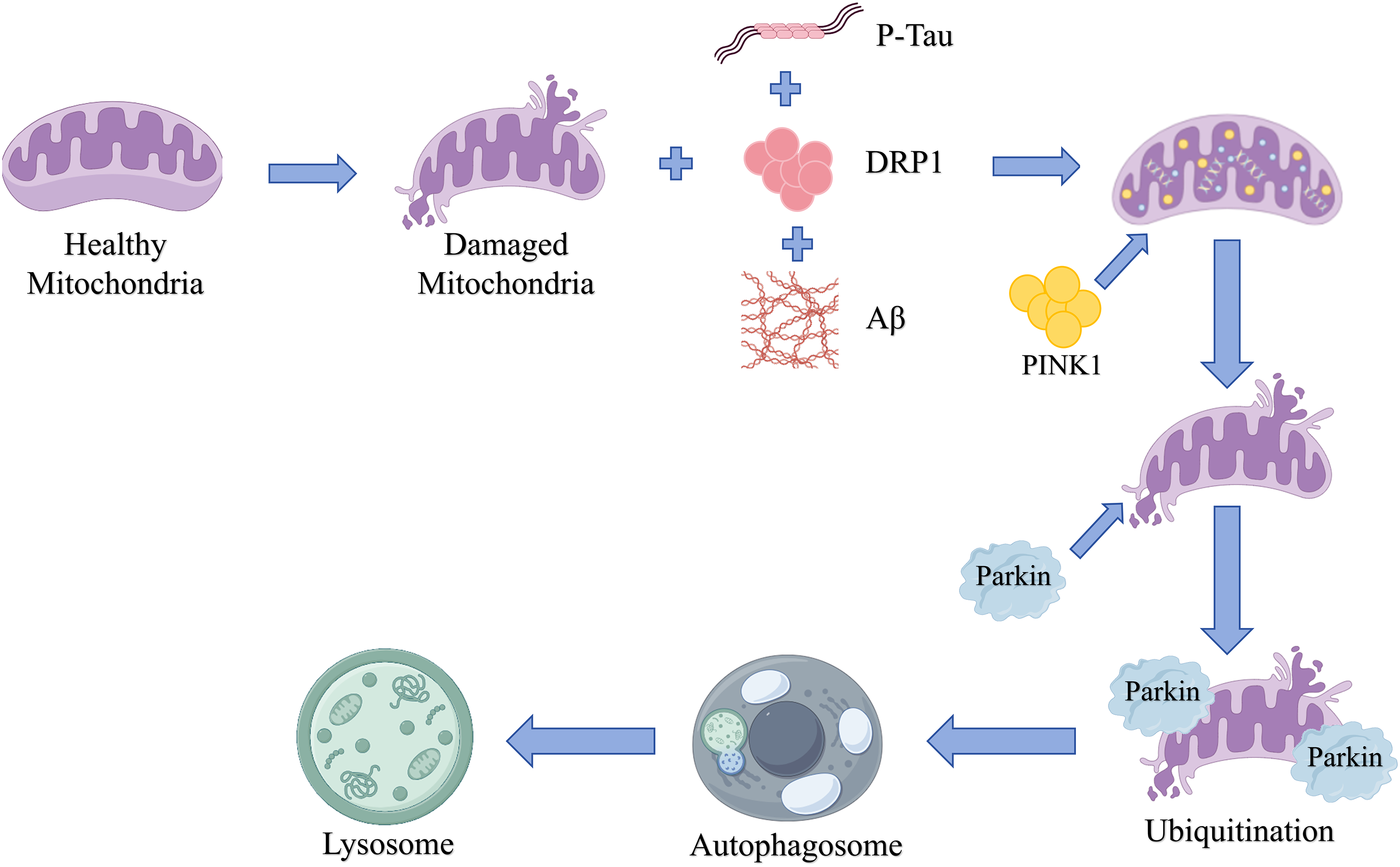
A brief overview of mitophagy events in AD with key toxic players like Aβ, P-Tau, and DRP1 inducers that progress the activation of PINK1/Parkin-mediated abnormal mitochondrial dynamics. P-Tau: phosphorylated tau; DRP1: Dynamin-related protein 1; Aβ: amyloid-β protein; PINK1: PTEN-induced putative kinase 1.
The accumulation of dysfunctional mitochondria has dire consequences. Firstly, these mitochondria become potent and chronic sources of ROS, exacerbating oxidative stress that further damages proteins, lipids, and nucleic acids. This oxidative milieu is known to promote both the production of Aβ and the hyperphosphorylation of tau. 31 Secondly, the bioenergetic deficit resulting from impaired mitochondria compromises vital neuronal functions, including synaptic vesicle recycling, ion pump activity, and axonal transport, all of which are energy-intensive processes. 32 Consequently, the failure of mitophagy directly contributes to the amplification of the very pathologies that caused it. Experimental evidence starkly illustrates this: loss-of-function mutations or knockdown of Parkin in AD models lead to accelerated accumulation of both Aβ and phosphorylated tau, coupled with more severe synaptic and cognitive deficits. 33
Given this central role, therapeutic strategies aimed at restoring or enhancing mitophagic flux have emerged as a promising avenue. These approaches target different nodes of the impaired pathway. Pharmacological inducers like UMI-77 have been designed to specifically enhance PINK1/Parkin-mediated clearance. 34 Natural compounds such as Urolithin A, a gut metabolite of ellagitannins, have demonstrated efficacy in improving mitochondrial health and promoting mitophagy in aged organisms, potentially through pathways parallel to PINK1/Parkin. 35 Non-pharmacological interventions, most notably physical exercise, have shown remarkable benefits in AD mouse models, upregulating pathways like the SIRT1-FOXO1/3 axis to boost mitophagy and reduce amyloid burden. 36 Furthermore, compounds like resveratrol alleviate tau pathology, an effect partially attributable to the restoration of mitochondrial quality control via enhanced mitophagy. 37 Collectively, these findings robustly underscore that rescuing mitophagic function is not just a correlative observation but a viable and potent therapeutic target with the potential to disrupt the core pathogenic cycle of AD.
Ferroptosis in Alzheimer’s disease
Core mechanisms and hallmarks of ferroptosis
Ferroptosis is a recently defined, iron-dependent form of regulated cell death that is morphologically, biochemically, and genetically distinct from apoptosis, necrosis, and autophagy. 38 It is characterized by the pervasive, iron-catalyzed peroxidation of phospholipids containing polyunsaturated fatty acids (PUFAs), leading to catastrophic membrane damage and cell lysis. 39 Morphological hallmarks, primarily observed via electron microscopy, include shrunken mitochondria with increased membrane density, reduction or disappearance of cristae, and outer mitochondrial membrane rupture, while the nucleus remains intact in the early stages. 40 The biochemical execution of ferroptosis is governed by the intricate interplay and failure of three core metabolic axes: the antioxidant defense system, iron homeostasis, and lipid metabolism (Figure 3).
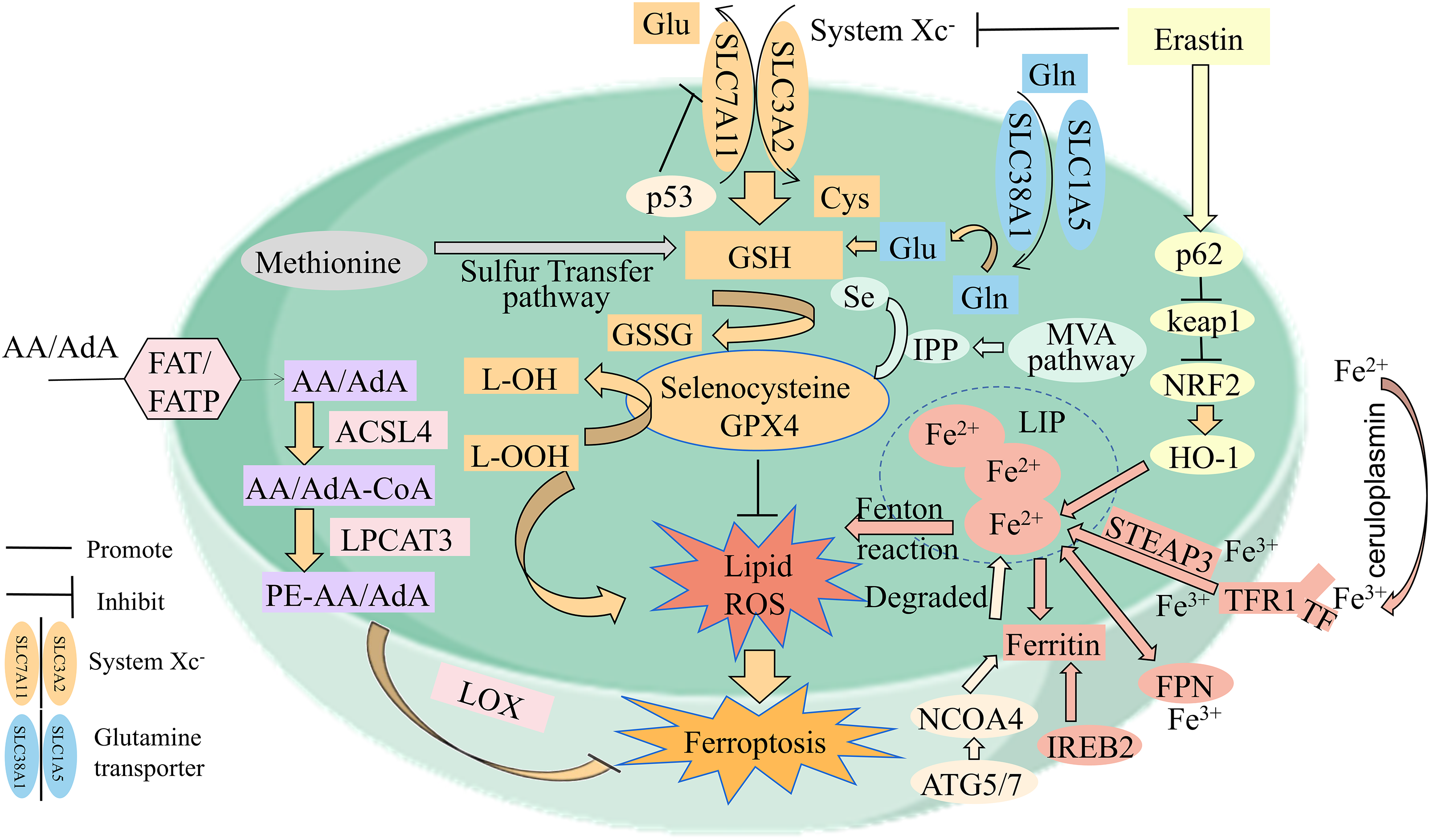
The mechanisms underlying ferroptosis. Various metabolic pathways interact and play a crucial role in ferroptosis. It is known that alterations in the metabolism of lipids, iron and amino acids can all contribute to the initiation of ferroptosis. In addition, the crosstalk between different metabolic pathways can further exacerbate this process. AA/AdA: Arachidonic acid/adrenic acid; AA/AdA-CoA: arachidonic acid/adrenic acidcoenzyme A; AA/AdA-PE: arachidonic acid/adrenic acid-phosphatidylethanolamine; ACSL4: acyl-coenzyme A synthetase long-chain family member 4; ATG5/7: autophagy 5/7; FAT: fatty acid translocase; FATP: fatty acid transport protein; FPN: ferroportin; Gln: glutamine; Glu: glutamic acid; GPX4: glutathione peroxidase 4; GSH: glutathione; GSSG: glutathione disulfide; HO-1: heme oxygenase-1; IPP: isopentenyl pyrophosphate; IREB2: iron responsive element binding protein 2; LIP: labile iron pool; Lipid ROS: lipid reactive oxygen species; LOX: lipoxygenase; LPCAT3: lysophosphatidylcholine acyltransferase 3; NCOA4: nuclear receptor coactivator 4; NRF2: nuclear factor-erythroid 2-related factor 2; STEAP3: six-transmembrane epithelial antigen of prostate 3; TFR1: transferrin receptor 1; Tf: transferrin.
The first and primary defensive barrier is the Glutathione-Based Antioxidant Axis (System Xc-/GSH/GPX4). This system maintains redox balance by regulating the synthesis of the major cellular antioxidant, glutathione (GSH). 41 The cystine/glutamate antiporter, System Xc- (composed of subunits SLC7A11 and SLC3A2), is responsible for importing extracellular cystine in exchange for intracellular glutamate. 42 Once inside the cell, cystine is rapidly reduced to cysteine, a rate-limiting precursor for GSH synthesis. 43 The selenoprotein glutathione peroxidase 4 (GPX4) is the central guardian against ferroptosis; it uniquely utilizes GSH to reduce toxic phospholipid hydroperoxides (PLOOHs) to their corresponding nontoxic phospholipid alcohols (PLOHs), thereby directly halting the lipid peroxidation chain reaction. Any disruption to this axis—whether through inhibition of System Xc- (e.g., by the compound erastin or transcriptional repression by p53), depletion of cellular cysteine/GSH pools, or direct inactivation of GPX4 (e.g., by covalent inhibitors like RSL3 or through selenium deficiency)—irreversibly releases the brake on lipid peroxidation, committing the cell to ferroptosis. 44
The second axis is the Iron Metabolism and Redox Cycling Axis. Ferroptosis is explicitly iron-dependent, requiring redox-active iron in its ferrous (Fe2+) state. 45 Cellular iron homeostasis is a tightly regulated process. Uptake occurs primarily via transferrin receptor 1 (TFR1)-mediated endocytosis of transferrin-bound iron. Within acidified endosomes, ferric iron (Fe3+) is reduced to Fe2+ by metalloreductases like STEAP3 and transported into the cytosol by divalent metal transporter 1 (DMT1), entering the labile iron pool (LIP). 46 Excess iron is safely sequestered within the ferritin nanocage. However, the cargo receptor NCOA4 can target ferritin for autophagic degradation in a process called ferritinophagy, releasing stored iron back into the LIP and increasing ferroptotic sensitivity. 47 Crucially, Fe2+ in the LIP catalyzes the Fenton reaction (Fe2+ + H2O2 → Fe3+ + •OH + OH−), generating highly reactive hydroxyl radicals (•OH) that directly abstract hydrogen atoms from PUFAs in membranes, initiating the lipid peroxidation cascade. 48 Thus, cellular iron overload, increased LIP availability, or enhanced ferritinophagy all create a pro-ferroptotic milieu.
The third axis is the Lipid Metabolism and Peroxidation Execution Axis. Not all lipids are equally susceptible; ferroptosis specifically targets phospholipids containing long-chain PUFAs such as arachidonic acid (AA) and adrenic acid (AdA). 49 The enzymatic machinery that prepares these PUFAs for incorporation into membranes is pro-ferroptotic. 50 Acyl-CoA synthetase long-chain family member 4 (ACSL4) esterifies free PUFAs to PUFA-CoA, and lysophosphatidylcholine acyltransferase 3 (LPCAT3) then incorporates them into membrane phospholipids, notably phosphatidylethanolamine (PE). 51 This enzymatic “tagging” enriches membranes with peroxidation-susceptible substrates. Once a lipid radical is generated (e.g., by •OH), a spontaneous, autocatalytic chain reaction of lipid peroxidation ensues, propagated by molecular oxygen and leading to the accumulation of lethal lipid hydroperoxides that GPX4 can no longer contain, resulting in loss of membrane integrity and function. 52
Execution of ferroptosis in Alzheimer’s disease pathogenesis
Converging lines of evidence strongly implicate the aberrant activation of this ferroptotic cascade in the pathogenesis of AD, positioning it as a significant contributor to the characteristic neuronal loss.53,54 Multiple aspects of the AD brain create a perfect storm that predisposes neurons to ferroptosis.
Histopathological and biochemical analyses reveal clear hallmarks of ferroptosis in the AD brain. 55 There is a well-documented increase in markers of lipid peroxidation, such as 4-hydroxynonenal (4-HNE) and malondialdehyde (MDA), in vulnerable brain regions like the hippocampus and cortex. 56 Concurrently, the expression and activity of the key anti-ferroptotic enzyme GPX4 are often found to be reduced. Perhaps most strikingly, quantitative MRI and histochemical studies have consistently demonstrated abnormal accumulations of iron in the AD brain, particularly within amyloid plaques and around neurofibrillary tangles, as well as in specific deep gray matter nuclei. 57 This pathological iron deposition provides the literal “fuel” for the Fenton reaction.
The AD pathological milieu actively cultivates a pro-ferroptotic environment. Both Aβ and hyperphosphorylated tau contribute to mitochondrial dysfunction and elevated oxidative stress, which depletes cellular GSH reserves and can inactivate GPX4. 58 Furthermore, these pathologies appear to directly and indirectly disrupt iron homeostasis. Aβ peptides may themselves have redox-active properties and can promote iron retention, while tau pathology is associated with alterations in iron-regulatory proteins. 59 This creates a self-perpetuating “iron dyshomeostasis cycle": iron promotes Aβ aggregation and tau phosphorylation via oxidative stress, and these pathological proteins, in turn, foster further iron accumulation and mislocalization. 60
Robust therapeutic proof-of-concept in preclinical models solidifies the causal role of ferroptosis in AD-related neurodegeneration. 61 Genetic or pharmacological strategies that bolster the antioxidant axis are protective. For instance, neuron-specific overexpression of GPX4 or dietary supplementation with the lipophilic radical-trapping antioxidant vitamin E (α-tocopherol) significantly attenuates cognitive decline and synaptic loss in AD mouse models. 62 Conversely, knocking out GPX4 in neurons accelerates neurodegeneration. Interventions targeting iron overload, such as the use of iron chelators (deferoxamine, deferiprone), reduce neuronal death and improve cognitive performance in AD animals. 63 Furthermore, several natural compounds with reported benefits in AD models, such as forsythoside A, have been shown to exert their effects, at least in part, by upregulating the Nrf2/SLC7A11/GPX4 signaling pathway, thereby enhancing the cellular defense against ferroptosis. 64
In summary, the integrated pathogenic cascade in AD can be conceptualized as follows: Upstream insults (Aβ, tau, genetic risk factors like APOE4) induce bioenergetic and oxidative stress, leading to mitochondrial dysfunction. This results in the dual collapse of the primary antioxidant defense (GSH/GPX4 depletion) and the dysregulation of iron homeostasis (iron overload). The consequent rise in catalytic Fe2+ then drives the uncontrolled peroxidation of PUFA-rich membranes via the Fenton reaction and enzymatic priming. The resulting flood of lipid peroxides leads to the physical rupture of cellular and organellar membranes, culminating in the iron-dependent, non-apoptotic demise of vulnerable neurons, synaptic failure, and the progressive cognitive decline that defines AD (Figures 4 and 5).
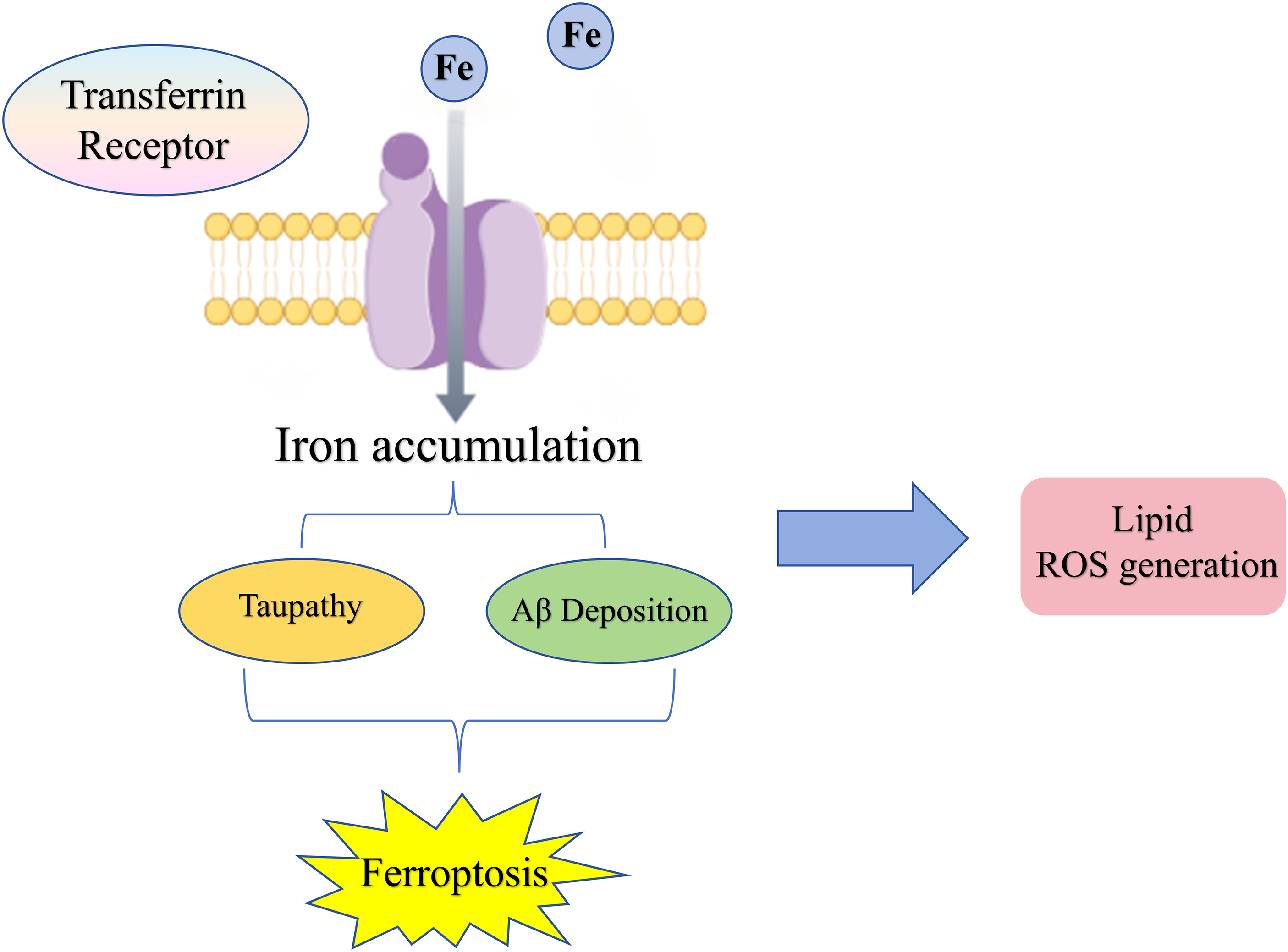
Ferroptosis in Alzheimer’s disease. All this evidence suggests that the fundamental mechanism of ferroptosis, lipid oxidative stress, is deeply engaged in the pathogenic progression of AD.
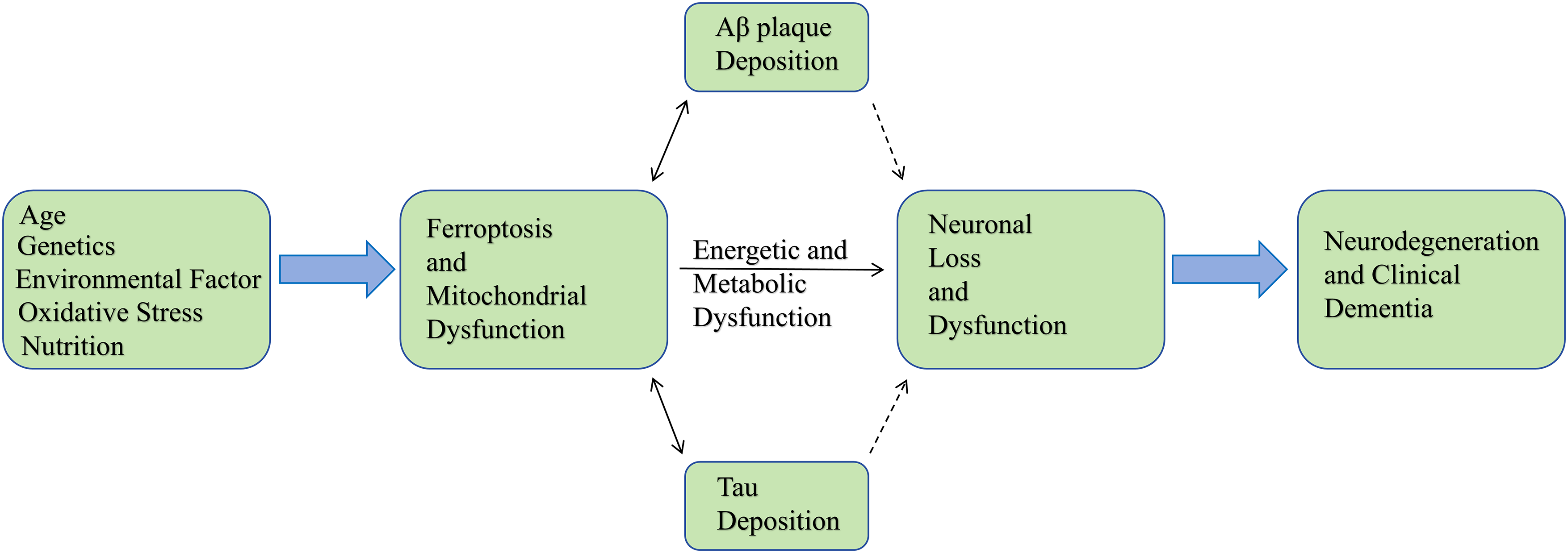
Mechanisms of ferroptosis involved in AD. Summarizes the pathogenic chain of Alzheimer's disease. This process begins with risk factors such as age, genetics, environment, oxidative stress, and nutrition. These factors trigger abnormalities in key cellular mechanisms, including ferroptosis and mitochondrial dysfunction.
The interplay between mitophagy and ferroptosis in Alzheimer’s disease
Having established the individual pathways and their dysregulation in AD, it is crucial to recognize that mitophagy and ferroptosis do not operate in isolation. 65 Instead, they engage in a dynamic, often synergistic crosstalk that forms a self-amplifying pathological network, likely acting as a central engine driving neurodegeneration in AD. 66
Shared molecular bridges: ROS and iron as central couplers
The interconnection between mitophagy and ferroptosis is most evident at the level of two fundamental biochemical entities: ROS and redox-active iron. 67 These serve as the primary molecular bridges that couple the quality control failure of mitochondria to the execution of lipid peroxidation-driven cell death.
ROS, particularly of mitochondrial origin (mtROS), functions as a critical bidirectional signaling molecule and damage agent. Under physiological conditions, a key function of functional mitophagy is to limit mtROS by removing its major source—damaged, electron-leaky mitochondria. 68 In AD, however, the impairment of mitophagy leads to the accumulation of such dysfunctional organelles. This results in a chronic overproduction of mtROS. This excess ROS is not merely a passive byproduct; it serves as the primary initiating force for the lipid peroxidation chain reaction that defines ferroptosis. 69 The peroxidation of PUFA-rich membranes, especially in sensitive compartments like mitochondria themselves and synaptic terminals, is directly fueled by this ROS surge. 70 Conversely, the process of ferroptosis itself generates a massive oxidative burst from the peroxidation reactions, which can inflict further damage on surrounding mitochondria, impairing their function and potentially overwhelming or inhibiting the already-fragile mitophagic machinery. 71 This establishes a devastating positive feedback loop: mitophagy failure → mtROS increase → lipid peroxidation/ferroptosis initiation → more mitochondrial damage → exacerbated mitophagy failure.
Iron homeostasis represents the second, equally critical functional link. As detailed in Chapter 2, the labile iron pool (LIP) is the essential catalyst for ferroptosis. 72 Mitophagy, through a specific subtype known as ferritinophagy, plays a direct role in regulating the LIP. The cargo receptor NCOA4 targets iron-storage ferritin for autophagic degradation, thereby controlling the release of bioavailable iron. Efficient mitophagy, therefore, contributes to iron homeostasis. 73 In AD, defective mitophagy extends to dysregulated ferritinophagy. This impairment can lead to either inadequate iron release (affecting normal metabolism) or, more pertinently, untimely and excessive release, causing a pathological elevation of the LIP. 74 This increased pool of catalytic Fe2+ dramatically lowers the threshold for ferroptosis by supercharging the Fenton reaction, making lipids vastly more susceptible to peroxidation even at moderate levels of ROS. 75 Thus, mitophagic dysfunction directly “primes” the cell for ferroptosis by mismanaging the cellular iron catalyst.
Key proteins with dual roles at the interaction node
Beyond shared small molecules, several key proteins sit at the intersection of both pathways, acting as molecular nodes that integrate signals and determine cellular fate. 76
The PINK1/Parkin system exemplifies this duality. While its primary and best-characterized role is initiating the ubiquitin-dependent clearance of damaged mitochondria, its dysfunction has far-reaching consequences that extend into the ferroptosis pathway. 77 Preliminary studies indicate that loss of Parkin not only leads to the expected accumulation of defective mitochondria and associated ROS but may also be associated with a decrease in intracellular GSH levels and a downregulation of GPX4 expression.78,79 This suggests a potential ‘double-hit’ scenario, whereby PINK1/Parkin dysfunction could simultaneously impair mitochondrial quality control and weaken key anti-ferroptotic defenses, potentially coupling mitochondrial failure to increased ferroptotic susceptibility. However, the direct causal role and molecular details of PINK1/Parkin in modulating ferroptosis in AD neurons require further validation.
The Ub-independent mitophagy receptors, particularly FUNDC1 and BNIP3/NIX, also exhibit context-dependent regulatory effects on ferroptosis. 80 Generally, BNIP3/NIX-mediated mitophagy is considered protective against ferroptosis, primarily through its efficient removal of ROS-generating mitochondria, thus reducing the initiating stimulus. 81 FUNDC1, however, presents a more complex and paradoxical picture. Under some conditions, FUNDC1-mediated mitophagy promotes cell survival by mitigating mitochondrial stress. Emerging evidence from studies in hepatic fibrosis models suggests that in certain severe pathological contexts, heightened FUNDC1 activity might contribute to the degradation of GPX4, potentially via an autophagy-dependent mechanism. 8 Whether this FUNDC1-GPX4 regulatory axis operates in AD brains and contributes to neuronal ferroptosis remains to be directly investigated, but it presents a plausible mechanistic link worthy of future exploration. 82 This highlights that not all mitophagy is universally anti-ferroptotic; its outcome depends on the specific receptors involved and the cellular context.
Furthermore, upstream signaling hubs like AMP-activated protein kinase (AMPK) can act as master regulators coordinating the response. 83 AMPK, activated by energy stress (common in AD), can phosphorylate and activate ULK1 to promote mitophagic initiation. Simultaneously, AMPK can phosphorylate and stabilize the transcription factor Nrf2, a master regulator of antioxidant response genes, including those involved in GSH synthesis (GCL) and GPX4 expression. 84 Thus, AMPK activation can theoretically promote both the clearance of damaged mitochondria (via mitophagy) and the bolstering of defenses against lipid peroxidation (via Nrf2), positioning it as a potential integrative node for therapeutic intervention aimed at restoring homeostasis. 85
The context-dependent paradox: guardian versus accomplice
The relationship between mitophagy and ferroptosis is not static but represents a profound biological paradox that is critically dependent on the cellular context, the severity of stress, and the stage of disease. 86 Under conditions of mild metabolic or oxidative stress, or in the early phases of AD pathology, a functional, well-regulated mitophagy system acts as a guardian. 87 By promptly eliminating mildly damaged mitochondria, it proactively removes the major sources of ROS and prevents iron-mediated toxicity, thereby upholding cellular redox and iron homeostasis and effectively suppressing any drift towards the ferroptotic threshold. 88
However, in the chronic, severe, and multifaceted stress environment of advanced AD, this protective relationship can break down and even reverse, with mitophagy becoming an accomplice to cell death. 89 This can occur through two distinct, but not mutually exclusive, mechanisms:
From catastrophic failure to ferroptosis. The complete collapse or severe impairment of core mitophagic machinery (e.g., profound loss of PINK1 or Parkin activity) leads to the massive, unmanageable accumulation of utterly dysfunctional mitochondria. 90 This results in an unsustainable burden of ROS production and severe disruption of iron metabolism, creating an intracellular environment where ferroptosis is virtually inevitable.
From dysregulated excess to ferroptosis. Alternatively, the hyperactivation or dysregulation of specific mitophagy pathways under extreme duress can have deleterious effects. 91 For instance, pathological overexpression or overactivation of receptors like FUNDC1 might lead to excessive or non-selective autophagic degradation. This could inadvertently target crucial survival proteins for destruction, such as GPX4. 92 In this scenario, an overactive but misdirected clearance mechanism actively depletes the cell's primary defense against ferroptosis, thereby directly facilitating cell death.
This central paradox resolves much of the seemingly contradictory evidence in the literature. It underscores that the net outcome of the mitophagy-ferroptosis interplay—whether it culminates in neuroprotective adaptation or in neurodegenerative execution—is not predetermined but hinges critically on the stage of AD progression, the specific neuronal or glial cell type affected, the integrity of upstream signaling networks (e.g., AMPK, Nrf2), and the intensity and duration of the pathological insult. Understanding this delicate balance and its contextual determinants is essential for designing stage-specific and pathway-selective therapeutic strategies aimed at breaking this vicious cycle in AD.
Specific role of interactions in AD pathogenesis
The interaction between mitophagy and ferroptosis functions as a core driver within the AD pathological process, rather than a passive concomitant phenomenon. This intricate interactive network synergistically amplifies core pathology through multidimensional, multi-tiered pathways, disrupting neural circuit homeostasis and profoundly influencing disease progression velocity and severity. At the molecular level, initial mitochondrial damage triggered by Aβ oligomers and hyperphosphorylated tau proteins constitutes the starting point of the pathological cascade. 93 The resulting mtROS serve both as key signals activating protective mitophagy via pathways such as PINK1/Parkin or FUNDC1, and as potent executors driving lipid peroxidation that subsequently induces ferroptosis. 94 When mitophagy functions properly, it efficiently clears damaged organelles, maintaining redox and iron homeostasis to suppress ferroptosis. 95 However, under sustained pathological stress in AD, the mitophagy system itself may become dysfunctional or experience compensatory failure. This leads to massive accumulation of damaged mitochondria, explosive mtROS production, and, through the Fenton reaction coupled with depletion of the GSH-GPX4 antioxidant defense system, collectively “transduces” molecular-level toxic stress into an irreversible ferroptosis program, ultimately resulting in neuronal loss. 96
More cutting-edge insights reveal this interaction functions as a “bridge” and “amplifier” linking and magnifying the two core pathologies of Aβ and tau. Ferroptosis is not a silent endpoint. For instance, lipid peroxidation products like 4-HNE can directly modify tau and AβPP, further propagating AD pathology. but the highly reactive lipid peroxidation end-products it generates (such as 4-HNE) can directly modify proteins, providing “seeds” for abnormal Aβ aggregation and activating kinase systems to promote excessive tau phosphorylation. This forms a self-perpetuating vicious cycle: “Aβ/tau → mitochondrial damage → ferroptosis → new Aβ/tau pathology”. 97 This positive feedback loop significantly accelerates the disease's progression from localized lesions to whole-brain dissemination. At the synaptic level, the destructive impact of this interaction manifests as a dual assault on synaptic plasticity, targeting both “energy” and “structure”. 98 The post-synaptic membrane, rich in polyunsaturated fatty acids (PUFAs), is particularly sensitive to ferroptosis-mediated lipid peroxidation. Once its integrity is compromised, synaptic function directly deteriorates. 99 Concurrently, the synaptic “energy crisis” induced by tau pathology obstructing mitochondrial axonal transport synergizes with ferroptosis’ structural disruption of synaptic membranes, jointly undermining the foundations of neural signal transmission. 100 This offers novel mechanistic insights into early-stage cognitive decline in AD. As synthesized in Figure 6, the mitophagy-ferroptosis axis forms a self-amplifying loop that is central to AD progression. This model illustrates several critical interactions: First, mtROS from damaged mitochondria not only directly oxidizes lipids to initiate ferroptosis but also impairs mitophagy efficiency (e.g., by inactivating PINK1/Parkin), creating a positive feedback cycle (①→②→①). Second, lipid peroxidation products like 4-HNE can further propagate toxicity by modifying tau and AβPP, thereby fueling the upstream drivers of the cycle (③). Third, key signaling nodes such as GSK-3β are positioned to integrate these stresses, exacerbating both tau pathology and mitochondrial dysfunction (④). This model underscores the necessity of dual-targeting strategies to simultaneously enhance mitophagic clearance and inhibit ferroptotic execution.
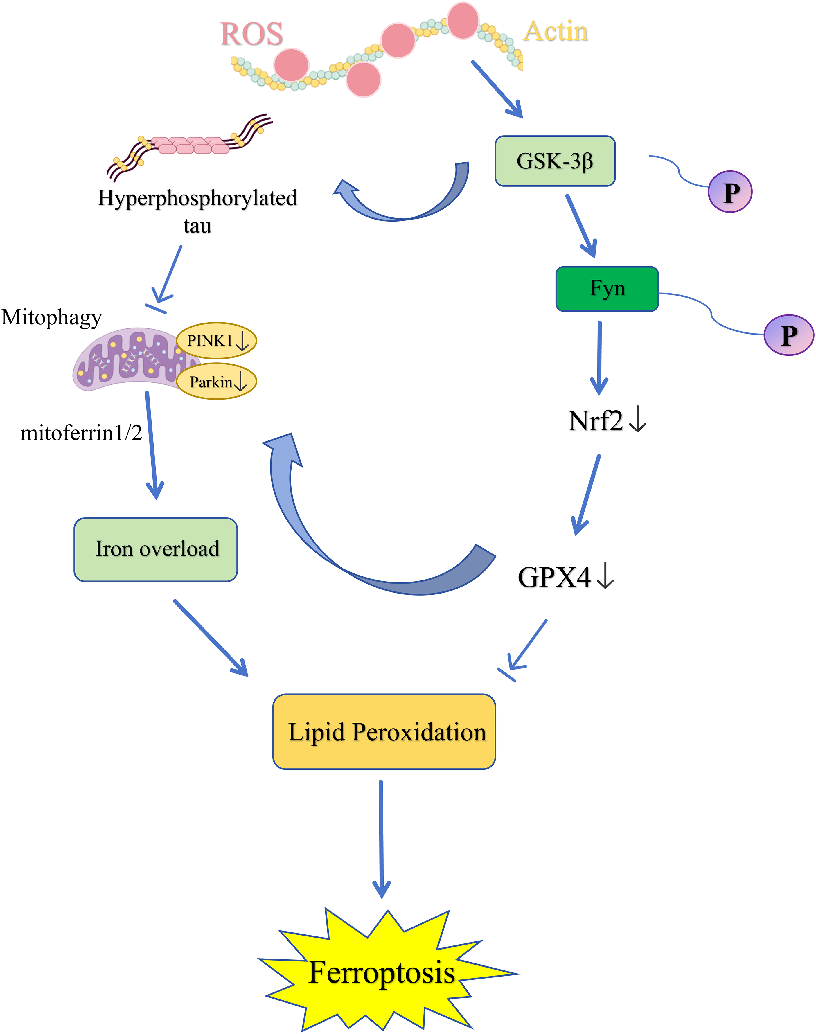
The Interplay between mitochondrial dysfunction and ferroptosis in Alzheimer’s disease. GSK-3β: glycogen synthase kinase-3β; Fyn: Fyn yes-related new; Nrf2: Nuclear factor erythroid 2-related factor 2; GPX4: glutathione peroxidase 4; PINK1: PTEN-induced putative kinase 1.
The pathological impact of this interaction is also markedly evident in the neuroinflammatory dimension, constituting an often-overlooked “accelerator” in the AD process. For instance, in microglia, impaired mitophagy leads to mtROS accumulation, which promotes NF-κB activation and pro-inflammatory cytokine release, thereby exacerbating neuronal ferroptosis. The mitophagy-ferroptosis axis within microglia and astrocytes is abnormally activated in AD. 101 Persistently activated microglia, if unable to effectively clear mitochondrial damage, may shift towards a pro-inflammatory phenotype or even undergo ferroptosis, releasing substantial inflammatory factors that further deteriorate the microenvironment. 102 Similarly, increased astrocyte susceptibility to ferroptosis compromises their antioxidant defense and glutamate clearance functions, indirectly exacerbating neuronal excitotoxicity and oxidative damage. 103 These non-neuronal interactions reveal the complexity and cell-type specificity of AD pathology, offering novel perspectives for immunometabolic therapeutic targeting.
Based on these mechanisms, future intervention strategies must shift from a “one-size-fits-all” approach to context-dependent, precision-dynamic regulation. Crucially, the implementation of such precision medicine is entirely dependent on the availability of biomarkers capable of delineating an individual's position within the mitophagy-ferroptosis vicious cycle. In the early stages of AD, when pathological damage is relatively mild and mitophagy remains plastic, mitophagy inducers such as uric acid A should be employed to “prevent problems before they arise” by enhancing endogenous cellular clearance capacity. 104 Conversely, in the late stages of disease when ferroptosis cascades are extensively activated, therapeutic focus must decisively pivot towards potent ferroptosis inhibition. This may involve iron chelators, GPX4 stabilizers, or novel radical-scavenging antioxidants. 105 The most promising strategy may involve sequential or synergistic combination therapies. For instance, mitophagy inducers could first ‘clear’ moderately dysfunctional mitochondria to reduce overall oxidative stress levels, followed immediately by ferroptosis inhibitors. This creates a critical time window for neuronal repair and remodelling. 106 To realize this precision medicine vision, developing multimodal biomarker systems reflecting in vivo interaction dynamics is paramount. This encompasses (1) neuroimaging assessments of cerebral iron deposition (e.g., via quantitative susceptibility mapping), (2) detection of mitochondrial DNA (mtDNA) and lipid peroxidation derivatives (e.g., 4-HNE, MDA) in bodily fluids like cerebrospinal fluid and blood, and (3) analysis of exosome-derived specific proteomic or genomic profiles (e.g., mitochondrial proteins, NCOA4). Such approaches enable tailored optimal neuroprotective strategies for patients at different stages, ultimately achieving effective disruption of this vicious cycle driving AD progression.
A summary of representative compounds and interventions that modulate mitophagy, ferroptosis, or both pathways, as discussed in current preclinical research, is provided in Table 1. This compilation underscores the diversity of potential therapeutic agents and highlights the need for context-dependent application as outlined above.
Classification of compounds/interventions targeting mitophagy and ferroptosis for Alzheimer’s disease treatment.

Critical analysis and unresolved controversies
While the interplay between mitophagy and ferroptosis presents a compelling narrative for AD pathogenesis, a critical examination of the literature reveals significant controversies, conflicting data, and fundamental gaps that must be acknowledged and addressed to advance the field beyond correlation toward mechanistic certainty and therapeutic translation.
The dual role of mitophagy: guardian or accomplice to ferroptosis?
The most salient controversy centers on the context-dependent outcome of mitophagy induction. Mitophagy can be either protective or detrimental. However, the precise conditions that dictate this switch remain poorly defined, leading to seemingly contradictory findings.
A robust body of work demonstrates that enhancing mitophagy, particularly via the PINK1/Parkin axis or through exercise mimetics, reduces Aβ and p-tau burden, improves mitochondrial function, and rescues cognitive deficits in AD models.26,36 In this paradigm, mitophagy is an unequivocal guardian, preventing the accumulation of the dysfunctional organelles that drive ferroptosis.
Conversely, emerging studies suggest that excessive or dysregulated mitophagy can actively promote ferroptosis. For instance, in non-AD models, hyperactivation of the mitophagy receptor FUNDC1 has been linked to the autophagic degradation of GPX4, the master inhibitor of ferroptosis.8 This presents a paradoxical scenario where the overzealous clearance of mitochondria inadvertently dismantles the cell’s primary defense against lipid peroxidation. Whether this “mitophagy-dependent GPX4 degradation” occurs in AD neurons is a critical unanswered question. If confirmed, it would establish a direct mechanistic link where one form of autophagy (mitophagy) facilitates another form of cell death (ferroptosis).
Reconciling the Conflict: This discrepancy may stem from disease stage, experimental model, and the specific mitophagy pathway engaged. Interventions in early or moderate AD models may successfully boost a deficient clearance system. In contrast, in late-stage disease with severe proteostatic collapse, the same induction might overwhelm an already compromised system or activate non-canonical, detrimental pathways like FUNDC1. Future research must move beyond binary assessments of “more” or “less” mitophagy, and instead develop tools to measure mitophagic flux quality and selectivity in vivo.
Inconsistencies in key molecular nodes and cellular context
The molecular links proposed earlier require rigorous validation, as current evidence is often preliminary or conflicting.
While PINK1/Parkin dysfunction is consistently associated with mitochondrial defects, its direct regulation of ferroptosis defenses (e.g., GSH, GPX4) in AD is inferred rather than proven.78,79 Is the observed downregulation of GPX4 in Parkin-deficient models a direct transcriptional consequence, a result of increased oxidative stress, or an indirect effect of rampant mitochondrial dysfunction? Establishing a direct molecular cascade from PINK1/Parkin to the System Xc-/GPX4 axis is a paramount gap.
A Largely Ignored Dimension: Nearly all mechanistic discussions focus on neurons. However, as hinted above glial cells are deeply implicated. The mitophagy-ferroptosis axis likely operates differently in microglia and astrocytes. For example, microglial mitophagy failure may promote a pro-inflammatory phenotype that externally exacerbates neuronal ferroptosis via cytokine release.101,102 In contrast, astrocytic ferroptosis could impair neurosupportive functions, creating a toxic milieu. 103 This cell non-autonomous regulation is severely understudied. Does enhancing neuronal mitophagy protect glia, or vice versa? Could a therapy beneficial for neurons inadvertently harm glial cells? These questions highlight a critical oversimplification in the current literature.
Major gaps in the current literature
Several fundamental gaps impede translational progress: (1) The Human Data Chasm: The entire proposed vicious cycle is built on preclinical models. We lack longitudinal human data demonstrating that a defect in mitophagic biomarkers precedes and predicts the onset of ferroptotic damage and cognitive decline in AD patients. Correlative post-mortem findings are insufficient to prove causality. (2) The Iron Homeostasis Black Box: While iron accumulation is a hallmark, the precise mechanism linking specific mitophagy defects (e.g., loss of PINK1 versus FUNDC1 overexpression) to specific disruptions in iron metabolism (e.g., ferritinophagy, iron export) in AD remains opaque. The field needs a more nuanced understanding of which form of mitophagic dysfunction leads to which type of iron dyshomeostasis. (3) The Biomarker Chasm and Staging Challenge: The context-dependency creates a potential therapeutic trap. A drug that potently induces mitophagy (e.g., a Urolithin A analog) could be beneficial in pre-symptomatic or early AD by clearing initial damage. However, the same drug administered in advanced AD, where ferroptosis is already rampant and GPX4 levels may be critically low, could theoretically accelerate GPX4 degradation and worsen outcomes. We currently lack the biomarkers to stratify patients into “mitophagy-deficient” versus “ferroptosis-dominant” stages to guide such precision therapy.
Addressing these controversies and gaps is not merely an academic exercise; it is essential for designing the next generation of targeted, effective, and safe interventions aimed at breaking the mitophagy-ferroptosis cycle in AD.
Conclusions and outlook
The synthesis presented in this review portrays a compelling, albeit predominantly descriptive, model of how mitophagy failure and ferroptosis execution can entwine to drive AD pathology. While the weight of preclinical evidence supports this interactive network, its translation into human therapeutics is fraught with complexities born from the context-dependent and often paradoxical nature of the underlying biology, as critically analyzed in the previous section. Therefore, the outlook must not only highlight promise but also confront these challenges.
This review systematically demonstrates that mitophagy and ferroptosis form a complex interactive network, mediated by shared molecules including ROS, iron, and key signaling proteins (e.g., PINK1/Parkin, FUNDC1, AMPK), throughout the pathological progression of AD. This network maintains a delicate equilibrium under physiological conditions. However, under sustained pathological stress in AD, its dysregulation fosters a vicious cycle: mitophagy dysfunction leads to the accumulation of damaged mitochondria, triggering iron homeostasis disruption and ROS surges, which significantly heighten neuronal susceptibility to ferroptosis. Concurrently, lipid peroxidation products and inflammatory mediators generated during ferroptosis further exacerbate mitochondrial damage, forming a self-amplifying pathological positive feedback loop that collectively drives neurodegeneration. Elucidation of this mechanism provides a novel integrative perspective for understanding the complex pathogenesis of AD.
Current understanding of mitophagy-ferroptosis interplay remains limited by several factors: (1) the lack of human longitudinal data validating causal relationships; (2) potential off-target effects and undetermined pharmacokinetics of chemical inducers (e.g., Urolithin A) in the brain; and (3) the frequent oversight of sex-specific differences in AD models. To address these gaps and translate mechanistic insights into therapy, future research should focus on:
Firstly, resolving context-dependency. It is essential to thoroughly map the dynamics of this interaction across different AD stages and cell types. Advanced models, such as human iPSC-derived neurons, microglia, and astrocytes carrying AD-related mutations, combined with single-cell multi-omics, can clarify cell-type-specific dialogue and its role in neuroinflammatory circuits.
Secondly, developing translational biomarkers. The field urgently requires multimodal biomarker platforms. This includes: (1) Molecular imaging probes, such as PET tracers targeting mitochondrial membrane potential or lipid peroxidation, to non-invasively visualize mitophagy and ferroptosis dynamics in vivo; (2) Liquid biopsy assays detecting mtDNA, NCOA4, or lipid peroxidation derivatives (e.g., 4-HNE adducts) in exosomes isolated from cerebrospinal fluid or blood. Such tools are indispensable for patient stratification and monitoring therapeutic responses.
Finally, designing precision therapeutic strategies. Future interventions must evolve from single-target to context-dependent, sequential combination therapies. A promising paradigm could be ‘priming and protecting’: using a mitophagy inducer (e.g., a brain-penetrant Urolithin A analog) in early-stage AD to first ‘clear’ moderately dysfunctional mitochondria and reduce overall oxidative stress, followed by or co-administered with a ferroptosis inhibitor (e.g., a GPX4 stabilizer) in later stages to create a critical window for neuronal repair. The challenge lies in fine-tuning this sequence and dosage based on disease stage biomarkers to avoid the paradoxical effects of excessive mitophagy. By integrating mechanistic research, biomarker development, and staged therapeutic design, we can envision a future where targeting the mitophagy-ferroptosis axis provides effective neuroprotection against AD progression.
Footnotes
Acknowledgements
The authors have no acknowledgments to report.

Author contribution(s)
Funding
The authors disclosed receipt of the following financial support for the research, authorship, and/or publication of this article: This study was supported by the Approval of the Innovative Scientific Research Project for Postgraduate Students at Heilongjiang University of Chinese Medicine (2025yjscx005).
Declaration of conflicting interests
The authors declared no potential conflicts of interest with respect to the research, authorship, and/or publication of this article.