



Abstract
Decline in mitochondrial quality is a prominent pathological feature of Alzheimer's disease (AD), manifested by impaired energy metabolism, disrupted mitochondrial biogenesis, abnormal mitochondrial dynamics, and defective mitophagy. Increasing evidence indicates that mitochondrial dysfunction contributes to the exacerbation of amyloid-β (Aβ) deposition and tau protein hyperphosphorylation, thereby accelerating AD pathogenesis. Of particular interest, physical exercise has been shown to effectively enhance mitochondrial quality and help prevent or slow the progression of AD, largely through the activation of key signaling pathways such as adenosine monophosphate-activated protein kinase (AMPK) and sirtuin 1 (SIRT1). However, regular physical activity may not be feasible for individuals in the prodromal or clinical stages of AD. In this context, exercise mimetics—compounds that pharmacologically simulate the molecular effects of exercise—have emerged as a promising alternative intervention. This review analyzes the mechanistic roles of exercise mimetics in improving mitochondrial quality under AD conditions, with a focus on their regulation of mitochondrial homeostasis via key signaling pathways. It further aims to provide theoretical insight for the development of mitochondria-targeted exercise mimetics and offer a potential strategy for addressing the growing global burden of AD.
Keywords
Introduction
Alzheimer's disease (AD) is the most prevalent neurodegenerative disorder, characterized by progressive cognitive decline. With the global population aging, the incidence of AD is steadily rising, and it is projected that the number of individuals affected by dementia will reach 152.8 million by 2050. 1 The pathological hallmarks of AD are extracellular amyloid-β (Aβ) plaques and intracellular neurofibrillary tangles composed of hyperphosphorylated tau protein. According to the amyloid cascade hypothesis, amyloid-β protein precursor (AβPP) is sequentially cleaved by β-secretase and γ-secretase to produce Aβ42 oligomers, which gradually aggregate into insoluble fibrils and plaques. These aggregates induce neuroinflammation and disrupt neuronal architecture, thereby impairing neuronal function and ultimately leading to cognitive dysfunction. Current research suggests that timely and effective clearance of Aβ42 can help prevent or delay the onset of AD. Conversely, excessive production or insufficient clearance of Aβ42 promotes the formation of oligomers and fibrils, triggering a cascade of downstream pathological events, including neuroinflammation, neuronal structural damage, and mitochondrial dysfunction. 2 However, therapeutic strategies based solely on the amyloid cascade have shown limited clinical efficacy, prompting increased attention toward alternative pathogenic mechanisms and the identification of novel therapeutic targets.
Mitochondria, often described as the cellular “powerhouses”, are essential for neuronal survival by generating adenosine triphosphate (ATP) through oxidative phosphorylation (OXPHOS). Given the high energy demands of neurons, maintaining mitochondrial integrity is critical for proper neuronal function. Substantial evidence has demonstrated that mitochondrial dysfunction is an early event in the pathogenesis of AD, potentially preceding the accumulation of Aβ plaques and hyperphosphorylated tau. 3 This has led to the formulation of the mitochondrial cascade hypothesis, which posits that mitochondrial dysfunction acts as an upstream trigger of AD, driven by both genetic and environmental factors that compromise mitochondrial homeostasis and subsequently exacerbate Aβ and tau pathologies. 4 Supporting this hypothesis, maternal inheritance of mitochondrial DNA (mtDNA) variants has been associated with increased AD risk, and studies have shown that metabolic deficits may emerge prior to the appearance of amyloid plaques and tau phosphorylation.5,6 In AD brains, mitochondrial abnormalities in neurons include impaired energy metabolism, elevated oxidative stress, imbalanced mitochondrial dynamics, and compromised mitophagy. 7 Notably, these mitochondrial defects are interrelated and often synergistically accelerate disease progression. In addition, Aβ has been shown to directly impair mitochondrial function, while mitochondrial dysfunction, in turn, exacerbates Aβ accumulation forming a vicious cycle that amplifies AD pathology. 4
Regular physical exercise has been extensively demonstrated as an effective non-pharmacological intervention with significant benefits for brain health and cognitive function. 8 However, for elderly individuals in the AD stage, the presence of comorbidities often makes it difficult to engage in sustained and effective exercise regimens. In 2008, the concept of “exercise mimetics” was first introduced by Narkar. 9 Exercise mimetics refer to a class of bioactive compounds that can simulate the physiological benefits of physical activity without actual physical exertion. 10 Some of these mimetics can target key signaling pathways, such as adenosine monophosphate-activated protein kinase (AMPK) and sirtuin 1 (SIRT1), which are central to the mitochondrial adaptations induced by exercise.
Previous reviews have extensively detailed the mitochondrial pathology of AD, the cognitive benefits of physical exercise, or the neuroprotective effects of specific agents like metformin and resveratrol. However, regular physical activity is often unfeasible for AD patients due to mobility constraints. A holistic perspective integrating these findings within the concept of exercise mimetics remains lacking. Although recent works have discussed exercise mimetics, they have predominantly focused on peripheral muscle function, with limited discussion on neurodegenerative diseases such as AD. Even when AD is addressed, the focus remains largely on neuroinflammation, often overlooking the critical role of mitochondria as the central mediator. Therefore, this review aims to elucidate recent advances in the regulation of mitochondrial quality by exercise mimetics in the context of AD. Specifically, we dissect the molecular mechanisms through which various mimetics enhance mitochondrial quality, with the goal of providing a theoretical foundation for mitochondria-targeted strategies in the prevention and treatment of AD (Table 1).
Comparison of the research focus between previous reviews and the current review.

Mitochondrial quality and AD
A decline in mitochondrial quality accompanies the entire course of AD and is closely linked to its classical pathological hallmarks, namely Aβ deposition and tau protein hyperphosphorylation. A substantial body of research has demonstrated that both the structural integrity and physiological function of mitochondria are significantly impaired at the early stages of AD, preceding the formation of Aβ plaques and hyperphosphorylated tau aggregates.4,16 Notably, reductions in cerebral energy metabolism, such as decreased glucose utilization, have been detected decades before the onset of clinical symptoms, indicating that impaired mitochondrial quality may occur during the preclinical phase of the disease. 17 These findings suggest that mitochondrial dysfunction may not only serve as an early pathological event in AD but also represent a potential biomarker and therapeutic target. Consequently, targeting mitochondrial quality offers a novel strategic direction for the early diagnosis and intervention of AD.
Abnormalities in mitochondrial dynamics
Mitochondrial fusion and fission are tightly regulated physiological processes that determine mitochondrial morphology, size, and intracellular distribution, and are essential for maintaining mitochondrial function. Mitochondrial fission is primarily driven by dynamin-related protein 1 (Drp1). Upon activation of fission signaling, outer mitochondrial membrane (OMM) proteins such as mitochondrial fission protein 1 (FIS1) and mitochondrial fission factor (Mff) recruit cytoplasmic Drp1 to the mitochondrial surface. There, Drp1 assembles into ring-like structures and, through guanosine triphosphate (GTP) hydrolysis, generates the mechanical constriction forces required to divide the mitochondria. 18 In contrast, mitochondrial fusion involves the coordinated action of mitofusin 1 and 2 (Mfn1/2) and optic atrophy 1 (OPA1). Mfn1/2 are GTPases localized to the OMM. They mediate outer membrane fusion by forming homotypic or heterotypic dimers between adjacent mitochondria. Following outer membrane fusion, OPA1, located in the inner mitochondrial membrane (IMM), facilitates inner membrane fusion by forming oligomeric complexes in a GTP-dependent manner. Once both membranes have fused, matrix contents such as mtDNA and proteins are allowed to mix, diluting damaged components and enabling mitochondrial repair. 19
In AD patients, the balance between mitochondrial fusion and fission is disrupted, typically characterized by excessive fission and impaired fusion. Drp1 expression and activity are elevated, while the levels and functions of Mfn1/2, and OPA1 are reduced, leading to mitochondrial fragmentation and decreased ATP production efficiency (Figure 1A). 20 Aβ and hyperphosphorylated tau have been shown to directly disturb mitochondrial dynamics. Specifically, Aβ promotes the translocation of Drp1 from the cytosol to the mitochondria, increases Drp1 phosphorylation and its GTPase activity. Thereby enhancing abnormal mitochondrial fission and resulting in increased fragmentation and synaptic damage.21–23 Moreover, Drp1 may contribute to AD pathogenesis by upregulating beta-site amyloid precursor protein cleaving enzyme 1 (BACE1) via the extracellular signal-regulated kinase (ERK)/Drp1 signaling pathway, further exacerbating Aβ production and accumulation in HEK293 cells. 24 Pharmacological or genetic inhibition of Drp1 has been shown to reverse memory deficits in AD mice models. 25 Collectively, these findings suggest that targeted suppression of pathological mitochondrial fission may represent a viable therapeutic strategy for ameliorating mitochondrial dysfunction and neurodegeneration in AD.
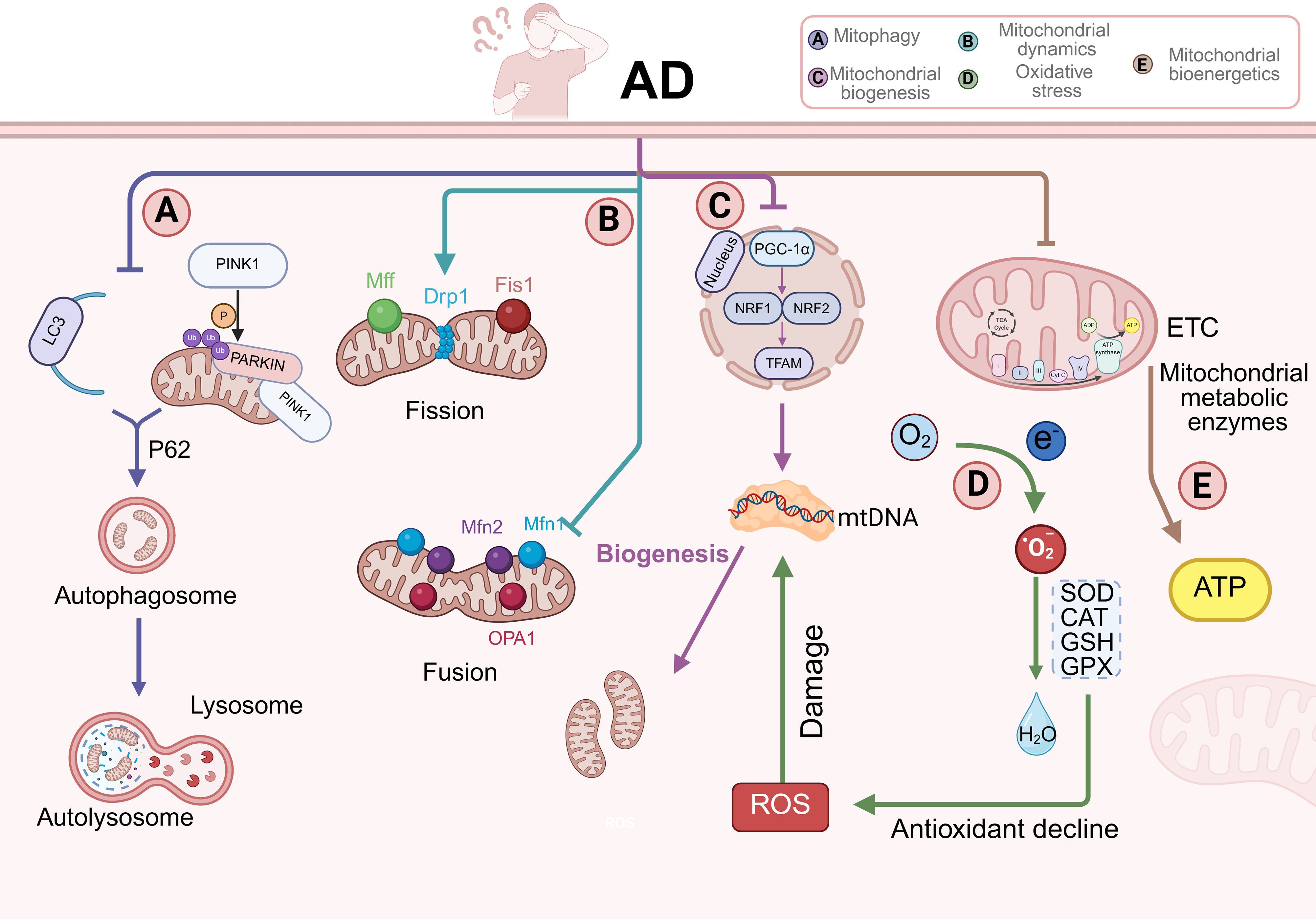
Mechanisms underlying impaired mitochondrial quality in AD. Under pathological conditions of AD, mitochondrial quality and function are severely compromised across multiple dimensions. (A) Mitophagy is impaired. The activities of key mitophagy-initiating proteins—PINK1, Parkin, are inhibited, and the expression of LC3 is downregulated. As a result, the formation of mitophagosomes is suppressed, leading to the intracellular accumulation of dysfunctional mitochondria that cannot be efficiently cleared; (B) Mitochondrial dynamics become imbalanced. Fission-related proteins, including Drp1, Mff, and FIS1, are excessively activated, while fusion-related proteins such as OPA1 and Mfn1/2 exhibit reduced expression and activity. This imbalance causes extensive mitochondrial fragmentation and network disruption; (C) Mitochondrial biogenesis is impaired. PGC-1α, the central regulator of mitochondrial biogenesis, is markedly downregulated, along with its downstream transcription factors—NRF1/2 and TFAM. This leads to inhibited mtDNA replication and transcription, resulting in insufficient mitochondrial generation; (D) Oxidative stress is exacerbated. The activity of ETC complexes is diminished, increasing electron leakage. Leaked electrons react with oxygen to generate large amounts of ROS, including superoxide anions and hydrogen peroxide. Meanwhile, the antioxidant defense system, comprising SOD and other key enzymes, is suppressed, reducing the clearance of free radicals and accelerating oxidative damage to cellular macromolecules such as mitochondrial DNA; (E) Mitochondrial energy metabolism is compromised. The activities of ETC complexes and various metabolic enzymes are reduced, impairing ATP synthesis. This energy deficiency weakens neuronal functions including synaptic transmission, plasticity, and survival.
Impairment of mitophagy
Mitophagy is a selective form of autophagy that removes dysfunctional, damaged, or senescent mitochondria through the encapsulation by double-membrane autophagosomes followed by lysosomal degradation. This process is essential for maintaining cellular health by preventing the accumulation of reactive oxygen species (ROS) and preserving mitochondrial quality. Mitophagy can be triggered by multiple stress signals, including elevated ROS levels, energy deficiency, and increased mitochondrial membrane permeability.
Mitophagy is broadly categorized into two major pathways: ubiquitin-dependent and ubiquitin-independent. The ubiquitin-dependent pathway is primarily mediated by the PTEN-induced putative kinase 1 (PINK1)/E3 ubiquitin ligase Parkin signaling cascade. In contrast, the ubiquitin-independent pathway is regulated by specific receptor proteins anchored in the outer mitochondrial membrane, such as BCL2/adenovirus E1B 19 kDa interacting protein 3 (BNIP3), NIP3-like protein X (NIX), and FUN14 domain-containing 1 (FUNDC1). These receptors interact directly with autophagy-related proteins via their light chain 3 (LC3)-interacting region (LIR) motifs to initiate mitophagy. The PINK1/Parkin pathway represents one of the canonical mechanisms of mitophagy. Under physiological conditions, PINK1 is imported into the IMM through the translocase of the outer membrane (TOM) and translocase of the inner membrane (TIM) complexes and is subsequently degraded. However, when mitochondria become depolarized, PINK1 is no longer translocated into the IMM and instead accumulates on the OMM, where it undergoes autophosphorylation. This accumulation leads to the recruitment and phosphorylation-based activation of Parkin, which ubiquitinates numerous outer membrane proteins, forming polyubiquitin chains that serve as degradation tags. These ubiquitinated substrates are recognized by autophagy receptors such as optineurin, NDP52, and p62. These receptors interact with microtubule-associated protein LC3 on the growing autophagosome membrane. The autophagosome then engulfs the damaged mitochondrion and fuses with lysosomes, leading to its complete degradation. 26
In AD patients, mitophagy mediated by the PINK1/Parkin pathway is significantly suppressed. In hippocampal neurons, the expression levels of key mitophagy-related proteins—including PINK1, Parkin, and UNC-51-like kinases 1 (ULK1)—are consistently downregulated. This leads to reduced clearance efficiency of damaged mitochondria and subsequent accumulation of dysfunctional organelles (Figure 1B). Mitochondrial accumulation further contributes to neuronal impairment and synaptic dysfunction. 23 Both Aβ and tau proteins are considered critical upstream suppressors of mitophagy. Experimental studies in mice have shown that intracellularly accumulated Aβ directly binds to cytosolic Parkin, preventing its translocation to depolarized mitochondria and thereby inhibiting mitophagic initiation. 27 Moreover, hyperphosphorylated tau can physically embed into mitochondrial membranes, increasing mitochondrial membrane potential and blocking the depolarization process. 28 This disruption hinders the stabilization of PINK1 on the outer mitochondrial membrane and impedes Parkin recruitment, ultimately impairing mitophagosome formation and lysosomal degradation. Importantly, impaired mitophagy not only limits the removal of dysfunctional mitochondria, but also aggravates Aβ aggregation and tau hyperphosphorylation, forming a deleterious feedback loop. Restoration of mitophagy represents a promising strategy for interrupting this pathological cycle. Recent studies have demonstrated that mitophagy-inducing agents are capable of alleviating Aβ and tau pathology and improving memory and cognitive performance in mouse hippocampal neurons.29,30
Impairment of mitochondrial biogenesis
Mitochondrial biogenesis is a self-renewing process by which cells generate new mitochondria through mtDNA replication, protein synthesis, and membrane structure assembly. This process ensures that mitochondrial quantity and function are dynamically regulated in response to shifting cellular energy demands and environmental stress.
Mitochondrial biogenesis is primarily regulated by peroxisome-proliferator-activated receptor gamma coactivator 1α (PGC-1α). It does not bind directly to specific DNA sequences but functions as a coactivator that enhances the transcriptional activity of various transcription factors. PGC-1α interacts with nuclear respiratory factors 1 and 2 (NRF1/2) to activate mitochondrial transcription factor A (TFAM), thereby initiating mtDNA transcription and replication, leading to an increase in mitochondrial number. 31 The activity of PGC-1α is modulated by several upstream signaling pathways. For instance, during exercise, increased ATP consumption raises the AMP/ATP ratio, which activates AMPK. AMPK phosphorylates PGC-1α, promoting mitochondrial biogenesis to meet elevated energy demands. 32 Additionally, SIRT1 activates PGC-1α through deacetylation, and cyclic adenosine monophosphate (cAMP) response element-binding protein (CREB) enhances PGC-1α transcription by binding to its promoter region.33,34
In AD, mitochondrial biogenesis is significantly suppressed. A study analyzing hippocampal tissues from 15 AD patients revealed that PGC-1α mRNA levels were reduced by approximately 50%, accompanied by downregulation of NRF1, NRF2α/β, and TFAM to varying extents (Figure 1C). 35 These changes were negatively correlated with dementia severity and Aβ pathology, suggesting that Aβ may interfere with upstream regulators of PGC-1α. 36 In vitro experiments have shown that exposing neuronal cells to Aβ42 oligomers for 24 h reduces total mitochondrial quality and lowers protein levels of SIRT1 and PGC-1α. This exposure alters their nucleocytoplasmic distribution, thereby disrupting the formation of the SIRT1–PGC-1α complex and suppressing PGC-1α activity.37,38 Importantly, a bidirectional relationship appears to exist between Aβ and PGC-1α. While Aβ suppresses PGC-1α activity, overexpression of PGC-1α has been found to reduce Aβ levels by downregulating BACE1, a key enzyme in Aβ generation.39–41
Impairments in energy metabolism
Mitochondria serve as the core powerhouses of neurons, generating ATP primarily through the tricarboxylic acid (TCA) cycle and OXPHOS to support the energy-intensive activities of neuronal cells. Although the brain accounts for only about 2% of total body weight, it consumes nearly 20% of the body's oxygen and 25% of its glucose. 17 Thus, efficient mitochondrial energy metabolism is vital for maintaining normal brain function. Once energy metabolism is impaired, it can severely compromise synaptic transmission, neuronal plasticity, and cell survival. 42 In the early stages of AD patients, impaired energy metabolism is characterized by markedly reduced glucose utilization in specific brain regions, particularly the prefrontal cortex and posterior cingulate cortex, accompanied by lactate accumulation.17,43,44 This suggests that defects in OXPHOS force neurons to rely more heavily on anaerobic glycolysis for compensation.45,46 The underlying mechanism is closely linked to mitochondrial electron transport chain (ETC) dysfunction, where the activities of Complexes I, IV, and V are generally reduced, with Complex IV being especially vulnerable to inhibition by Aβ oligomers.47,48 As a result, the mitochondrial proton gradient is disrupted, electron transfer efficiency declines, and ATP production is compromised—ultimately undermining the overall capacity of OXPHOS. Furthermore, key enzymes in the TCA cycle are concurrently impaired in AD brain tissues. For example, studies in AD patients have shown that the activity of pyruvate dehydrogenase complex is reduced by approximately 41%, isocitrate dehydrogenase activity by 27%, and α-Ketoglutarate Dehydrogenase Complex activity by 57% (Figure 1D). 49 These impairments severely limit the generation of nicotinamide adenine dinucleotide (NADH) and flavin adenine dinucleotide (FADH2), further reducing substrate availability for OXPHOS. Crucially, deficiencies in mitochondrial quality control also contribute to metabolic dysfunction. Abnormal mitochondrial dynamics, such as excessive fragmentation due to fission-fusion imbalance, disrupt cristae architecture and reduce mitochondrial membrane potential, directly impairing ATP synthesis efficiency. 50 Defects in PINK1/Parkin-mediated mitophagy lead to the accumulation of damaged mitochondria, which continuously generate reactive ROS, exacerbating oxidative stress and further suppressing ATP production. 51 In addition, impairment of the PGC-1α/NRFs signaling axis compromises mitochondrial biogenesis, limiting the replenishment of functional mitochondria required to meet increasing energy demands. 52
Exacerbation of oxidative stress
Oxidative stress arises from an imbalance between pro-oxidant and antioxidant systems, resulting in damage to macromolecules such as lipids, mtDNA, and proteins. 53 Mitochondria are the primary sites of oxidative stress, as most ROS are generated as byproducts of the ETC. Under normal physiological conditions, ROS are efficiently neutralized by endogenous antioxidant systems including superoxide dismutase (SOD), catalase (CAT), and glutathione (GSH). However, this redox equilibrium is disrupted in AD patients. 54 In AD patients, the impaired function of the ETC leads to reduced activity of mitochondrial complexes and impaired electron transfer, causing electron leakage. These leaked electrons react with molecular oxygen to produce superoxide anions, which are subsequently converted into hydrogen peroxide and hydroxyl radicals, contributing to excessive ROS generation. 55 Simultaneously, antioxidant defenses in the AD patients brain are diminished, as evidenced by decreased activities of key enzymes such as SOD, CAT, and GSH.56,57 This dual impairment results in insufficient ROS clearance, promoting their intracellular accumulation and triggering widespread oxidative damage (Figure 1E). The accumulation of ROS initiates oxidative modifications of lipids, proteins, and mtDNA, ultimately leading to mitochondrial dysfunction, neuronal apoptosis, and accelerated AD progression.58,59
Aβ is considered a key inducer of oxidative stress in AD. 60 Studies in AD patients have demonstrated a positive correlation between Aβ levels in the hippocampus and cortex and oxidative damage to intracellular macromolecules. 61 Aβ can activate NADPH oxidase, 62 disrupt mitochondrial membrane potential, and inhibit Complex IV activity. 63 Moreover, Aβ localized within the mitochondrial matrix can bind to amyloid-binding alcohol dehydrogenase (ABAD), resulting in the accumulation of NADH and depletion of NAD+. This interaction impairs mitochondrial membrane permeability, suppresses the activity of respiratory chain enzymes, and further increases ROS production. 64 In addition, Aβ oligomers can bind to cyclophilin D (CypD), promoting its translocation to the inner mitochondrial membrane and triggering the opening of the mitochondrial permeability transition pore (mPTP), thereby increasing mitochondrial permeability, dissipating membrane potential, and exacerbating oxidative stress. 65
Exercise-mediated regulation of mitochondrial quality effectively prevents AD progression
At present, therapeutic options for AD remain limited. The most commonly used medications—acetylcholinesterase inhibitors and N-methyl-D-aspartate (NMDA) receptor antagonists, can alleviate cognitive symptoms to some extent but are unable to halt or reverse the pathological course of the disease. 66 Thus, developing long-term and effective intervention strategies has become a major focus in AD research. In this context, exercise, as a non-pharmacological intervention, has drawn growing attention due to its cost-effectiveness, safety, and broad applicability. In addition, given the pronounced clinical and pathological heterogeneity in AD, by the time clinical symptoms appear, irreversible neuronal loss and functional impairments in the brain have often already occurred. Therefore, initiating lifestyle interventions, particularly exercise from the early stages of disease holds great significance. A growing body of epidemiological evidence supports the protective role of exercise in reducing AD risk. Compared with sedentary individuals or those with low levels of physical activity, people who maintain a high level of physical activity show a significantly lower risk of developing AD. 67 A large-scale meta-analysis including nearly 1.5 million participants revealed that individuals engaging in high-intensity physical activity had a 26% lower risk of developing AD compared to those with the lowest activity levels. 68 Considering that aging is one of the most significant risk factors for AD, studies focusing on older adults further suggest that regular physical activity may be a key behavioral factor for preventing AD in populations aged 65 and above. 69 More importantly, exercise not only lowers the risk of developing AD, but also demonstrates the ability to enhance cognitive function in patients already diagnosed with the disease. A growing number of randomized controlled trials have demonstrated that physical exercise significantly enhances executive function, processing speed, and memory, while exerting beneficial effects on both brain structure and function. 70 These clinical observations are supported by compelling evidence from animal studies, which provide mechanistic insights at the molecular level. Specifically, long-term regular exercise has been shown to elevate the expression of brain-derived neurotrophic factor (BDNF) in the hippocampus and cortex of AD model rats, 71 attenuate imbalances in mitochondrial dynamics, upregulate α-secretase activity, 72 and suppress β-secretase activity. 73 These effects collectively reduce the generation of neurotoxic Aβ and improve cognitive functions, particularly learning and memory. 74 Although a minority of studies have reported inconsistent results, the overall body of evidence strongly supports physical exercise as an effective non-pharmacological strategy for both the prevention and intervention of AD.75,76
One of the core mechanisms underlying the neuroprotective effects of exercise in AD is its ability to regulate multiple aspects of mitochondrial quality. It includes the activation of mitophagy, maintenance of mitochondrial dynamics, enhancement of mitochondrial biogenesis, attenuation of oxidative stress, and improvement of mitochondrial energy metabolism.77,78 These mechanisms together play a critical role in preventing and slowing the progression of AD. In AD pathology, exercise has been shown to activate key energy-sensing pathways such as AMPK and SIRT1.79,80 This activation enhances the activity of PGC-1α, which subsequently upregulates downstream factors such as NRF1/2 and TFAM. These processes promote mtDNA replication and transcription, ultimately increasing the number of functional mitochondria in the brain (Figure 2B). 81 Notably, PGC-1α also induces the expression of fibronectin type III domain-containing protein 5 (FNDC5), which facilitates the synthesis of BDNF, enhancing synaptic plasticity and alleviating Aβ-induced cognitive deficits. 79 Exercise has further been shown to restore the balance of mitochondrial dynamics in AD. In APPswe/PS1ΔE9 (APP/PS1) transgenic mice, 12 weeks of treadmill training significantly corrected the imbalance between mitochondrial fission and fusion. 82 This was evidenced by a downregulation of the fission-related proteins Drp1 and Mff and an upregulation of the fusion-related proteins Mfn1/2 and OPA1, thereby reducing Aβ levels and enhancing neuroplasticity (Figure 2D).83,84 Interestingly, some studies have also reported that exercise induces upregulation of Drp1 in the cortex of aged mice. This suggests that moderate enhancement of mitochondrial fission may promote the turnover and redistribution of damaged mitochondria within neurons, thereby facilitating their clearance through mitophagy in AD. 85 Moreover, exercise activates the AMPK/ULK1 signaling pathway and suppresses mammalian target of rapamycin (mTOR) activity, 86 thereby enhancing PINK1/Parkin-mediated mitophagy to eliminate damaged mitochondria and accumulated Aβ in APP/PS1 mice. 81 Exercise also promotes mitophagy via the SIRT1-transcription factors forkhead box proteins O1/O3 (FOXO1/3) axis in mice. By increasing the NAD+/NADH ratio, exercise activates SIRT1, which deacetylates FOXO1/3, leading to upregulation of PINK1 expression and subsequent activation of mitophagy (Figure 2A). 87 Exercise also alleviates oxidative stress associated with AD in mice.88,89 Mechanistically, it suppresses kelch-like ech-associated protein 1 (Keap1), thereby activating transcription factor nuclear factor erythroid-2-related factor 2 (Nrf2) and increasing the expression of various antioxidant enzymes, including heme oxygenase-1 (HO-1) and glutathione peroxidase 4 (GPX4), which protect neurons from oxidative damage (Figure 2E). 90 Lastly, exercise directly improves mitochondrial respiratory function, elevating ATP levels in the hippocampus and other brain regions of AD model mice. 91 These improvements are associated with increased activities of complex I, complex IV, and ATP synthase, which together enhance mitochondrial OXPHOS capacity (Figure 2C). 92
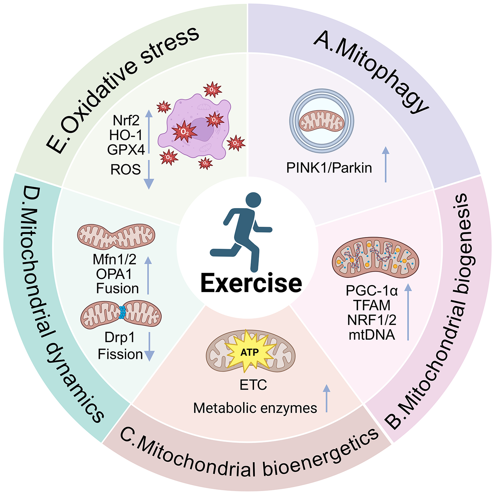
Mechanisms by which exercise reverses impaired mitochondrial quality in AD. Exercise enhances mitochondrial function under AD conditions through multiple mechanisms. (A) Mitophagy: Exercise induces mitophagy by activating the AMPK/ULK1/mTOR pathway and the SIRT1–FOXO1/3 axis, which together enhance the PINK1/Parkin-mediated clearance of dysfunctional mitochondria; (B) Mitochondrial Biogenesis: Exercise activates upstream regulators such as AMPK and SIRT1, which promote the activity of PGC-1α. PGC-1α binds to NRFs, facilitating the transcription and replication of mtDNA; (C) Mitochondrial Energy Metabolism: Exercise enhances mitochondrial respiratory function by increasing the activity of ETC complexes and oxidative metabolism-related enzymes, thereby boosting ATP production; (D) Mitochondrial Dynamics: Exercise helps restore mitochondrial dynamics by downregulating fission-related proteins (Drp1) and upregulating fusion-related proteins (Mfn1/2, OPA1), promoting the dynamic balance between fission and fusion; (E) Mitochondrial Oxidative Stress: Exercise reduces oxidative stress by activating Nrf2, which upregulates key antioxidant enzymes and suppresses ROS production, thereby mitigating mitochondrial oxidative damage.
In summary, exercise exerts a systemic regulatory effect on brain mitochondrial function primarily through the activation of core signaling pathways such as AMPK, SIRT1, and PGC-1α. This activation leads to improved bioenergetic output, enhanced antioxidant defense capacity, and reinforced mechanisms of mitochondrial quality control. These concerted effects collectively enhance mitochondrial integrity and functionality, thereby increasing neuronal resilience to AD–related pathological insults.
Enhancing mitochondrial quality via exercise mimetics: A novel strategy for the prevention and treatment of AD
Regular physical exercise has been established as an effective non-pharmacological strategy to prevent and delay the onset and progression of AD. Elucidating the molecular mechanisms by which exercise improves mitochondrial function has not only deepened our understanding of disease modulation, but also opened up novel avenues for therapeutic target discovery and drug development. However, in clinical practice, AD patients often experience significant cognitive decline and impaired motor coordination, which limit their ability to adhere to sustained, structured physical activity regimens. This practical challenge underscores the need for alternative approaches that can mimic the beneficial effects of exercise at the molecular level. Exercise mimetics, which activate intracellular signaling pathways associated with physical activity, have therefore emerged as a promising strategy for the prevention and treatment of AD. Recent studies have demonstrated that several exercise mimetics can delay AD progression and improve mitochondrial function, synaptic plasticity, and cognitive performance. 93 Although the precise mechanisms remain incompletely defined, increasing evidence suggests that their effects are closely related to the modulation of core AD pathological processes, including Aβ accumulation, tau hyperphosphorylation, and mitochondrial dysfunction. 94 Mechanistically, these compounds exert their effects by activating key neuronal signaling pathways such as AMPK, SIRT1, and PGC-1α. Through these pathways, exercise mimetics promote mitochondrial biogenesis, enhance oxidative metabolism, and maintain mitochondrial homeostasis, ultimately improving mitochondrial quality and exerting neuroprotective effects in the context of AD. In the following section, we focus on several representative exercise mimetics and examine their mechanisms of action in restoring mitochondrial quality in AD (Table 2).
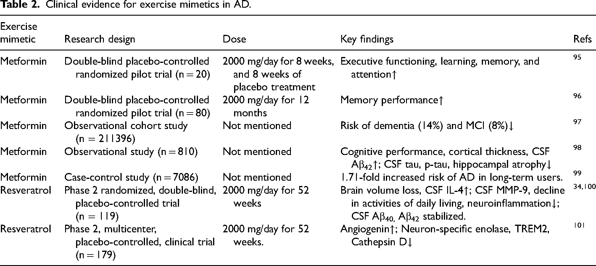
Clinical evidence for exercise mimetics in AD.
Metformin
Metformin is a first-line therapeutic agent for type 2 diabetes, primarily functioning by inhibiting hepatic gluconeogenesis and promoting hepatic glycogen synthesis, thereby enhancing glucose uptake and utilization in peripheral tissues. 102 As a classical AMPK activator, metformin exerts its action in part by inhibiting complex I of the mitochondrial respiratory chain, leading to reduced ATP production and an elevated intracellular AMP/ATP ratio, which subsequently activates the AMPK signaling pathway. 103 Nevertheless, this mechanism remains under debate, as the direct inhibition of complex I generally requires suprapharmacological concentrations of metformin—levels that are difficult to achieve under clinical conditions. 102 Alternative evidence in mice suggests that metformin may inhibit hepatic gluconeogenesis through suppression of mitochondrial glycerol-3-phosphate dehydrogenase, thereby indirectly activating AMPK and exerting glucose-lowering effects. 104 Once AMPK is activated, it phosphorylates and inhibits acetyl-coA carboxylase (ACC), resulting in suppressed fatty acid synthesis and enhanced fatty acid β-oxidation. 105 These actions reflect a broader metabolic shift favoring energy preservation and improved mitochondrial efficiency, mechanisms increasingly recognized as relevant in the context of neurodegenerative diseases such as AD.
Metformin can cross the blood–brain barrier (BBB), and its distribution has been detected in plasma, cerebrospinal fluid (CSF), and various brain regions following oral administration, supporting its potential action within the central nervous system. 106 In recent years, metformin has been found to exert beneficial effects on AD animals, 107 including the suppression of Aβ accumulation, 108 reduction of tau hyperphosphorylation, 109 upregulation of cerebral glucose transporter (GLUT) expression, 110 enhancement of the TCA, and promotion of mitochondrial respiration. 111 Mitochondrial complex I has been identified as a key molecular target of metformin. 112 By inhibiting complex I activity, metformin induces a slight increase in ROS production. 113 Although it may elevate ROS to some extent, multiple studies have demonstrated that metformin reduces overall oxidative stress. For instance, in a high-fat diet-induced mouse model of insulin resistance, metformin was shown to reverse the elevated ROS levels in brain mitochondria. 114 This apparent paradox can be explained by the concept of mitohormesis, which suggests that mild mitochondrial stress, such as modest inhibition of complex I or transient ROS elevation, can activate adaptive stress responses within the cell. These responses enhance intrinsic antioxidant defenses, improve mitochondrial function, and potentially prolong lifespan.115,116 Therefore, metformin may act as a subtle metabolic stressor, initiating downstream protective signaling pathways such as AMPK, 117 protein kinase B (AKT), 118 Nrf2. 119 This activation stimulates endogenous antioxidant systems, ultimately mitigating oxidative stress (Figure 3E). 111 For example, in a streptozotocin-induced AD rats model, metformin was able to reverse Aβ-induced Nrf2 downregulation, attenuate oxidative damage, and improve memory performance. 120

Targeting of upstream mechanistic axes by different exercise mimetics to improve mitochondrial quality in AD. Metformin, AICAR, and Irisin are grouped as agents that primarily initiate their neuroprotective effects through the AMPK activation axis. Resveratrol mainly targets the SIRT1 signaling pathway, while BDNF functions as a direct agonist of the TrkB receptor. Despite these distinct upstream initiation mechanisms, activation of these axes converges to enhance mitochondrial quality and neuronal health, ultimately contributing to the reduction of AD pathology.
Metformin restores mitochondrial quality primarily via the AMPK signaling axis. It promotes mitochondrial biogenesis via the AMPK/PGC-1α pathway, effectively reversing the downregulation of NRF1 and TFAM observed in an Aβ-induced human neural stem cell model of AD. 121 Of particular note, metformin may also promote the synthesis of irisin via the AMPK/PGC-1α pathway in C2C12 cells, contributing to its neuroprotective effects.122,123 However, some studies report that chemical inhibitors or siRNA-mediated knockdown of the AMPK pathway do not block metformin-induced irisin secretion. 124 This discrepancy suggests that metformin may engage multiple regulatory mechanisms, and that its actions may not rely exclusively on AMPK signaling. Under conditions of AMPK inhibition, compensatory activation of alternative pathways may occur to preserve its downstream effects. Furthermore, studies reveal that metformin activates the AMPK/ULK1 pathway to initiate mitophagy, subsequently upregulates PINK1/Parkin signaling, and ultimately promotes mitochondrial autophagic clearance in 3xTg model AD mice. Nevertheless, conflicting findings exist.125,126 In a model of doxorubicin-induced cardiotoxicity, metformin has been shown to protect against mitochondrial damage by suppressing excessive mitophagic flux. The study found that doxorubicin caused mitochondrial depolarization and triggered excessive degradation of mitochondria, whereas metformin effectively inhibited the overactivation of mitophagy induced by doxorubicin. 127 This indicates that metformin may act as a biphasic regulator: it promotes mitophagy under conditions of mitochondrial dysfunction to facilitate the clearance of damaged organelles, while suppressing mitophagy when overactivation becomes deleterious. Additionally, studies have shown that metformin activates the AMPK signaling pathway, leading to the inhibition of Drp1 activity and simultaneous upregulation of Mfn2 and OPA1 in mouse hippocampal neurons. These molecular changes reduce mitochondrial fragmentation, alleviate oxidative stress and neuronal apoptosis, ultimately improving cognitive function (Figure 3D).128,129 However, some studies have reported divergent findings. For instance, metformin significantly downregulated the expression of Drp1 and FIS1 in the hippocampus of AD mice but exerted no significant effect on the levels of OPA1 and Mfn2, suggesting that its primary regulatory role may lie in the suppression of mitochondrial fission rather than in promoting fusion. 130
While metformin has demonstrated robust efficacy in preclinical models, its translational value for AD remains debated. A recent large-scale observational cohort study utilizing the UK Clinical Practice Research Datalink, which enrolled 211,396 adults aged 40 or older with type 2 diabetes, identified an association between metformin use and a reduced risk of dementia. However, this potential protective effect was absent in individuals aged 80 years and older. 97 This aligns with corroborating observational evidence indicating that metformin users exhibit superior cognitive performance and healthier biomarker profiles compared to non-users. 98 Although these findings suggest that early metabolic intervention may attenuate dementia risk in diabetic populations, evidence for a direct benefit in AD is conflicting. A recent meta-analysis found no statistically significant improvement in cognitive function or AD prevention. 131 Furthermore, a case-control study has reported that long-term metformin administration might even be associated with a marginally increased risk of AD. 99 These conflicting results highlight the limitations of current evidence and suggest that the neuroprotective potential of metformin may be obscured by population-specific factors, particularly the confounding presence of diabetes.
Since metformin is a typical medication for diabetes, a critical question remains: does it attenuate dementia risk indirectly by ameliorating metabolic dysregulation, or does it possess intrinsic neuroprotective properties? A pilot randomized controlled trial (RCT) involving 80 patients demonstrated that memory performance was superior to placebo following one year of treatment. 96 Similarly, another RCT in 20 non-diabetic patients with mild cognitive impairment (MCI) or early AD reported that metformin treatment resulted in significantly better executive function compared to placebo. 95 Taken together, these pilot trials provide preliminary evidence supporting the direct neuroprotective potential of metformin in non-diabetic cohorts, distinct from its glucose-lowering effects.
Nevertheless, pharmacokinetic limitations constitute a significant barrier. Although metformin can cross the BBB, its permeability is restricted, achieving CSF concentrations of only approximately 20% of plasma levels. 132 Consequently, therapeutic efficacy in AD may necessitate higher dosages or prolonged administration regimens. Thus, the true clinical utility of metformin in AD warrants further validation through rigorous, large-scale systematic trials.
5-Aminoimidazole-4-carboxamide ribonucleoside (AICAR)
AICAR is one of the most extensively utilized AMPK agonists. Within cells, it is phosphorylated into 5-aminoimidazole-4-carboxamide ribonucleotide (ZMP), an AMP analog that binds to the γ subunit of AMPK, inducing conformational changes that facilitate its activation. Unlike AMP accumulation induced by energetic stress, AICAR activates AMPK without significantly altering the intracellular AMP/ATP ratio, thereby initiating downstream signaling cascades. 133 Through this mechanism, AICAR exerts multiple biological effects, including enhanced fatty acid oxidation, anti-inflammatory responses, and improved mitochondrial function.134,135
As an exercise mimetic, AICAR exerts potential neuroprotective effects in AD by activating key exercise-responsive signaling pathways. 136 In diabetic AD mouse models, AICAR enhances insulin-degrading enzyme (IDE) activity via AMPK activation, thereby reducing Aβ deposition and suppressing tau hyperphosphorylation.137–139 Studies using streptozotocin-induced AD rats models similarly demonstrated that AICAR restores mitochondrial membrane potential and ATP levels, upregulates the activity of mitochondrial Complex I and SOD, reduces ROS levels, and inhibits neuronal apoptosis (Figure 3C). These effects collectively result in improved spatial memory, suggesting that AICAR may ameliorate AD pathology by restoring mitochondrial function. 140 Accordingly, AICAR-mediated AMPK activation to mitigate mitochondrial damage represents a promising therapeutic strategy for AD intervention.
AICAR, as a typical AMPK pathway agonist, exerts its mitochondrial improvement effects primarily by activating the AMPK pathway. In aging mouse models, it promotes mitochondrial biogenesis and improves mitochondrial cristae structure via the AMPK/PGC-1α axis. Notably, while both AICAR and exercise improved mitochondrial quality, only the exercise group enhanced mitochondrial respiratory function. This indicates that AICAR partially mimics the effects of exercise on mitochondrial remodeling. 141 Furthermore, exogenous AICAR administration into the bilateral paraventricular nuclei can activate the AMPK/Nrf2 signaling pathway, which induces the upregulation of antioxidant enzymes such as SOD, and concurrently suppresses the expression of tumor necrosis factor-α (TNF-α), interleukin-1β (IL-1β), and ROS. 120 In H9C2 cells, AICAR can inhibit the expression of mitochondrial fission proteins Mff and FIS1, while promoting the expression of mitochondrial fusion proteins Mfn1/2. 142 Interestingly, under certain pathological conditions, such as diabetic polyneuropathy, AICAR has been reported to increase the expression of Drp1, ULK1, and LC3-II via AMPK activation. This suggests that AICAR may promote mitochondrial fission to facilitate the selective removal of damaged mitochondria through mitophagy. 143
It is also important to note that AICAR is widely employed as a metabolic exercise mimetic, capable of enhancing skeletal muscle performance by activating peripheral AMPK pathways. Recent studies suggest that such peripheral activation may exert indirect neuroprotective effects on the aging brain. In an aging mouse model, intraperitoneal injection of AICAR (500 mg/kg/day for 14 days) significantly improved learning and memory as well as motor coordination. These behavioral improvements were accompanied by the upregulation of mitochondrial-related genes in skeletal muscle and increased expression of neurogenesis and synaptic plasticity-related genes in the hippocampus. However, these benefits were entirely abolished in muscle-specific AMPKα2 mutant mice, indicating that the beneficial effects of AICAR depend critically on muscle AMPK signaling. 144 Given AICAR's limited permeability across the BBB, 145 these findings collectively support the notion that AICAR may modulate central nervous system function through a muscle–brain axis, wherein enhanced skeletal muscle metabolism via peripheral AMPK activation leads to improved neural outcomes.
Regrettably, despite its therapeutic potential, the clinical translation of AICAR for AD has stalled. No clinical trials have yet evaluated its effects in patients. Currently, AICAR serves primarily as a pharmacological probe for elucidating AMPK mechanisms in research settings, offering mechanistic support for the utility of AMPK activation.
Resveratrol
Resveratrol is a naturally occurring polyphenolic compound found widely in plants such as grapes and blueberries. It is also recognized as a classic activator of SIRT1. Numerous studies in mice have demonstrated that resveratrol can alleviate AD-related pathological features. For example, resveratrol has been shown to reduce ROS production in the hippocampus, upregulate intracellular antioxidants such as GSH, and thereby mitigate oxidative stress. In addition, resveratrol inhibits β-secretase, reducing the cleavage of AβPP into toxic Aβ fragments, 146 and suppresses tau protein hyperphosphorylation,147,148 thereby preserving neuronal function. Furthermore, long-term resveratrol intervention has been shown to activate AMPK and SIRT1, reduce Aβ deposition, upregulate mitochondrial complex IV levels, and enhance mitochondrial respiration. 149 However, some studies have found that AMPK activity is abnormally elevated in hippocampal neurons following Aβ treatment, and that resveratrol may suppress this overactivation, thereby preventing neuronal death. 150 These findings suggest that AMPK activity in AD is not unidirectional, as both excessive and insufficient activation may lead to neuronal damage. Resveratrol appears to dynamically modulate AMPK activity, exerting a neuroprotective effect. 151
Resveratrol exhibits a wide range of biological activities, with particularly notable antioxidant and anti-inflammatory properties. On one hand, resveratrol can directly inhibit the production of ROS, thereby mitigating oxidative stress-induced cellular damage. 152 On the other hand, it indirectly exerts antioxidant effects by upregulating endogenous antioxidant enzymes, including SOD, glutathione peroxidase (GSH-Px), and HO-1. 153 Interestingly, resveratrol has also been shown to exert pro-oxidative effects in certain cancer models, where it induces excessive ROS accumulation, activates mitophagy, and promotes apoptosis in tumor cells. 154 In the Aβ1−42-induced PC12 cell, resveratrol can exert its antioxidant effects via the phosphatidylinositol-3-kinases (PI3 K)/AKT/Nrf2 pathway, upregulating HO-1, SOD, and GSH levels, downregulating malondialdehyde (MDA) levels, and significantly attenuating oxidative stress. 155 In addition, SIRT1 is also a key molecular target in the antioxidant actions of resveratrol. In both APP/PS1 transgenic mice and primary neurons treated with Aβ, resveratrol activates SIRT1 signaling, reduces multiple oxidative stress markers, and inhibits amyloid plaque formation. Notably, inhibition of SIRT1 activity significantly weakens these protective effects, further confirming the central role of SIRT1 in the regulation of oxidative stress by resveratrol. 156
Distinct from metformin and AICAR discussed previously, resveratrol primarily exerts its beneficial effects on mitochondrial quality by activating the SIRT1 signaling pathway. PGC-1α is likely a critical downstream effector mediating the neuroprotective effects of resveratrol. 157 Evidence indicates that resveratrol promotes mitochondrial biogenesis via the SIRT1/PGC-1α axis in both APP/PS1 mice and Aβ-induced rat primary neurons, thereby providing neuroprotection. Notably, in neuronal models treated with Aβ, inhibition of SIRT1 activity using suramin did not abolish the protective effects of ZLN005, a selective PGC-1α activator, on reducing cell apoptosis. In contrast, SIRT1 activation via resveratrol failed to significantly reduce apoptosis when PGC-1α was silenced using siRNA. 158 Another study offers a plausible explanation for alternative activation pathways. Resveratrol may also activate PGC-1α via the sirtuin 3 (SIRT3)/FOXO3 pathway, thereby promoting mitochondrial biogenesis in TCMK-1 cells. 159 Furthermore, in Aβ-induced N2a cell models of AD, resveratrol significantly reduced Drp1 expression while upregulating Mfn1, Mfn2, and OPA1, thereby promoting mitochondrial network integrity and significantly enhancing neurite outgrowth. Another study in mice elucidated the mechanism regarding dynamics. Resveratrol, through SIRT1 activation, reduces the acetylation level of PGC-1α, facilitating its binding to the Drp1 promoter region and downregulating Drp1 transcription. This mechanism effectively inhibits excessive mitochondrial fission. Additionally, in a PC12 cell-based in vitro AD model, resveratrol intervention was shown to promote mitophagy. 160 Further research in neuronal cells suggests that resveratrol may trigger mitophagy via AMPK rather than SIRT1, as the mitophagic effect was abolished following treatment with an AMPK inhibitor.161,162
Based on findings from animal studies, clinical trials have begun to evaluate the therapeutic potential of resveratrol in AD patients. A Phase II randomized controlled trial involved 119 participants with mild-to-moderate AD. 34 It demonstrated that 52 weeks of resveratrol treatment stabilized Aβ40 levels in both CSF and plasma. This contrasted with the significant decline observed in the placebo group. Such stabilization suggests that resveratrol may impede the pathological deposition of Aβ within the brain parenchyma. Notably, neuroimaging revealed greater brain volume atrophy in the resveratrol group compared to placebo. Further investigation offered a plausible explanation. Resveratrol likely reduces matrix metalloproteinase-9 (MMP-9) levels. 100 This action repairs the damaged BBB and resolves inflammatory edema. Consequently, this resolution manifests as an apparent reduction in brain volume. Furthermore, another biomarker analysis corroborated this anti-inflammatory mechanism. 101 It found that CSF TREM2 levels were significantly reduced post-treatment. This indicates that pathological microglial overactivation was inhibited, effectively alleviating neuroinflammation.
Despite resveratrol's favorable oral absorption profile, and the detection of both the parent compound and its metabolites in plasma and CSF, which suggests its ability to cross the BBB, its systemic bioavailability remains relatively low, largely due to extensive first-pass hepatic metabolism. 10 To address this limitation, several novel brain-targeted delivery strategies have been developed in recent years. Mounting evidence suggests that mitochondria-targeted antioxidant therapy holds promise for ameliorating AD-associated pathology. 163 For example, encapsulating resveratrol into nanostructured lipid carriers coated with red blood cell membranes enables it to cross the BBB and selectively accumulate in neuronal mitochondria, where it alleviates mitochondrial oxidative stress and significantly improves learning and memory deficits in APP/PS1 transgenic mice. 164
Irisin
Irisin is a myokine cleaved from the membrane protein FNDC5 and is known to exert multiple physiological functions, including anti-inflammatory and neuroprotective effects, attenuation of oxidative stress, cardiovascular protection, osteoporosis mitigation, promotion of energy expenditure, and improvement of insulin sensitivity. 165 Irisin was first reported by Boström et al. in 2012, who demonstrated that exercise-induced upregulation of PGC-1α in skeletal muscle leads to increased FNDC5 expression. FNDC5 is then proteolytically cleaved and secreted into the circulation in the form of irisin. 166 Importantly, irisin is capable of crossing the blood–brain barrier and activating the ERK signaling pathway, 167 which upregulates the expression of BDNF in the hippocampus. 168 BDNF binds to its high-affinity receptor tropomyosin receptor kinase B (TrkB), initiating a phosphorylation cascade that activates CREB. This in turn enhances PGC-1α expression, promotes mitochondrial biogenesis, and improves mitochondrial function in neurons affected by AD, thereby contributing to improved memory and learning capacity.169,170 Notably, irisin is not only a downstream effector of the AMPK/PGC-1α signaling axis but can also serve as an upstream activator of AMPK signaling. Exercise has been shown to suppress aberrant mitochondrial fission, enhance mitochondrial respiratory efficiency, and inhibit neuronal apoptosis through the irisin/AMPK/SIRT1 signaling pathway in mice. The use of irisin antagonists significantly attenuates exercise-induced upregulation of AMPK and SIRT1, suggesting that irisin mediates, at least in part, the neuroprotective effects of physical activity. 171
In recent years, irisin has emerged as a key modulator in the therapeutic landscape of AD. 172 While clinical trials using exogenous irisin for AD have not yet been conducted, observational studies strongly suggest its physiological relevance. In a study of 39 participants, AD patients exhibited significantly lower irisin levels in CSF, which positively correlated with BDNF, Aβ42 and cognitive function. 173 These findings were recently substantiated by a clinical cohort study, which confirmed significant reductions in both plasma and CSF irisin in patients with AD and MCI. 174 In mice, genetic deletion of FNDC5/irisin in the brain results in severe impairment in novel object recognition memory. Conversely, intracerebroventricular administration of exogenous irisin restores cognitive function and reverses these deficits. 93 Additionally, irisin enhances the activity of mitochondrial complexes I, II, and IV, 165 improves mitochondrial respiration, and reduces tau hyperphosphorylation and TNF-α levels in hippocampal tissues. 175
Irisin improves mitochondrial quality via multiple pathways. Experimental evidence indicates that intracerebroventricular injection of recombinant irisin improves neuronal mitochondrial biogenesis in mice. 176 Further mechanistic studies indicate that irisin activates the SIRT1/PGC-1α pathway in the microglia of P301S mice, restoring mitochondrial OXPHOS and inhibiting microglial senescence. 177 Importantly, the role of irisin in promoting mitochondrial biogenesis may not be entirely dependent on PGC-1α. A study conducted in H9C2 rat cardiomyocytes showed that irisin was still able to upregulate mRNA levels of cytochrome c oxidase subunit 4, a key gene involved in mitochondrial biogenesis, even under conditions in which PGC-1α expression was suppressed. The authors hypothesized that this may involve a compensatory mechanism mediated by PGC-1β. 178 In addition, in mice, irisin can also promote mitophagy via the AMPK pathway. Regarding mitochondrial dynamics, irisin improves dynamics via multiple pathways. 179 In mice, it has been shown to activate the AMPK, Nrf2/SIRT3, and AKT/ERK1/2 pathways, which collectively inhibit Drp1 phosphorylation and promote the expression of mitochondrial fusion proteins.180–183
Intrahippocampal injection of exogenous irisin into the dentate gyrus of rats has been shown to reduce levels of MDA and total oxidative status (TOS), while elevating GSH, SOD, and total antioxidant capacity (TAC) levels. These changes correlate with improved learning and memory performance and attenuated oxidative stress in the brain. 184 A key mechanism involves uncoupling protein 2 (UCP2). Irisin increases UCP2 expression, which reduces the mitochondrial proton gradient, activates the AMPK pathway, and ultimately lowers ROS production from the mitochondrial ETC, thereby improving mitochondrial function and alleviating oxidative stress.176,185 Moreover, irisin exerts antioxidant effects via upregulation of Nrf2 and suppression of the nuclear factor kappaB (NF-κB) pathway. This modulation leads to increased expression of HO-1, SOD, and GSH, as well as reduced production of high mobility group box-1 protein (HMGB1). Ultimately, these changes enhance endogenous antioxidant defense capacity, preventing excessive hydrogen peroxide generation and mitigating oxidative damage in mice.186–188 Furthermore, irisin also reduces oxidative stress via activation of the PI3 K/AKT/mTOR signaling axis in RAW 264.7 mouse macrophages. 180
BDNF
BDNF is one of the most extensively studied neurotrophins and is the most abundantly expressed neurotrophic factor in the central nervous system. It plays critical roles in a variety of physiological processes, including neuronal survival, differentiation, and synaptic plasticity, and exerts notable neuroprotective effects. BDNF is initially synthesized in its precursor form, proBDNF, which is subsequently cleaved by proteases to generate biologically active mature BDNF. Its primary receptor is TrkB, which mediates downstream signaling upon BDNF binding to regulate cellular proliferation and survival. 189
Numerous studies have demonstrated that BDNF levels decline significantly during the preclinical stages of AD, and the extent of this reduction correlates positively with the degree of cognitive impairment.190,191 Mechanistically, Aβ has been shown to suppress CREB activity, thereby inhibiting BDNF gene transcription and reducing its expression.121,192,193 The downregulation of BDNF further activates the Janus kinase 2 (JAK2)/signal transducer and activator of transcription 3 (STAT3) signaling pathway, which in turn enhances δ-secretase activity, facilitating Aβ production and tau hyperphosphorylation, thereby establishing a vicious cycle that accelerates AD progression. 194 Exercise interventions have been shown to elevate hippocampal BDNF levels, 71 thereby improving mitochondrial dysfunction, enhancing synaptic plasticity, and ameliorating cognitive deficits. 195 Moreover, exogenous BDNF supplementation has been shown to replicate the beneficial cognitive effects of exercise in AD mice models. 196 Mechanistically, it functions by upregulating α-secretase activity and reducing the production of neurotoxic Aβ species. 72 Furthermore, it promotes hippocampal neurogenesis, and enhancing both synaptic plasticity and mitochondrial function.192,197
Upon binding to TrkB, BDNF activates protein kinase A (PKA), leading to phosphorylation and nuclear translocation of CREB, which subsequently binds to the promoter region of the PGC-1α gene, enhancing its transcription. PGC-1α then co-activates transcription factors such as NRF1/2, upregulating TFAM expression and initiating the transcription of nuclear-encoded mitochondrial genes, thereby promoting mitochondrial biogenesis. 198 These processes are essential for the formation and maintenance of hippocampal dendritic structures and synaptic networks. 199 Importantly, PGC-1α is not only a downstream effector of BDNF signaling but also contributes to a positive feedback loop: it can counteract Aβ-induced suppression of BDNF expression. 200 This suggests a synergistic regulatory mechanism whereby BDNF upregulates PGC-1α, enhancing mitochondrial biogenesis, while PGC-1α in turn reinforces BDNF signaling, jointly promoting neurotrophic support and mitochondrial function in the AD brain.
Although the precise mechanisms by which BDNF regulates mitochondrial respiration have not been fully elucidated, existing studies suggest its potential to enhance mitochondrial respiratory function. Experimental evidence indicates that exogenous BDNF can stimulate mitochondrial respiration in neurons isolated from brain tissue. 201 Notably, BDNF selectively enhances coupled respiration related to mitochondrial complex I, while its effect on complex II is less pronounced. 202 This suggests that its role in mitochondrial energy metabolism enhancement may primarily depend on the regulation of complex I activity.
In rat cortical neurons, BDNF binds to its high-affinity receptor TrkB on the neuronal membrane, promoting the production of nitric oxide (NO), which in turn activates soluble guanylyl cyclase (sGC), increasing the intracellular level of cyclic guanosine monophosphate (cGMP). This elevation activates cGMP-dependent protein kinase (PKG). Importantly, BDNF not only activates PKG through cGMP, but also directly upregulates PKG expression. A key downstream target in this pathway is NF-κB. Activation of PKG facilitates the nuclear translocation of NF-κB p65/p50 subunits, upregulating the transcription of Sestrin2, thereby reducing ROS production and exerting both antioxidant and neuroprotective effects. 203 In addition, BDNF activates the mitogen-activated protein kinases (MAPK)/ERK and PI3 K/AKT signaling pathways in rats, which promote the nuclear translocation of Nrf2, a transcription factor regulating cellular antioxidant defense. This leads to increased expression of antioxidant enzymes such as HO-1 and SOD, thereby lowering ROS levels and alleviating oxidative damage.204,205
In neurons, BDNF also facilitates the mitochondrial translocation of TrkB, resulting in increased cAMP levels. This activates PKA, which forms a signaling complex with D-AKAP1, a mitochondria-anchoring protein. The complex phosphorylates the inhibitory site Ser637 of Drp1, thereby preventing its recruitment from the cytosol to the outer mitochondrial membrane. This process inhibits mitochondrial fission and promotes mitochondrial fusion. Inhibition of PKA abrogates BDNF-induced mitochondrial fusion in neuronal dendrites. 201 Furthermore, BDNF triggers the hypoxia-inducible factor-1alpha (HIF-1α)/BNIP3 pathway via TrkB activation, significantly increasing LC3-II levels and reducing p62 expression, which indicates enhanced mitophagy in mice. 206 Additionally, BDNF can activate the AMPK signaling cascade, promoting PINK1/Parkin-dependent mitophagy, which helps maintain mitochondrial quality control in brain microvascular endothelial cell. 207
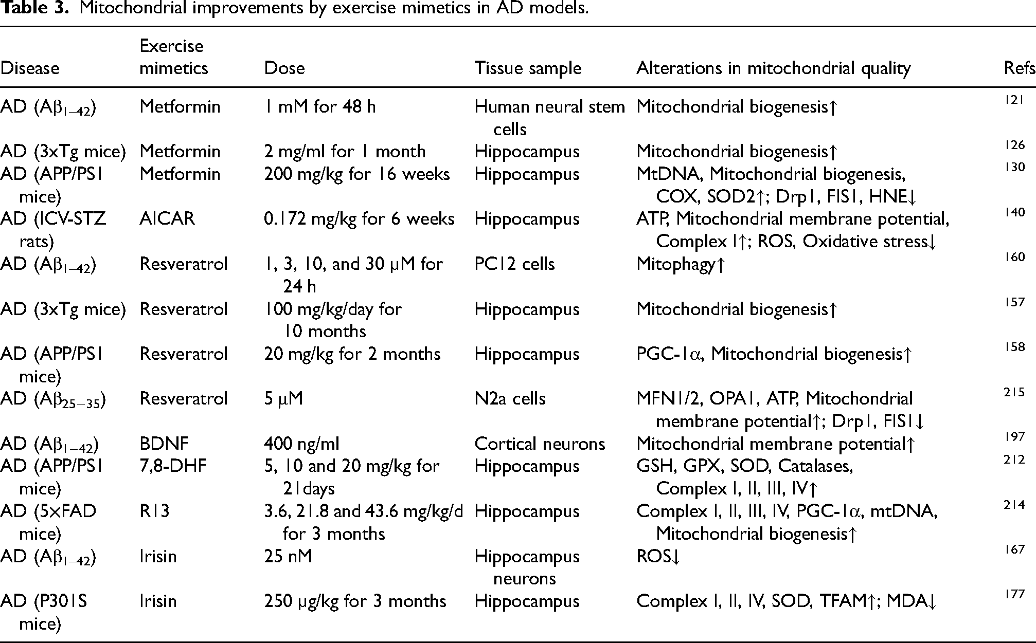
To date, no interventional clinical trials have employed exogenous BDNF to treat AD. However, research on physical activity supports its potential as an exercise mimetic. Multiple studies indicate that exercise increases BDNF levels and correlates with cognitive improvement. For example, a trial involving 55 elderly patients with MCI reported positive results. In that study, a 16-week combined aerobic and resistance training program elevated BDNF levels and enhanced neurocognitive function. 208 Furthermore, Despite the significant neuroprotective potential of BDNF in AD, its clinical application is limited by several pharmacokinetic barriers. These include its high molecular weight, short plasma half-life, and poor permeability across the BBB. Consequently, researchers have proposed a range of strategies to enhance the central bioavailability of BDNF or to activate its downstream signaling pathways via alternative means. One such approach involves the upregulation of endogenous BDNF expression. For instance, irisin is capable of crossing the BBB and has been shown to stimulate BDNF production within the brain. In doing so, it indirectly activates the BDNF/TrkB signaling pathway, thereby exerting neuroprotective effects and mitigating AD-associated cognitive deficits. 209 Another promising strategy lies in the development of small-molecule BDNF mimetics with high BBB permeability. Among these, 7,8-dihydroxyflavone (7,8-DHF) is a notable TrkB agonist that can directly bind to and activate the TrkB receptor, thereby initiating downstream signaling cascades comparable to those triggered by endogenous BDNF. 210 Experimental studies have demonstrated that 7,8-DHF can reduce oxidative stress, enhance mitochondrial function, inhibit BACE1 activity, and decrease Aβ generation, ultimately improving cognitive function in AD models animals.211,212 To further improve its oral bioavailability and pharmacokinetic stability, a prodrug form of 7,8-DHF, known as R13, has been developed. In 5XFAD transgenic mouse models, long-term oral administration of R13 effectively activates TrkB signaling in hippocampal neurons, suppresses Aβ deposition and tau hyperphosphorylation, and ameliorates memory impairments in a dose-dependent manner (Table 3; Figure 4).213,214
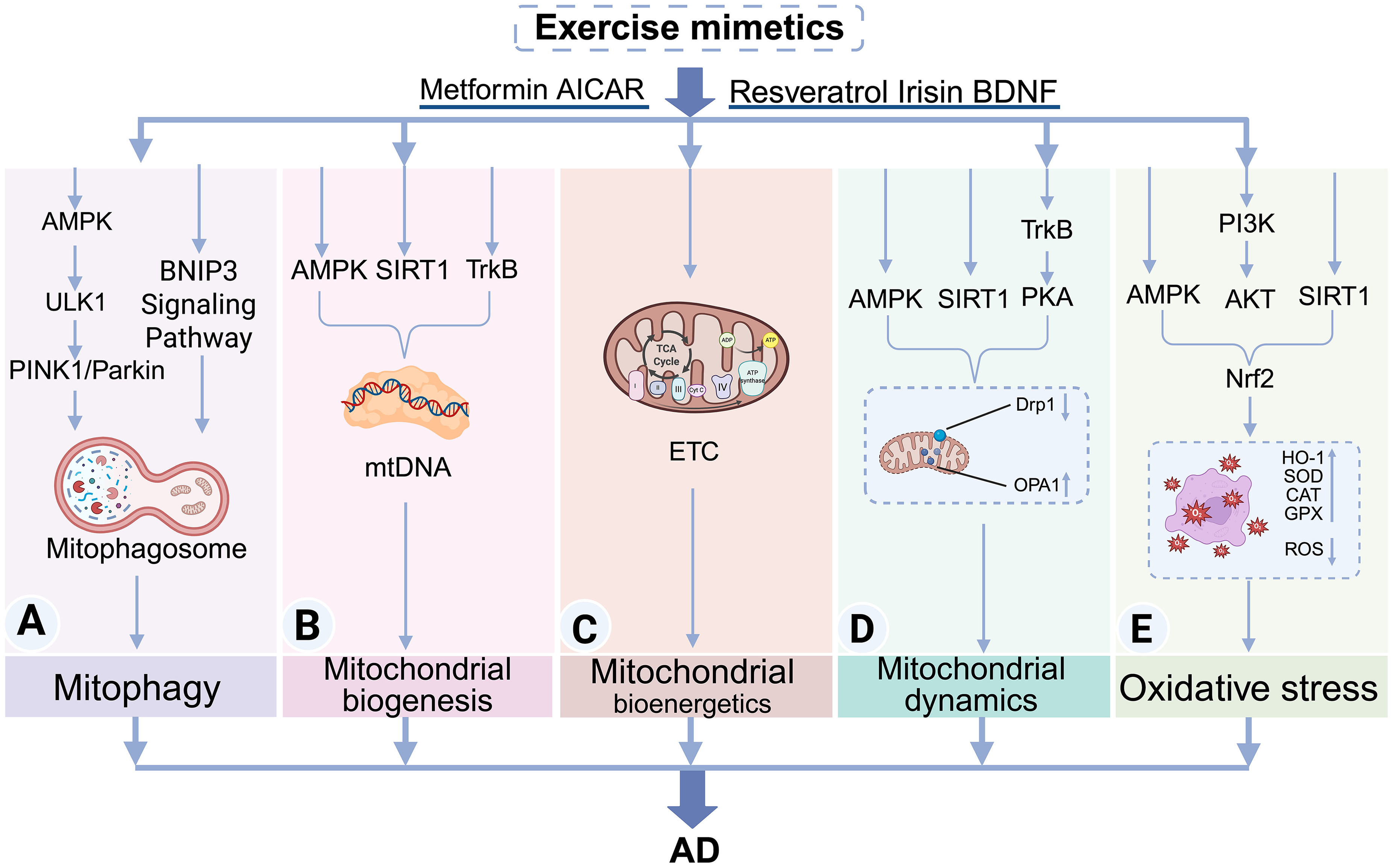
Mechanisms of exercise mimetics in ameliorating impaired mitochondrial quality in AD. Exercise mimetics have been shown to enhance mitochondrial quality in AD through several interconnected mechanisms, each targeting a key aspect of mitochondrial regulation: (A) Mitophagy: Exercise mimetics activate the AMPK–PINK1/Parkin signaling axis as well as the BNIP3-dependent pathway, thereby initiating selective mitophagy and promoting the clearance of damaged mitochondria; (B) Mitochondrial Biogenesis: Through activation of AMPK, SIRT1, and TrkB, exercise mimetics upregulate PGC-1α, which binds to downstream transcription factors NRFs and TFAM, thereby facilitating mtDNA replication and transcription and stimulating mitochondrial biogenesis; (C) Mitochondrial Energy Metabolism: By enhancing the activity of mitochondrial ETC complexes, exercise mimetics promote OXPHOS and increase ATP production, improving neuronal bioenergetic capacity under AD pathology; (D) Mitochondrial Dynamics: Exercise mimetics restore mitochondrial dynamic balance by upregulating AMPK, SIRT1, and PKA. This leads to inhibition of the fission protein Drp1 and activation of fusion-related proteins OPA1 and Mfn1/2, thus promoting mitochondrial network integrity; (E) Oxidative Stress Regulation: Exercise mimetics mitigate mitochondrial oxidative stress by activating AMPK, SIRT1, and the PI3 K/AKT pathway, which in turn upregulate the expression of Nrf2. This transcription factor induces the production of key antioxidant enzymes such as HO-1 and SOD, thereby reducing ROS accumulation and protecting mitochondrial and neuronal integrity.
Mitochondrial improvements by exercise mimetics in AD models.
Combination therapies and multimodal approaches
AD is the most common neurodegenerative disorder characterized by a complex etiology. Regrettably, current clinical medications fail to reverse the pathological progression of the disease. Therefore, combination therapy represents a promising direction for future research. Specifically, the combined application of different exercise mimetics, or the integration of exercise mimetics with clinically common drugs, may offer better therapeutic outcomes for AD patients.
There is an inherent biological connection between these exercise mimetics. Upstream regulators, such as metformin and resveratrol, can stimulate the secretion of irisin, which subsequently increases BDNF levels. Therefore, a combinatorial approach could potentially compound these beneficial effects. Indeed, experiments in high-fat diet-fed mice have demonstrated that the combined treatment of metformin and resveratrol significantly ameliorates insulin resistance. 216 Given that insulin resistance is closely linked to AD pathology, this combination could theoretically improve AD outcomes, although further experimental verification is required.
Current mainstream drugs, such as donepezil, can alleviate symptoms. Importantly, emerging evidence suggests that their therapeutic efficacy may be enhanced when combined with exercise mimetics. In vitro studies utilizing human blood samples have demonstrated that combining donepezil with metformin significantly potentiates acetylcholinesterase inhibition and suppresses Aβ aggregation compared to donepezil alone. 217 Furthermore, in vivo studies using an AD rat model reveal a synergistic effect between donepezil and resveratrol. Compared to monotherapy, this combination significantly reduced microglial proliferation, increased astrocyte numbers, and diminished Aβ deposition.218,219 Additionally, it alleviated oxidative stress, as evidenced by decreased MDA levels and elevated SOD activity. 57
Prospective and conclusion
This review focuses on the issue of impaired mitochondrial quality in AD, emphasizing its importance as a novel therapeutic target that complements traditional approaches centered on Aβ and tau proteins. Given its multifaceted benefits in improving mitochondrial quality, physical exercise exhibits considerable neuroprotective potential. In recent years, exercise mimetics have emerged as a promising intervention strategy for AD, with preclinical studies demonstrating their ability to attenuate AD pathology and improve cognitive function. However, the clinical translation of this strategy still faces multiple challenges. A primary hurdle is the inconsistency of therapeutic outcomes. Taking metformin as an example, while most studies support its anti-AD effects, preclinical research has identified a potential adverse mechanism wherein metformin may upregulate BACE1 transcription via AMPK activation, thereby leading to increased Aβ generation. 220 This preclinical finding offers a plausible explanation for why metformin use has been associated with an increased risk of AD in certain clinical studies.
To guide clinical translation, it is essential to evaluate the safety, dosage, and distinct profiles of these mimetics. Currently, clinical data are largely limited to metformin and resveratrol. Metformin, an antidiabetic agent with a long history of clinical use, is characterized by its high cost-effectiveness, favorable safety profile, and well-established data. Its clinical adverse reactions are primarily limited to gastrointestinal discomfort. Furthermore, even in normoglycemic individuals, metformin administration does not induce hypoglycemia. Currently, the commonly used clinical dosage is 500 mg/day, a dose that demonstrates good tolerability. 221 Consequently, long-term administration of 500 mg/day of metformin has been proposed as a strategy to reduce the risk or delay the onset of AD and related dementias. 222 However, notably, this approach requires further evaluation. Regarding resveratrol, clinical interventions have utilized doses up to 2000 mg/day with good tolerability, reporting only mild adverse effects such as nausea and diarrhea. 34 However, its clinical utility is severely hampered by poor oral bioavailability. To address this, advanced delivery systems, including liposomes and nanoparticles, have been developed to improve BBB penetration and enhance cognitive outcomes in animal models.223,224 In contrast, emerging candidates such as AICAR, Irisin, and BDNF have demonstrated therapeutic potential in preclinical experiments. However, clinical trial data for these agents are currently unavailable. This precludes a comprehensive assessment of their safety dosages and toxicological profiles in humans, representing a major barrier to translation.
Finally, it must be acknowledged that even though exercise mimetics offer a promising pharmaceutical alternative, they may not fully replicate the systemic, pleiotropic benefits of physical exercise. For agents with existing clinical data, specifically metformin and resveratrol, the primary challenge lies in their pharmacokinetic limitations. These include weak BBB permeability and low bioavailability. Future research should prioritize the development of advanced delivery technologies, such as nanoparticle-based carriers, to address these issues. For emerging candidates without established clinical data, rigorous clinical studies are urgently needed to validate their safety and efficacy in humans. Furthermore, the exploration of exercise mimetics should not be confined solely to mitochondrial mechanisms. Research must broaden its scope to integrate other neuroprotective pathways. Consequently, moving beyond monotherapy toward multi-target combination therapies represents a critical strategic direction for maximizing therapeutic efficacy and arresting disease progression.
Footnotes
Acknowledgements
The authors have no acknowledgments to report.

Author contribution(s)
Funding
The authors disclosed receipt of the following financial support for the research, authorship, and/or publication of this article: This work was supported by the Natural Science Foundation of Hubei Province of China (grant number 2023AFB978).
Declaration of conflicting interests
The authors declared no potential conflicts of interest with respect to the research, authorship, and/or publication of this article.