



Abstract
Background
Growing evidence links periodontitis to Alzheimer's disease (AD), yet the specific links between periodontitis severity gradients and brain functional alterations remain poorly understood.
Objective
To investigate brain functional alterations quantified by functional connectivity density (FCD) and regional homogeneity (ReHo) across periodontitis severity gradients, include microbiota measures as explanatory variables, and assess correlations between these functional alterations and cognitive impairment.
Methods
Clinical periodontal data, subgingival plaque, cognitive tests, and brain MRI data were collected from all 89 participants, including community-recruited normal cognition (NC) and patients with amnestic mild cognitive impairment (aMCI) and AD from a hospital neurology department. According to periodontal examination, participants were categorized into mild, moderate, and severe groups. FCD and ReHo were compared among different periodontal condition groups and subgroups. Correlation analyses were conducted to explore the relationship among FCD, ReHo, periodontal indices, and cognition.
Results
With increasing severity of periodontitis, the FCD of bilateral middle frontal gyrus (MFG.R, MFG.L), right inferior frontal gyrus, triangular part (IFGtriang.R), and the ReHo of IFGtriang.R all decreased. These regions are commonly associated with executive control, working memory, and attention. These changes were more strongly correlated with overall periodontal inflammatory burden and cognitive performance than to the abundance of specific taxa in the subgingival plaque microbiota.
Conclusions
Periodontitis severity is associated with reduced prefrontal FCD/ReHo and cognitive decline, and these associations appear to be more strongly driven by overall periodontal inflammatory burden than by specific subgingival taxa.
Introduction
Periodontitis, a chronic infectious disease affecting the periodontal tissues, ranks among the most prevalent chronic inflammatory diseases worldwide, characterized by the progressive impairment of periodontal tissues, such as the gingival soft tissue and alveolar bone, ultimately resulting in tooth loss.1,2 With the in-depth study of the systemic impact of periodontitis, more and more evidence shows that its chronic inflammatory state extends beyond the local oral cavity and could potentially induce systemic inflammatory reaction through blood circulation, neural pathway or immune activation, and then participate in the onset and development of various systemic diseases, acting as a mutual risk factor for various systemic diseases,3–5 including Alzheimer's disease (AD), cardiovascular diseases, rheumatoid arthritis, diabetes, hypertension, nonalcoholic fatty liver disease, inflammatory bowel disease, and osteoporosis.6–12
AD, a chronic neurodegenerative disorder, is marked by the accumulation of amyloid-β plaques and neurofibrillary tangles in the brain with primary clinical manifestations including progressive memory decline, cognitive impairment, and various psychiatric symptoms and behavioral disturbances.13,14 Amnestic mild cognitive impairment (aMCI), recognized as the prodromal phase of AD, is characterized by neurocognitive dysfunction, but not to the extent of dementia. 15 However, the etiology of AD is still unclear, and recent studies have increasingly revealed a strong connection between periodontitis and AD.16–18 A recent large-scale retrospective cohort study identified clinical and microbial markers of periodontitis as being associated with both AD incidence and mortality. 19 Additionally, a 6-year longitudinal cohort study revealed a statistically significant relationship between periodontitis and cognitive decline. 20 Our recent research also demonstrated that subgingival plaque microbiota and periodontitis were associated with brain grey matter volume change and cognitive impairment. 21 The potential mechanism may involve: (1) microorganisms and their metabolites in dental plaque enter the brain through blood flow or peripheral nerves, activating inflammatory pathways in the central nervous system, leading to neuroinflammation and neuronal dysfunction; (2) Periodontitis induces systemic inflammation, leading to elevated levels of pro-inflammatory cytokines in the serum, resulting in sustained low-grade systemic inflammation. These pro-inflammatory molecules disrupt the blood-brain barrier and enter the brain, causing neuronal damage.18,22–24 Emerging evidence, particularly implicating Porphyromonas gingivalis (P. gingivalis)—the primary pathogen in chronic periodontitis—supports this hypothesis. P. gingivalis DNA and gingipains have been detected in the brains of AD patients. 18
Resting-state functional magnetic resonance imaging (rs-fMRI), a non-invasive brain imaging method, is used to evaluate spontaneous brain activity through the blood oxygen-level-dependent (BOLD) signal. In rs-fMRI, regional homogeneity (ReHo) and functional connectivity density (FCD) are often employed to evaluate grey matter function. ReHo is used to determine the extent of functional synchronization between a specified voxel and the voxels in its vicinity, 25 while FCD, a graph-theory measure based on data-driven methods, detects functional hubs within the brain at the voxel level. 26 Voxels exhibiting higher FCD display a greater number of functional connections with other voxels, signifying their critical role in information processing. However, there have been no comprehensive studies on changes in brain FCD and ReHo associated with different phases of periodontitis.
The primary objectives of the current study were to explore whether periodontitis and subgingival plaque dysbiosis are associated with brain function alterations in FCD and ReHo, and whether these changes correlate with cognitive impairment. To address these objectives, the study was conducted in two phases: (1) exploring brain function changes (FCD and ReHo) among different periodontitis severity groups, and (2) analyzing the relationships among clinical indicators of periodontitis, subgingival plaque microbiota, cognitive test scores and brain function.
Methods
This cross-sectional observational study was designed and reported in accordance with the STROBE (Strengthening the Reporting of Observational Studies in Epidemiology) guidelines. 27 A completed STROBE checklist, with section references for each item, is provided in the Supplementary Material.
Participants
The current study enrolled 92 right-handed participants. But three individuals were excluded because of a poor quality of the imaging data. The final sample included 89 individuals, including 40 normal cognition (NC) participants, 27 aMCI patients and 22 AD patients. The diagnosis of AD was based on the criteria set by the National Institute of Neurological and Communicative Disorders and Stroke-Alzheimer's Disease and Related Disorders Association (NINCDS-ADRDA) for “probable” or “possible” AD.28,29 Structural magnetic resonance imaging (MRI) in AD patients revealed medial temporal lobe atrophy.29,30 The diagnosis of aMCI followed Petersen criteria.15,31 All aMCI patients had a Clinical Dementia Rating (CDR) score of 0.5, reported self-perceived memory deficits verified through neuropsychological tests, and showed no clinical signs of dementia. Participants in the NC group met the following inclusion criteria: (1) normal overall physical health, (2) normal overall cognitive function, demonstrated by MMSE scores between 24 and 30 (adjusted for education level), (3) a CDR score of 0, and (4) no subjective memory complaints. Exclusion criteria for all participants encompassed: a history of other neurological or psychiatric disorders or head injuries resulting in loss of unconsciousness, sedative drug use within 24 h prior to neuropsychological evaluation, drug or alcohol dependence, and prior chronic exposure to neurotoxic substances. The NC were recruited from community, while aMCI and AD were enrolled from the Department of Neurology at the First Affiliated Hospital of Anhui Medical University in Hefei, Anhui Province, China. All participants were sourced from the same study base, ensuring uniform examination procedures and experimental protocols. The study was approved by the Ethics Committee of the First Affiliated Hospital of Anhui Medical University and conducted in accordance with the Declaration of Helsinki (ethics approval registration number: PJ 2024-09-28). The Ethics Approval Letter is provided in the Supplemental Material. Written informed consent was obtained from all participants before examination and evaluation. The written informed consent form followed standardized nationally approved templates. All participants underwent brain MRI scanning, periodontal examination and completed a battery of cognitive assessments.
Examination of dental status
The periodontal examination was performed as described in our previous study. 32 The clinical indicators included the number of teeth present (NTP), bleeding on probing (BOP), probing depth (PD), and clinical attachment level (CAL). All participants were diagnosed as periodontitis according to the conditions: interdental CAL was detectable at ≥2 non-adjacent teeth, or buccal or oral CAL ≥ 3 mm with PD > 3 mm was detectable at ≥2teeth, And the observed CAL cannot be ascribed to non-periodontal causes such as: 1) gingival recession of traumatic origin; 2) dental caries extending in the cervical area of the tooth; 3) the presence of CAL on the distal aspect of a second molar and associated with malposition or extraction of a third molar, 4) an endodontic lesion draining through the marginal periodontium; and 5) the occurrence of a vertical root fracture. 33 The severity of periodontitis was classified based on the 2018 World Workshop Classification System: stage I (mild or initial periodontitis), interdental CAL 1–2 mm at site of greatest loss with PD ≤ 4 mm; stage II (moderate periodontitis), interdental CAL 3–4 mm at site of greatest loss with PD ≤ 5 mm and no teeth loss due to periodontitis; stage III/stage IV (severe periodontitis), interdental CAL ≥5 mm at site of greatest loss with PD ≥ 6 mm, with II-IV furcation involvement, history of periodontal tooth loss, tooth hypermobility due to secondary occlusal trauma and the sequelae of tooth loss: posterior bite collapse and drifting.33,34 Periodontal examinations were performed on all participants, revealing 22 cases of mild periodontitis, 34 cases of moderate periodontitis, and 33 cases of severe periodontitis.
Cognitive assessment
Each participant underwent cognitive tests, including the Mini-Mental Status Examination (MMSE) and Montreal Cognitive Assessment (MoCA) to evaluate general cognitive function, the Auditory Verbal Learning Test (AVLT) to evaluate episodic memory and Digit Span Test (DST) to evaluate working memory and attention function.
Image acquisition
MRI scanning was performed using a 3.0-Tesla system (Discovery MR750w, General Electric, Milwaukee, WI, USA) equipped with a 24-channel head coil. The specific scanning procedures and parameters are detailed in the Supplemental Material.
fMRI data preprocessing
SPM12 and Data Processing & Analysis for Brain Imaging (DPABI, http://rfmri.org/dpabi) were utilized for preprocessing rs-fMRI BOLD data. The specific processing steps are detailed in the Supplemental Material.
ReHo and FCD analysis
The ReHo and FCD were calculated using the approach described in previous studies.25,26 The specific analytical methods are detailed in the Supplemental Material.
Microbiome sample collection and sequencing
Among the 89 participants included in the neuroimaging analysis, subgingival plaque microbiota samples were successfully collected from 66. The specific processing steps of the sample collection, storage, DNA purity testing, quantification, amplification, analysis, library construction, sequencing, and ASV abundance table construction are detailed in the Supplemental Material.
Statistical analysis
All statistical analyses were conducted using SPSS 26.0 (SPSS, Chicago, IL, USA). Normally distributed variables were compared among multiple groups using one-way analysis of variance (ANOVA) with post hoc tests for pairwise comparisons, while non-normally distributed variables were compared among groups using the Kruskal-Wallis H-test or Mann-Whitney U-test. Sex ratios were compared by chi-square test.
Brain images were analyzed using DPABI within MATLAB. A general linear model was established and one-way ANOVA was performed with age, sex, years of education, total intracranial volume (TIV), and framewise displacement (FD) as covariates. A non-parametric permutation test with threshold-free cluster enhancement (TFCE, 5000 permutations, family-wise error (FWE) < 0.05) was adopted to identify the brain regions with a significant difference among the three groups. TFCE (5000 permutations, FWE < 0.05) method was also used in the two-sample t-test afterwards. Compared to traditional voxel-wise or cluster-wise inferences, TFCE balances detection sensitivity and false positive control.35,36 In subsequent analyses, we adopted stratified analyses to account for the influence of cognitive diagnosis—analyses were conducted separately in the NC, aMCI, and AD subgroups.
Brain regions with significant group differences in FCD and ReHo were selected as ROIs, and partial correlations were conducted among brain function changes, cognitive ability, periodontal indices and dental plaque microbial composition. p < 0.05 was considered as statistically significant.
Results
Participant characteristics
Demographics, clinical, and neuropsychological scores are summarized in Table 1. The three periodontitis severity groups were well matched for sex distribution (chi-square test, χ2 = 1.497, p = 0.473), age (Kruskal-Wallis H-test, H = 5.586, p = 0.061), years of education (Kruskal-Wallis H-test, H = 1.456, p = 0.483), FD (Kruskal-Wallis H-test, H = 0.371, p = 0.831) and TIV (Kruskal-Wallis H-test, H = 0.679, p = 0.712), but there was significant difference in MMSE score (Kruskal-Wallis H-test, H = 8.209, p = 0.016). The group comparisons of demographic information in 66 participants who donated dental plaque samples were similar with above (Supplemental Table 1a). Meanwhile, we compared the key baseline characteristics (e.g., age, sex, NTP, periodontitis severity, among others) between participants who provided plaque samples and those who did not (Supplemental Table 1b). Demographic information for the NC, aMCI, and AD subgroups is detailed in Supplemental Table 2a-c.
Demographics, clinical characteristics and neuropsychological scores of whole sample (89 participants).

NC: normal cognition; aMCI: amnestic mild cognitive impairment; AD: Alzheimer's disease; FD: framewise displacement; TIV: total intracranial volume; MMSE: Mini-Mental Status Examination; MoCA: Montreal Cognitive Assessment; AVLT: Auditory Verbal Learning Test; DST: Digit Span Test; NTP: number of teeth present; BOP: bleeding on probing; PD: probing depth; CAL: s clinical attachment level. *p < 0.05, **p < 0.01, *** p < 0.001.
Group differences in FCD and ReHo
Whole group differences in FCD and ReHo
Significant differences were found among different periodontitis severity groups in right middle frontal gyrus (MFG.R), inferior frontal gyrus, triangular part (IFGtriang.R), and left middle frontal gyrus (MFG.L) in FCD, and in IFGtriang.R in ReHo after controlling for age, sex, years of education, TIV, and FD (Supplemental Table 3a and Figure 1). Post-hoc comparisons of FCD and ReHo values among the three groups of periodontitis patients were conducted. Compared with the mild periodontitis group, the moderate and severe periodontitis groups showed a decreasing trend in these brain regions (Figure 1).

Significant differences in FCD and ReHo among mild, moderate and severe periodontitis groups. FCD: functional connectivity density; ReHo: regional homogeneity; MFG.R: right middle frontal gyrus; MFG.LL: left middle frontal gyrus; IFGtriang.R: right middle frontal gyrus of triangular part.
NC/aMCI/AD subgroup differences in FCD and ReHo
In NC, significant differences were found among mild, moderate and severe periodontitis subgroups in ReHo of IFGtriang.R and in FCD of right insula (INS.R) after controlling for age, sex, years of education, TIV, and FD. As periodontitis severity increased, the FCD value of the INS.R showed an upward trend, while the ReHo value of IFGtriang.R showed a decreasing trend. Detailed information is provided in Supplemental Figure 1A and Supplemental Table 3b.
In aMCI, significant differences were found among mild, moderate and severe periodontitis subgroups in FCD of MFG.R after controlling for age, sex, years of education, TIV, and FD. As periodontitis severity increased, the FCD value of the MFG.R showed a decreasing trend. Detailed information is provided in Supplemental Figure 1B and Supplemental Table 3c. However, no significant ReHo differences were detected.
In AD, significant differences were found among mild, moderate and severe periodontitis subgroups in FCD of MFG.R, right superior frontal gyrus, medial (SFGmed.R), and right superior frontal gyrus, dorsolateral (SFGdor.R) after controlling for age, sex, years of education, TIV, and FD. As periodontitis severity increased, the FCD value of MFG.R, SFGmed.R, and SFGdor.R showed a decreasing trend. Detailed information is provided in Supplemental Figure 1C and Supplemental Table 3d. Again, no significant ReHo differences were found.
Correlations between FCD, ReHo and domains of cognition
After controlling for age, sex, years of education, TIV, and FD, MoCA, AVLT, and DST were positively correlated with brain function changes related to the severity of periodontitis in all participants (Figure 2). Detailed p and r values were described in Supplemental Table 4.
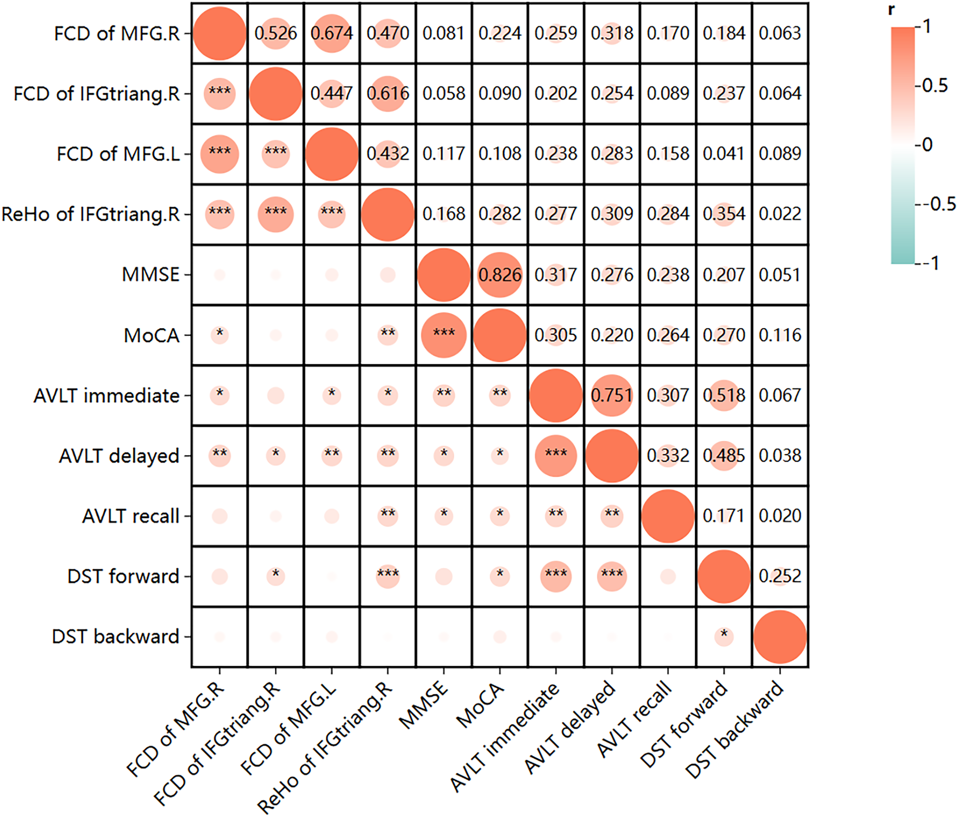
Heatmap of partial correlation analysis between the extracted FCD and ReHo values and cognition domains in all groups. FCD: functional connectivity density; ReHo: regional homogeneity; MFG.R: right middle frontal gyrus; MFG.L: left middle frontal gyrus; IFGtriang.R: right middle frontal gyrus of triangular part; MMSE: Mini-Mental State Examination; MoCA: Montreal Cognitive Assessment; AVLT: Auditory Verbal Learning Test; DST: Digit Span Test.
Correlations between FCD, ReHo and clinical periodontal indices
After controlling for age, sex, years of education, TIV, and FD, BOP was negatively correlated with ReHo of IFGtriang.R, FCD of MFG.R, IFGtriang.R and MFG.L, while PD = 1–3 mm (%) and CAL = 0–2 mm (%) was positively correlated with ReHo of IFGtriang.R, FCD of MFG.R, IFGtriang.R and MFG.L; PD = 4–5 mm (%) and CAL = 3–4 mm (%) was negatively correlated with ReHo of IFGtriang.R, FCD of MFG.R, IFGtriang.R and MFG.L(Figure 3). Detailed p and r values were described in Supplemental Table 5a. Except for FCD of INS.R, the correlation patterns between brain function (FCD and ReHo) and clinical periodontal indices in NC, aMCI, and AD subgroups were similar with above (Supplemental Figure 2A-C), with detailed p and r values in Supplemental Table 5b-d.

Heatmap of partial correlation analysis between the extracted FCD and ReHo values and clinical periodontal indices. FCD: functional connectivity density; ReHo: regional homogeneity; MFG.R: right middle frontal gyrus; IFGtriang.R: right middle frontal gyrus of triangular part; MFG.L: left middle frontal gyrus; NTP,: number of teeth present; BOP: bleeding on probing; PD: probing depth; CAL: clinical attachment level.
Correlations between FCD, ReHo and microbial flora of dental plaque
We compared the key baseline characteristics between participants with plaque samples (n = 66) and those without samples (n = 23) (Supplemental Table 1b). There were no significant differences between the two groups in sex, age, FD, TIV or periodontitis severity, but significant differences were observed in years of education, NTP and BOP(%). Therefore, NTP and BOP(%) were additionally included as covariates. In 66 samples with subgingival plaque microbiota data, after adjusting for age, sex, years of education, TIV, FD, NTP, and BOP(%), ReHo/FCD were found to be correlated with different subgingival plaque microbiota abundances. Specifically, ReHo/FCD were positively correlated with the abundance of the phylum Synergistota (class Synergistia, order Synergistales, family Synergistaceae, et al.) and were negative correlation with the abundance of the phylum Proteobacteria (class Gammaproteobacteria, order Pasteurellales, family Pasteurellaceae, et al.). Detailed information was described in Figure 4 and Supplemental Table 6.
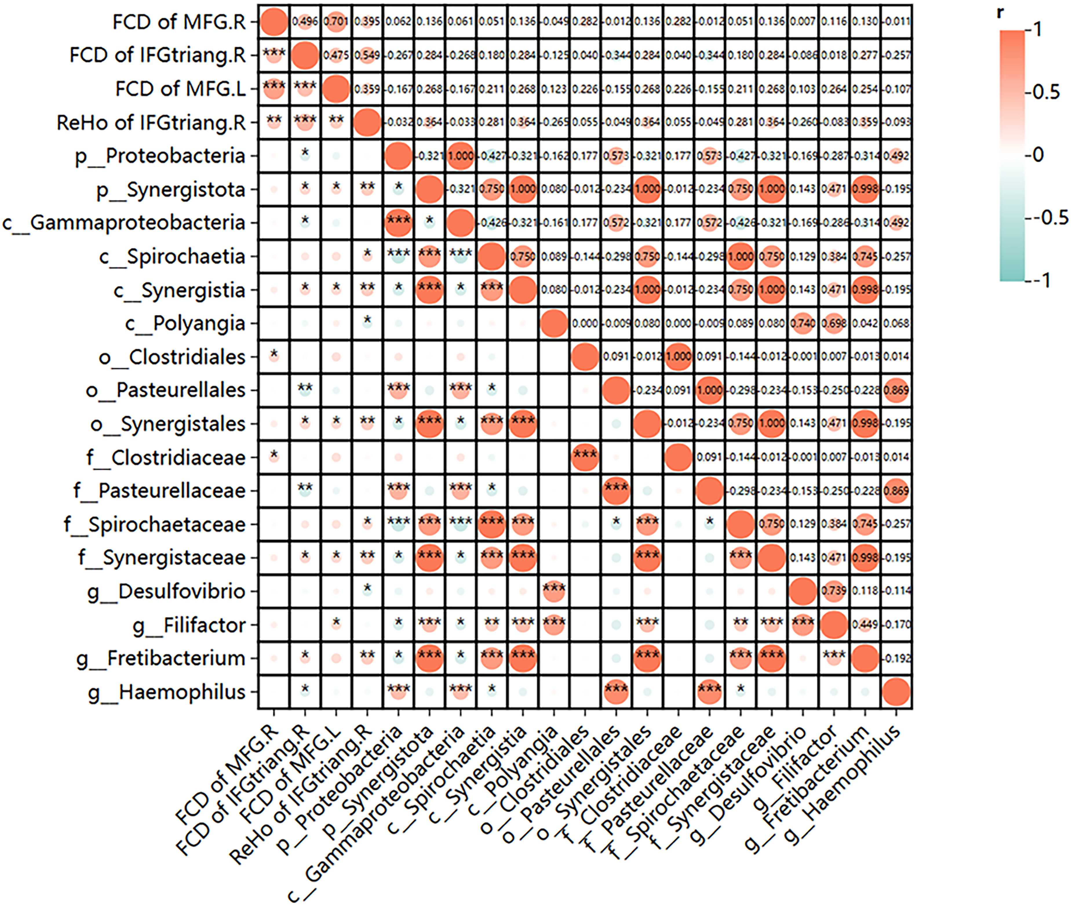
Heatmap of partial correlation analysis between the extracted FCD and ReHo values and subgingival plaque microbiota. FCD: functional connectivity density; ReHo: regional homogeneity; MFG.R: right middle frontal gyrus; MFG.L: left middle frontal gyrus; IFGtriang.R: right middle frontal gyrus of triangular part; p: phylum; c: class; o: order; f: family; g: genus.
Sensitivity analyses
When constructing the primary model, we only included age, sex, education, FD, and TIV as covariates, but excluded MMSE. This is because MMSE is not a typical confounder, but may function as a mediator in the hypothesized pathway (periodontitis → brain changes → cognition). Treating it as a standard covariate risks overadjustment bias, potentially underestimating or distorting the true effect of periodontal severity on brain measures. Therefore, we conducted sensitivity analyses in which MMSE was added as an additional covariate to the primary model (Supplemental Table 7a-d). Subsequently, we compared the results of the sensitivity analyses with those of the primary model to assess whether including MMSE altered the analytical results regarding the association between periodontitis severity and brain function. When MMSE was additionally included as a covariate, the overall pattern of results remained highly consistent with the primary model. However, in the AD group, the FCD in the SFGmed.R was no longer significant after MMSE adjustment. The SFGmed.R cluster in the primary model was very small (8 voxels), which may partly explain its reduced robustness in the sensitivity analysis. In addition, we also used different Gaussian smoothing kernels (full width at half-maximum [FWHM] of 4 and 8 mm) to verify the stability of the results, following the initial analysis with a 6 mm kernel. Major clusters observed in the primary model still reached significance (Supplemental Table 8a-9d).
Discussion
To our knowledge, the present study is the first to compare brain function changes in individuals with different severity of periodontitis from the perspective of FCD and ReHo. Two major findings were recorded in our study. First, with the progression of periodontitis severity, brain function changed with mainly a decreasing trend (i.e., ReHo of IFGtriang.R and FCD of MFG.R, IFGtriang.R, and MFG.L). Second, the differences in both FCD and ReHo among the different severity levels of periodontitis, were significantly correlated with the periodontal clinical indices, domains of cognition and microbial flora of dental plaque.
Our study revealed a potential association between periodontitis and brain function. Following the progression of periodontitis, FCD in the MFG.R, IFGtriang.R, MFG.L and ReHo in the IFGtriang.R were decreased. There was also a similar trend in the subgroups except for FCD in INS.R. The inferior frontal gyrus, particularly its triangular part, plays a crucial role in language comprehension, cognitive control, emotional regulation, and attention allocation.37–39 Damage to this region can impair language processing, executive functioning, and social skills. Studies have shown that in patients with aMCI and AD, the gray matter volume of the right inferior frontal triangular region is significantly reduced. 40 Furthermore, AD patients exhibit abnormal functional connectivity between the IFGtriang.R and other brain regions, such as the prefrontal cortex and hippocampus, resulting in diminished regulation of cognitive control and memory retrieva. 41 These structural changes are strongly linked to the deterioration of cognitive and language functions. Both the middle frontal gyrus and the superior frontal gyrus, dorsolateral, which are key in the dorsolateral prefrontal cortex, is involved in several higher-order cognitive functions, including executive functioning, attention regulation, working memory, and emotional modulation.42,43 In aMCI patients, both ALFF and ReHo are decreased in SFG.R and MFG.R. 44 In AD patients, the MFG.R exhibits significant grey matter atrophy, abnormality, and altered regional cerebral blood flow.45–47 In aMCI patients, the betweenness centrality of the MFG.L is decreased. 48 These structural and functional changes in these brain regions may be closely associated with the cognitive decline and behavioral impairments characteristic of AD. Thus, it is reasonable to conclude that more severe periodontitis is associated with greater neuropathological damage and cognitive-behavioral impairments. Our findings provide further support for strong associations among periodontitis, FCD and ReHo reduction, and cognitive dysfunction.
In addition, the FCD of INS.R shows an upward trend in NC subgroup. Our explanation for this is that the insula is a key brain area responsible for integrating visceral sensory cortex, emotion regulation, and pain perception. 49 In pain studies of chronic pain patients, it has been consistently found that the insular cortex is activated.50,51 The increase in FCD of INS.R may reflect the activation of compensatory activity in the insula due to chronic inflammation caused by periodontitis, in response to systemic inflammatory burden.
In addition, the current study reveals statistically strong associations among FCD/ReHo changes and the clinical indicators of periodontitis, subgingival plaque microbiota, cognitive deficits. We speculate that periodontitis, as a chronic inflammatory disease, releases inflammatory factors (such as IL-6 and TNF-α) that can cross the blood-brain barrier, induce activation of microglia in the brain, and may influence or exacerbate FCD, ReHo, and cognitive decline as observed in aMCI and AD.18,52,53 In this study, we found that the magnitudes of FCD and ReHo reduction were correlated with periodontitis severity, including negative correlations with BOP, PD 4–5 mm, and CAL = 3–4 mm as well as positive correlations with PD 1–3 mm and CAL 0–2 mm. In general, more severe periodontitis was associated with greater reductions in FCD and ReHo, consistent with our hypotheses.
We also found that the abundance of some bacteria in subgingival plaque microbiota was positively or negatively associated with certain brain regions, which may involve more complex mechanisms. Whether this association is protective or detrimental points to the potential influence of microbial exposure. 54 Porphyromonas gingivalis, Treponema denticola, and Tannerella forsythia —collectively termed the “red complex”—are the most strongly associated with periodontitis. Notably, although the red-complex species are key periodontal pathogens, we did not observe significant associations with FCD/ReHo in the current cohort. This finding may indicate that the observed brain functional alterations are more closely related to overall subgingival microbial dysbiosis and/or inflammatory burden rather than individual taxa. On the other hand, subtle associations might have been missed due to insufficient sample size, limited statistical power, or inherent limitations of species-level quantification in our study. Future studies with larger samples may further clarify these relationships.
Our study has several limitations that need to be considered. First, the sample size was relatively small, particularly for the subgroup analysis, larger samples are needed in future studies for validation purposes. Second, the cross-sectional design of the study cannot explore the causal relationships; thus, longitudinal and interventional studies are warranted in future research. Third, adjusting for MMSE may introduce overadjustment bias if cognition is on the causal pathway; therefore, MMSE-adjusted findings are presented and interpreted only as sensitivity analyses. Fourth, microbiome–brain function correlation analysis was based on a relatively small sample size, resulting in insufficient statistical power. Additionally, compared with participants without plaque samples, participants with plaque samples had slightly higher education levels, more remaining teeth, and lower BOP(%). This indicates that the microbiome sample may be subject to selection bias, with limited representativeness. Therefore, the microbiome-related findings of this study should be regarded as exploratory results, and further verification and replication in larger, more representative independent samples are required in the future. Fifth, detailed information on important grade modifiers for periodontitis, particularly smoking status and diabetes, was not systematically collected in this study. This may have led to residual confounding. Future studies evaluating periodontitis and its related outcomes should routinely record and control for these risk factors. Sixth, our patients were recruited from a single center, multi-center studies are necessary for validation. Finally, inflammatory cytokines were not explored in this study. Subgingival plaque microbiota might induce local and systemic inflammatory responses mediated by inflammatory cytokines, potentially affecting cognitive function—a hypothesis that requires further investigation in future research.
Conclusions
Our study reveals for the first time the association between the severity of periodontitis and specific brain function changes. The brain regions with altered FCD and ReHo were closely associated with clinical indicators of periodontitis, subgingival plaque microbiota, and cognitive impairment. These findings not only deepen our understanding of the links between periodontitis and brain function but also provide preliminary insights into potential connections between oral health and AD, offering a theoretical basis for further exploration of underlying mechanisms. Notably, given the cross-sectional design of the present study, these observations reflect associations rather than definitive causal relationships. Future longitudinal, interventional, and combined imaging–oral health follow-up studies are required to establish temporal directionality and potential causal mechanisms.
Supplemental Material
sj-docx-1-alz-10.1177_13872877261426906 - Supplemental material for Periodontitis associated with multi-region brain functional alterations and cognitive impairment: A cross-sectional study in normal cognition, amnestic mild cognitive impairment, and Alzheimer's disease
Supplemental material, sj-docx-1-alz-10.1177_13872877261426906 for Periodontitis associated with multi-region brain functional alterations and cognitive impairment: A cross-sectional study in normal cognition, amnestic mild cognitive impairment, and Alzheimer's disease by Ju Ma, Shanshan Zhou, Wanqiu Zhu, Yufei Tao, Wenrui Wang, Xiao Chen, Wei Ye, Yun Lu, Ziwen Gao, Xiaohu Li, Xiaoshu Li and Yongqiang Yu in Journal of Alzheimer's Disease
Footnotes
Acknowledgements
We strongly acknowledge the invaluable support of patients and their families, clinicians, and technicians.

Ethical considerations
The studies involving human participants were reviewed and approved by the Medical Research Ethics Committee of the first Affiliated Hospital of Anhui Medical University.
Consent to participate
Not applicable
Consent for publication
Not applicable
Author contribution(s)
Funding
The authors disclosed receipt of the following financial support for the research, authorship, and/or publication of this article: The work was supported by the Scientific Research Project of University in Anhui Province, China (grant number: 2022AH051138), National Natural Science Foundation of China (grant numbers: 81901726, 82071905).
Declaration of conflicting interests
The authors declared no potential conflicts of interest with respect to the research, authorship, and/or publication of this article.
Data availability statement
The data supporting the findings of this study are available on request from the corresponding author upon reasonable request.
Supplemental material
Supplemental material for this article is available online.
References
Supplementary Material
Please find the following supplemental material available below.
For Open Access articles published under a Creative Commons License, all supplemental material carries the same license as the article it is associated with.
For non-Open Access articles published, all supplemental material carries a non-exclusive license, and permission requests for re-use of supplemental material or any part of supplemental material shall be sent directly to the copyright owner as specified in the copyright notice associated with the article.
