



Abstract
Background
Alzheimer's disease (AD) is the leading cause of dementia in the elderly. Acetylcholinesterase inhibitors are the mainstay of symptomatic treatment, and vascular dysfunction is increasingly recognized as a key contributor to AD pathophysiology. While donepezil is a standard AD treatment, its effects on the vascular system remain poorly understood despite known neurovascular interactions.
Objective
To investigate whether donepezil treatment influences endothelial progenitor cell (EPC) populations and differentiation capacity in patients with AD.
Methods
EPCs were evaluated in healthy controls and patients with AD (n = 20 per group; N = 80 total): controls (Ctrl), patients initiating donepezil 5 mg (Dp_Start), patients receiving donepezil 5 mg for ≥6 months (Dp_5 mg), and patients escalated to 10 mg after ≥6 months of 5 mg treatment (Dp_10 mg). Peripheral blood samples were collected at baseline, 12 weeks, and 24 weeks. Circulating EPCs were quantified by flow cytometry, and EPC differentiation capacity was assessed by counting early and late EPC colony-forming units (CFUs).
Results
At baseline, EPC differentiation capacity was reduced in AD patients compared with controls. Circulating EPC levels did not show significant changes across groups or treatment durations. In contrast, both early and late EPC CFU counts were significantly increased in AD patients receiving donepezil, particularly during the first 12 weeks of treatment. This effect was pronounced in patients initiating donepezil therapy.
Conclusions
Donepezil enhanced EPC differentiation into early and late populations without altering circulating EPC levels. These findings suggest that donepezil improves EPC functional competence and vascular regenerative capacity beyond its established cognitive effects.
This is a visual representation of the abstract.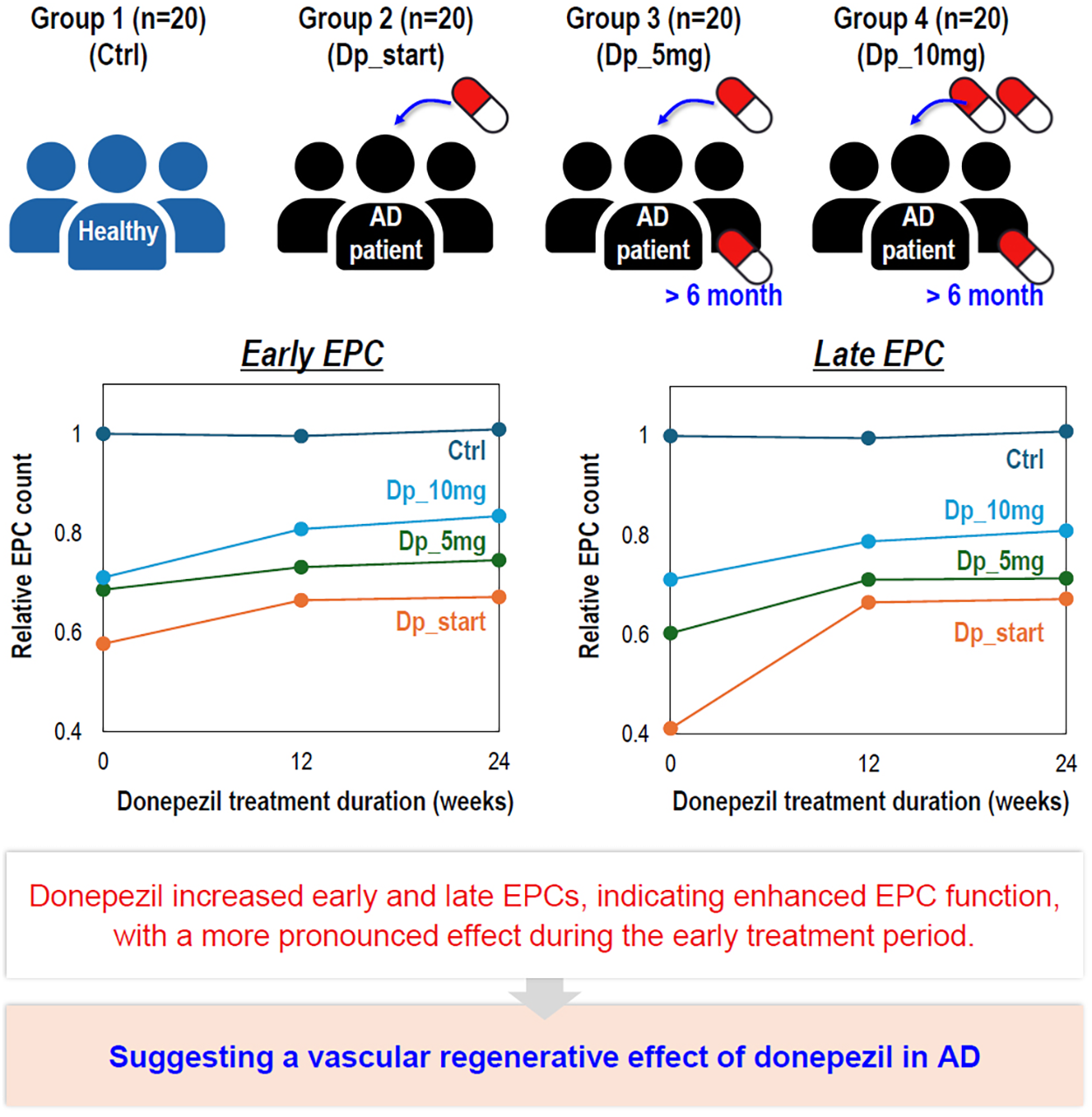
Keywords
Introduction
Alzheimer's disease (AD) is a progressive neurodegenerative disorder characterized by memory and cognitive decline accompanied by behavioral and psychological symptoms of dementia. 1 The pathological features of AD include amyloid-β (Aβ) accumulation and neurofibrillary tangles composed of hyperphosphorylated tau. 2 Beyond these classical pathological features, it is now well recognized that vascular dysfunction is an integral component of AD pathogenesis, with cerebral hypoperfusion, endothelial dysfunction, and disruption of the neurovascular unit.3,4
Acetylcholinesterase inhibitors (AChEIs) are the mainstay of symptomatic treatment for AD.5,6 Although their therapeutic effects are primarily attributed to enhancement of cholinergic neurotransmission,7–9 acetylcholine also exerts important regulatory actions on the vascular endothelium, including modulation of nitric oxide production, vascular tone, and inflammatory signaling.10–13 Consistent with this biology, emerging experimental and clinical evidence suggests that AChEIs may exert pleiotropic effects extending beyond neuronal networks, with potential implications for vascular function.14,15
Donepezil, a selective and centrally acting AChEI, is among the most extensively used pharmacological therapies for AD.16–18 Preclinical studies have reported that donepezil improves endothelial function and nitric oxide–dependent vasodilation, and attenuates vascular dysfunction under pathological conditions.19–22 Despite these observations, the vascular-related mechanisms of donepezil remain incompletely understood, and its therapeutic relevance in vascular dementia continues to be debated.23–26 Reflecting this lack of mechanistic evidence, the use of donepezil for vascular dementia has not been approved in South Korea since July 2019. 27
Endothelial progenitor cells (EPCs) are a heterogeneous population of circulating progenitor cells primarily derived from the bone marrow, defined by their capacity to differentiate into endothelial cells and to contribute to postnatal vasculogenesis.28–30 EPCs are considered a biological indicator of systemic vascular regenerative capacity, and reduced EPC levels have been associated with endothelial dysfunction and cognitive decline.31–33 Given that neurovascular dysfunction and vascular aging contribute to the progression of AD, impaired EPC-mediated vascular repair may represent a relevant link between vascular pathology and neurodegeneration.34–37 However, the effects of donepezil on EPC differentiation and maturation in patients with AD remain poorly characterized.
Therefore, we examined the impact of donepezil treatment on EPC populations in patients with AD, focusing on EPC differentiation and maturation, which are commonly characterized by distinct early and late EPC populations with differing proliferative and functional properties. We found that while circulating EPC numbers remained unchanged, donepezil treatment was associated with a significant increase in both early and late EPC populations. Notably, this effect was most pronounced in patients initiating donepezil therapy, whereas patients receiving long-term high-dose treatment exhibited evidence of response saturation. These findings suggest that donepezil may modulate vascular regenerative capacity through its effects on EPC differentiation, providing mechanistic insight into vascular contributions to AD pathophysiology and highlighting a potential therapeutic dimension of cholinergic treatment beyond symptomatic cognitive improvement.
Methods
Study population and ethics approval
This study was conducted in accordance with the principles of Good Clinical Practice and the Declaration of Helsinki. The study protocol was reviewed and approved by the Institutional Review Board of Seoul National University Hospital (IRB No. H-2101-194-1194). Written informed consent was obtained from all participants prior to enrollment.
A total of 80 subjects were enrolled, including 20 healthy controls and 60 patients diagnosed with AD. Healthy controls had no cognitive impairment and were not receiving donepezil treatment. AD patients were classified into four groups according to donepezil treatment status: (Ctrl) healthy control, (Dp_Start) AD patients initiating donepezil (treatment-naive at baseline), (Dp_5 mg) AD patients receiving donepezil 5 mg for ≥ 6 months, and (Dp_10 mg) AD patients escalated to 10 mg after ≥ 6 months of 5 mg treatment. Blood samples were collected at baseline (0 week), 12 weeks, and 24 weeks during routine clinical visits.
Blood collection and PBMNC isolation
Peripheral venous blood (10 mL) was collected in heparin-coated tubes from all participants. PBMNCs were isolated by density gradient centrifugation using Histopaque-1077 (Sigma-Aldrich, St Louis, MO, USA) according to the manufacturer's instructions. Isolated PBMNCs were washed with phosphate-buffered saline (PBS) and counted prior to downstream analyses.
Flow cytometric analysis of circulating EPCs
Circulating EPCs were quantified by flow cytometry based on surface marker expression. PBMNCs were detached using 0.25% trypsin and incubated for 20 min at 4 °C with fluorochrome-conjugated antibodies against CD34 (FITC), CD133 (APC), and KDR (PE). Isotype-matched antibodies were used as negative controls. At least 30,000 events were acquired per sample using a flow cytometer, and EPCs were defined as CD34+CD133+KDR+ cells within the monocytic gate. Circulating EPC counts were expressed as a proportion of total monocytic cells. All flow cytometric analyses were performed in a blinded manner.
Culture of early and late EPCs
For EPC culture assays, PBMNCs were seeded onto fibronectin-coated 100-mm culture plates at a density of 2.5 × 10⁶ cells/cm2. Plates were coated with fibronectin (R&D Systems, Minneapolis, MN, USA) and incubated at 37°C for 24 h prior to cell seeding. Cells were cultured in endothelial growth medium-2 (EGM-2; Lonza, Walkersville, MD, USA) supplemented with 5% fetal bovine serum.
The culture medium was changed daily for the first three days and every 48 h thereafter. After 7 days of culture, non-adherent cells were removed and early EPCs were identified based on spindle-shaped morphology and cluster formation. Continued culture up to 21 days resulted in the appearance of late EPCs (ECFCs) exhibiting a cobblestone-like morphology. Early and late EPCs were quantified by counting morphologically defined EPC populations under light microscopy.
Immunofluorescent staining of circulating EPCs
To confirm EPC identity and functional activity, immunofluorescent staining was performed on circulating EPCs. Cells were incubated with acetylated low-density lipoprotein (acLDL) and Ulex europaeus agglutinin I (Ulex-lectin). FITC-labeled antibodies were used to assess endothelial marker expression. Images were acquired using a fluorescence microscope, and endothelial identity and functional uptake were confirmed by dual positivity.
Statistical analysis
All experimental analyses were performed in a blinded manner with respect to clinical and demographic information. Data are presented as mean ± standard deviation. Statistical comparisons were conducted using one-way or two-way analysis of variance (ANOVA) followed by Tukey's post hoc test, as appropriate, using GraphPad Prism software. A p-value ≤ 0.05 was considered statistically significant. A post hoc power analysis was conducted using G*Power for within-group repeated-measures ANOVA across three time points. Assuming a moderate within-subject correlation (r = 0.5), nonsphericity correction ε=1, and α=0.05, a sample size of 20 participants per group provides approximately 80% power to detect an effect size of f≈0.30, corresponding to a moderate effect. Larger effects (f ≥ 0.40) would be detected with >95% power.
Results
Isolation and characterization of circulating and cultured EPCs
To isolate and characterize circulating and cultured EPCs, peripheral blood samples were collected from healthy donors and AD patients treated with donepezil and processed as outlined in Figure 1A. PBMNCs were isolated by density gradient centrifugation, and a subset of cells was used for flow cytometric and immunofluorescent analysis to quantify circulating EPCs, while the remaining cells were seeded under endothelial differentiation conditions to obtain early and late EPCs, by colony forming appearance.
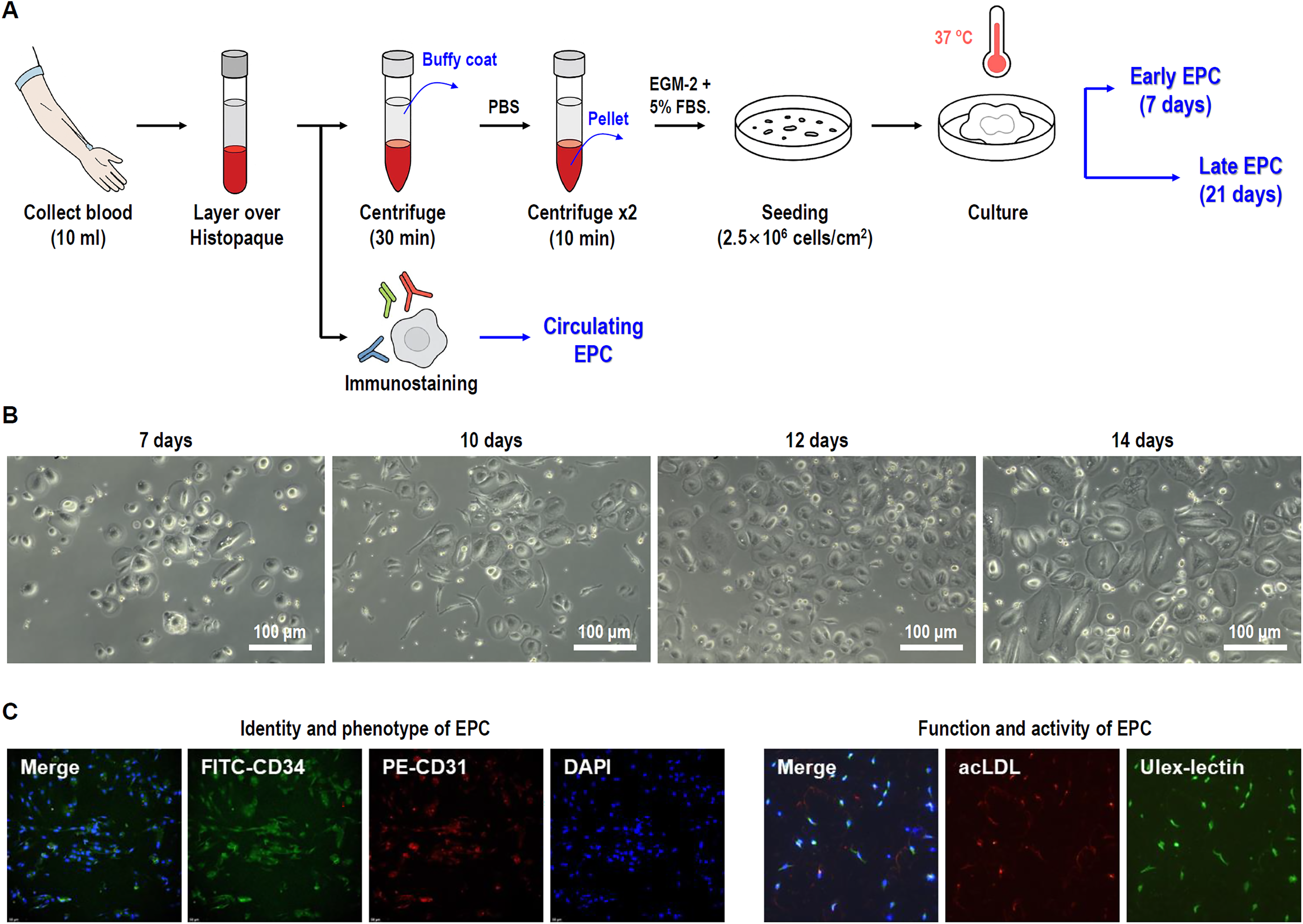
Isolation, culture, and characterization of circulating and cultured EPCs. (A) Schematic overview of the experimental workflow. Peripheral blood was collected and processed by centrifugation to isolate PBMNCs. (B) Representative morphology of cultured colonies of EPCs over time. EPCs were successfully isolated and cultured, exhibiting characteristic morphology, and endothelial colony-forming cells (ECFCs) further differentiated into mature endothelial cells. (C) Immunofluorescent staining of circulating EPCs. Endothelial identity and functional activity of EPCs were confirmed by marker expression (FITC) and functional uptake of acetylated LDL (acLDL) and binding of Ulex europaeus agglutinin I (Ulex-lectin).
Cultured EPCs exhibited time-dependent morphological changes consistent with endothelial differentiation (Figure 1B). After 7 days of culture, early EPCs with a spindle-shaped morphology and colony forming clusters of PBMNCs (peripheral blood mononuclear cells), representing the initial stage of differentiation, were observed. By day 14, cells exhibited a cobblestone-like morphology, a characteristic feature of late EPC colonies, which were mature form of endothelial cells.
The endothelial identity and functional activity of EPCs were further confirmed by immunofluorescent staining (Figure 1C). Circulating EPCs expressed endothelial markers and demonstrated functional uptake of acetylated LDL (acLDL) as well as binding of Ulex europaeus agglutinin I (Ulex-lectin), confirming that the isolated cells exhibited both phenotypic and functional features of EPCs.
Effect of donepezil on circulating EPCs
To evaluate whether donepezil administration alters circulating EPC levels, EPCs were quantified in peripheral blood at baseline (0 weeks) and after 12 and 24 weeks of treatment (Figure 2). No significant differences in circulating EPC populations were observed according to donepezil dose or treatment duration. Circulating EPC counts remained comparable across all experimental groups and time points, indicating that donepezil treatment did not significantly affect the number of EPCs present in the peripheral circulation. However, PBMNCs obtained at each time point (0, 12, and 24 weeks) were subsequently cultured under endothelial differentiation conditions. Early EPCs were assessed at day 7 of culture and late EPCs at day 21. In contrast to circulating EPC counts, cultured EPCs demonstrated group-dependent differences in colony formation, with variations observed in both early and late EPC populations.
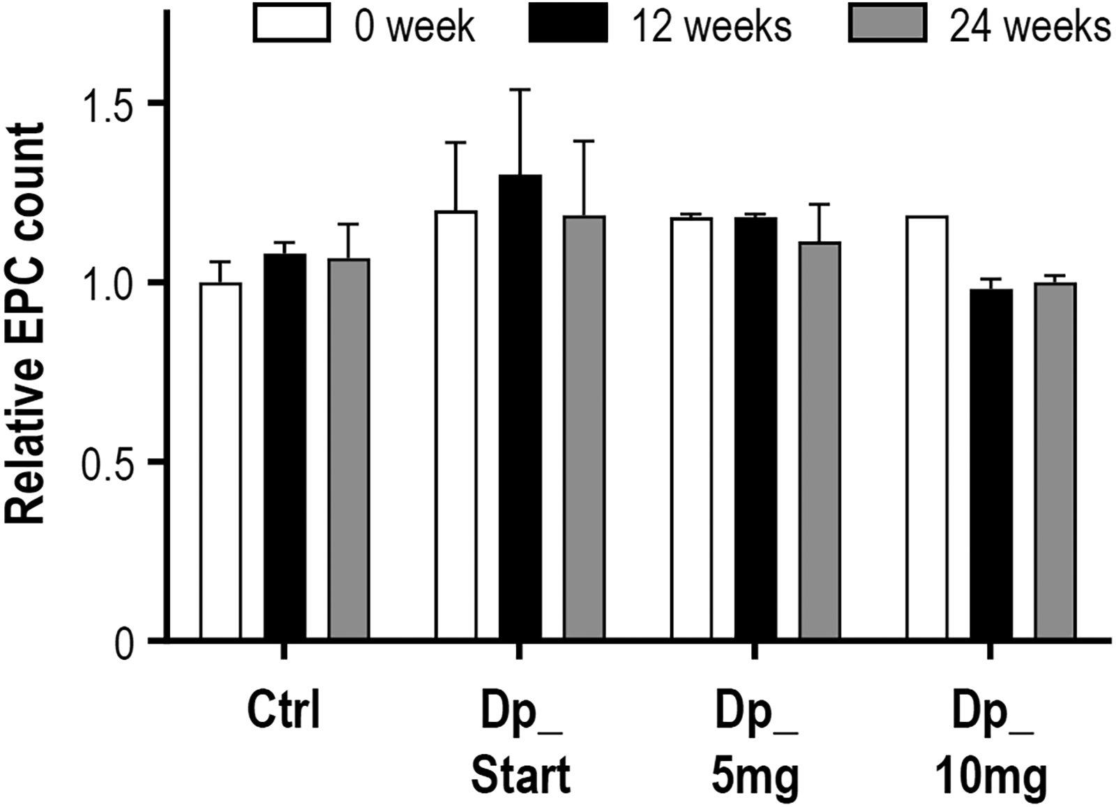
Quantitative analysis of circulating EPCs following donepezil treatment. Circulating EPCs were quantified in peripheral blood at baseline (0 weeks) and after 12 and 24 weeks of donepezil treatment. EPC counts were used as an indicator of endothelial regenerative potential, reflecting the degree of vascular injury and progenitor cell mobilization. Relative EPC counts were normalized to Ctrl at week 0. Data are presented as mean ± SD (n = 20 per group). Statistical significance was determined by one-way ANOVA followed by Tukey's multiple comparison test.
Morphological characterization of early and late EPCs
To investigate the morphological characteristics of cultured EPCs following donepezil treatment, early and late EPCs were examined at defined time points during culture (Figure 3). Early EPC colonies, observed at day 7, consisted of a central cluster of round cells surrounded by elongated spindle-shaped cells, a morphology indicative of proliferative capacity and paracrine-mediated differentiation.
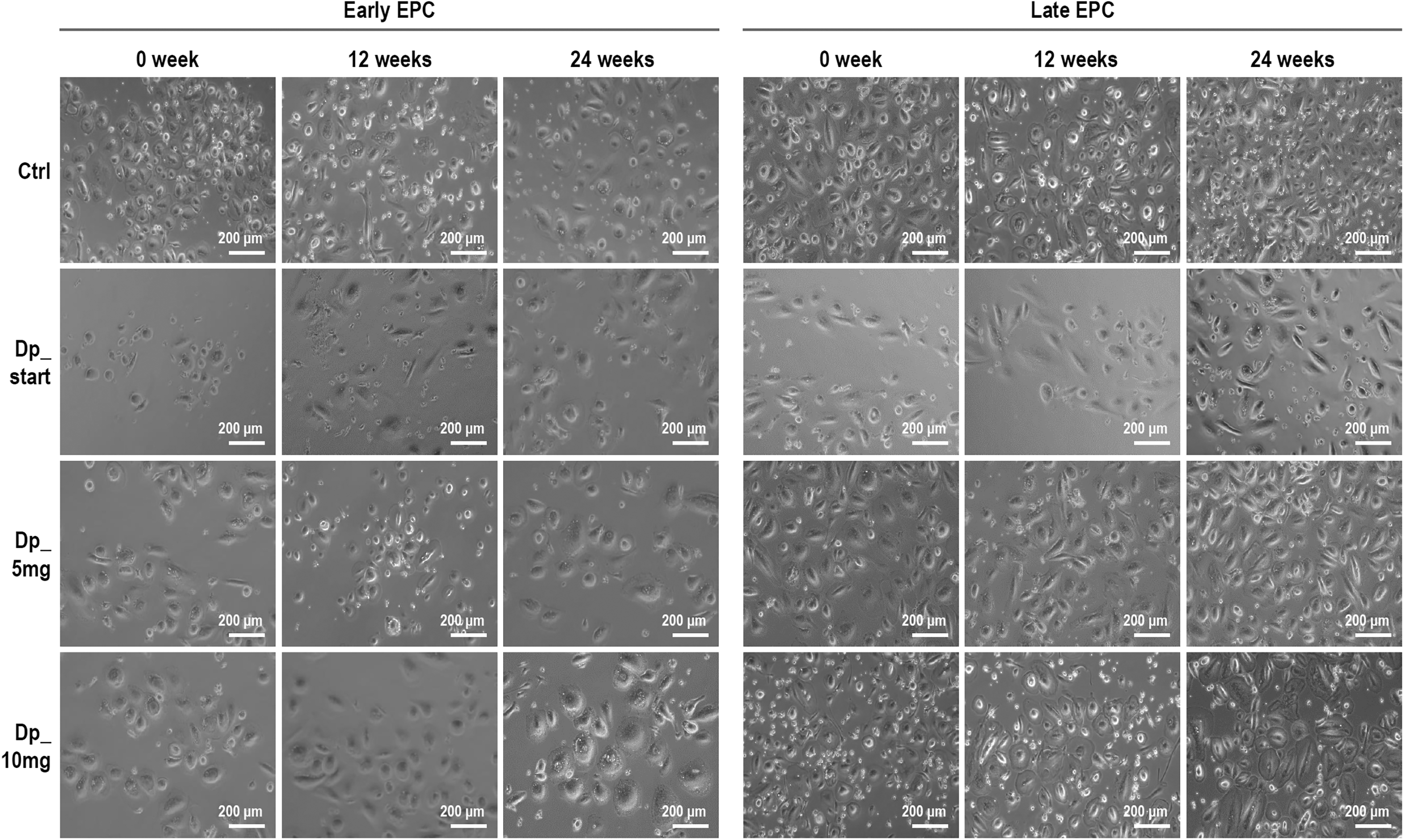
Morphology of early and late EPCs after donepezil treatment. Representative morphology of early EPC colonies at day 7 of culture. Early EPCs formed characteristic colonies consisting of a central cluster of round cells surrounded by elongated spindle-shaped cells, reflecting their proliferative and paracrine differentiation potential. Representative morphology of late EPC colonies (endothelial colony-forming unit cells, ECFCs) at day 21 of culture. Late EPCs exhibited a cobblestone-like morphology, indicating differentiation into mature endothelial lineage cells and potential for direct vascular regeneration.
In contrast, late EPCs, observed at day 21 of culture, exhibited a cobblestone-like morphology characteristic of ECFCs, which differentiate into mature endothelial lineage cells. These distinct morphological features confirm the successful differentiation of EPCs into early and late populations during in vitro culture.
Quantitative effects of donepezil on early and late EPC CFUs
To determine whether donepezil treatment influences the differentiation capacity of EPCs, early and late EPC counts were quantified from PBMNC cultures (Figure 4). Early EPC counts were significantly increased following donepezil treatment across all experimental groups, with a greater increase observed during the early treatment period (Figure 4A). However, this effect reached a plateau from 12 weeks onward, indicating saturation of the donepezil-induced response.

Quantitative analysis of early and late EPCs CFUs following donepezil treatment. (A) Quantification of early EPC CFU counts derived from PBMNC cultures. Early EPC CFU counts, reflecting the differentiation and paracrine capacity of circulating progenitor cells, increased in response to donepezil treatment across all experimental groups, with a greater increase observed during the early treatment period. The increase reached a plateau from 12 weeks onward. Relative EPC counts are normalized to Ctrl at 0 week. (B) Quantification of late EPC CFU counts. Late EPC CFU counts increased following donepezil treatment, with the most pronounced increase observed in the Dp_Start group during the early treatment period. No further substantial increase was observed after 12 weeks. Relative EPC counts are normalized to Ctrl at 0 week. EPC counts were measured at 0, 12, and 24 weeks of donepezil treatment. Data are presented as mean ± SD (n = 20 per group). Statistical significance was determined by one-way ANOVA followed by Tukey's multiple comparison test (*p < 0.05, **p < 0.01, ***p < 0.001).
Similarly, late EPC counts increased following donepezil administration (Figure 4B). Notably, the most pronounced increase in late EPCs was observed in the Dp_Start group during the early treatment period, while no further substantial increases were detected after 12 weeks. These findings indicate that although donepezil does not alter the number of circulating EPCs, it enhances the differentiation capacity of EPCs in vitro, with effects that are most evident during the early phase of treatment and subsequently reach saturation.
Discussion
In the present study, we investigated the effects of donepezil treatment on circulating, early, and late EPCs in patients with AD. Our findings demonstrate that while donepezil administration did not significantly alter the number of circulating EPCs in peripheral blood, it enhanced the differentiation capacity of EPCs into early and late EPC populations in vitro, particularly during the early phase of treatment. These results suggest that donepezil may modulate EPC function rather than mobilization, highlighting a potential vascular-regenerative mechanism beyond its established cholinergic effects.
Circulating EPCs are widely regarded as a surrogate marker of systemic endothelial regenerative potential and vascular injury.38,39 Previous studies have reported reduced EPC numbers and impaired EPC function in aging and neurodegenerative conditions, including AD, linking vascular dysfunction to cognitive decline.40–43 In this study, however, circulating EPC counts, defined as CD34+CD133+KDR+ cells, were not significantly affected by donepezil dose or treatment duration. This finding indicates that donepezil does not substantially influence EPC mobilization from the bone marrow into the circulation, at least within the observed timeframe. Instead, the lack of change in circulating EPC numbers suggests that the drug's effects may occur downstream of mobilization, at the level of EPC differentiation and functional maturation.
Consistent with this interpretation, we observed a clear increase in both early and late EPC counts derived from PBMNC cultures following donepezil treatment. Early EPCs, which are thought to contribute to vascular repair primarily through paracrine mechanisms and extracellular vesicle secretion,44,45 showed increased counts across all experimental groups. This increase was most evident during the early treatment period and appeared to plateau after 12 weeks, suggesting a saturation effect. A similar pattern was observed for late EPCs, also referred to as ECFCs, which possess the capacity for direct endothelial incorporation and vascular regeneration.46,47 Notably, the most pronounced increase in late EPC counts was observed in the Dp_Start group during the early treatment period, implying that initiation of donepezil may be particularly effective in restoring impaired EPC differentiation capacity.
The observed saturation of EPC counts after 12 weeks may reflect a ceiling effect in EPC differentiation under donepezil treatment, possibly limited by intrinsic progenitor cell availability or systemic factors associated with aging and AD pathology. Importantly, this plateau does not indicate a loss of effect but rather suggests that donepezil rapidly enhances EPC differentiation to a new steady state, which is then maintained with continued treatment. This interpretation is supported by the observation that patients receiving long-term donepezil treatment exhibited baseline EPC counts comparable to those observed after 24 weeks in the Dp_Start group, indicating sustained EPC-supportive effects without apparent exhaustion.
From a mechanistic perspective, donepezil may influence EPC differentiation through indirect pathways related to endothelial health, inflammation, or mitochondrial function rather than direct progenitor cell mobilization.19,48–50 Acetylcholinesterase inhibition has been reported to modulate inflammatory signaling and endothelial function in experimental settings, and improved systemic vascular environments may favor EPC survival and differentiation in vitro.48,51 Additionally, emerging evidence suggests that cholinergic signaling can modulate angiogenic pathways and endothelial cell metabolism, 49 which may contribute to the enhanced maturation of EPCs observed in this study. However, inflammatory cytokines or endothelial biomarkers were not directly measured in the present study, and therefore these interpretations remain speculative. Further mechanistic studies will be required to delineate the precise molecular pathways involved.
From a broader pathophysiological perspective, EPCs may play a critical role in counteracting endothelial activation and microvascular dysfunction, which are increasingly recognized as central contributors to AD and vascular dementia. 43 Endothelial activation in AD is characterized by impaired nitric oxide bioavailability, increased oxidative stress, upregulation of adhesion molecules (e.g., ICAM-1, VCAM-1), and disruption of blood–brain barrier integrity. Dysfunctional endothelium contributes to cerebral hypoperfusion, neuroinflammation, and impaired amyloid-β clearance, thereby amplifying neurodegenerative cascades.
Early EPCs are thought to exert predominantly paracrine effects through the secretion of pro-angiogenic and cytoprotective mediators, including vascular endothelial growth factor (VEGF), stromal cell–derived factor-1 (SDF-1), and extracellular vesicles containing regulatory microRNAs.44,52 These factors may promote endothelial survival, reduce inflammatory signaling, and enhance vascular repair. 53 Late EPCs (ECFCs), in contrast, possess proliferative and endothelial lineage–committed properties, enabling direct incorporation into damaged endothelium and restoration of vascular integrity. 54 Enhanced differentiation of EPCs, as observed following donepezil treatment in the present study, may therefore improve microvascular stability, support endothelial regeneration, and potentially mitigate vascular contributions to cognitive decline.19,55
Several limitations of this study should be acknowledged. First, EPC functional assays were limited to differentiation and morphological assessment; direct evaluation of angiogenic functions such as migration, tube formation, or in vivo vascular repair was not performed. In addition, mechanistic biomarkers related to cholinergic or endothelial signaling, including acetylcholinesterase activity or nitric oxide levels, were not measured, which limits direct mechanistic interpretation of the observed EPC changes. Second, although the study design included multiple time points and treatment groups, the observational nature of the clinical sampling limits causal inference. Third, while the total sample size was 80 participants, each treatment group included 20 individuals. Although post hoc power analysis indicated adequate power to detect moderate effects in repeated-measures comparisons, the study may have been underpowered to detect small effect sizes. In addition, the follow-up period was limited to 24 weeks, and longer longitudinal studies are needed to determine whether enhanced EPC differentiation translates into sustained vascular or cognitive benefits. Given that endothelial remodeling and vascular regenerative processes may require prolonged periods to manifest clinically meaningful effects, the relatively short duration of observation may limit interpretation of long-term treatment-related changes in AD and ADRD pathology.
Baseline demographic and clinical characteristics were generally comparable across groups (Supplemental Table 1); however, given the moderate group size and the primary focus on within-group repeated-measures analyses rather than between-group comparisons, additional multivariate adjustment was not performed and may yield unstable estimates in this cohort. Finally, the study population consisted primarily of older adults, and age-related EPC impairment may have influenced the magnitude of the observed effects.
Despite these limitations, our study provides novel evidence that donepezil enhances EPC differentiation capacity in AD patients without altering circulating EPC numbers. These findings suggest that the vascular benefits of donepezil may extend beyond symptomatic cognitive improvement and involve modulation of endogenous vascular repair mechanisms. Understanding the interplay between neurodegeneration, vascular dysfunction, and progenitor cell biology may offer new insights into disease-modifying strategies for AD and support the consideration of EPC-related endpoints in future therapeutic studies.
Conclusions
In conclusion, this study demonstrates that donepezil treatment enhances the differentiation capacity and functional potential of endothelial progenitor cells into early and late EPC populations in patients with AD. Rather than increasing the number of circulating EPCs, donepezil appears to improve EPC competence, leading to enhanced endothelial regenerative capacity at the cellular level. The effects were most evident during the early phase of treatment and reached a stable plateau with prolonged administration. Collectively, these findings suggest that donepezil may confer vascular-regenerative benefits beyond its established cognitive effects, providing new insight into the modulation of vascular repair mechanisms in AD.
Supplemental Material
sj-docx-1-alz-10.1177_13872877261444020 - Supplemental material for Donepezil increases angiogenic potential in patients with Alzheimer's disease
Supplemental material, sj-docx-1-alz-10.1177_13872877261444020 for Donepezil increases angiogenic potential in patients with Alzheimer's disease by Mijung Lee and Manho Kim in Journal of Alzheimer's Disease
Footnotes
Acknowledgements
The authors have no acknowledgments to report.

Ethical considerations
This study was conducted in accordance with the principles of the Declaration of Helsinki and was approved by the Institutional Review Board (IRB) of Seoul National University Hospital (Approval No. H-2101-194-1194).
Consent to participate
Written informed consent was obtained from all individual participants included in the study. In the case of participants with cognitive impairment who were unable to provide direct consent, written informed consent was obtained from their legally authorized representatives (e.g., family members or legal guardians) prior to the collection of blood samples and clinical data.
Consent for publication
Not applicable
Author contribution(s)
Funding
The authors disclosed receipt of the following financial support for the research, authorship, and/or publication of this article: This work has been supported by Esai. This work has also been supported by a grant of the Korea Dementia Research Project through the Korea Dementia Research Center (KDRC), funded by the Ministry of Health & Welfare and Ministry of Science and ICT, Republic of Korea (grant number: RS-2022-KH127855)
The funders had no role in study design, data collection and analysis, decision to publish, or preparation of the manuscript.
Ministry of Science and ICT, South Korea, Korea Dementia Research Center, (grant number RS-2022-KH127855, Korea Dementia Research Project).
Declaration of conflicting interests
The authors declared no potential conflicts of interest with respect to the research, authorship, and/or publication of this article.
Data availability statement
All data generated or analyzed during this study are included in this published article and its Supplemental Material.
Supplemental material
Supplemental material for this article is available online.
References
Supplementary Material
Please find the following supplemental material available below.
For Open Access articles published under a Creative Commons License, all supplemental material carries the same license as the article it is associated with.
For non-Open Access articles published, all supplemental material carries a non-exclusive license, and permission requests for re-use of supplemental material or any part of supplemental material shall be sent directly to the copyright owner as specified in the copyright notice associated with the article.
