



Abstract
Background
Alzheimer's disease (AD) is the most common form of dementia, characterized by the accumulation of amyloid-β (Aβ) plaques and hyperphosphorylated tau tangles. The locus coeruleus (LC) is among the first brain regions to show degeneration and tau pathology during the early stages of AD. Previous studies have demonstrated that short-term chemogenetic LC stimulation can improve memory performance in the TgF344-AD rat model, while long-term norepinephrine reuptake inhibition can worsen memory deficits in the ADLPTau mouse model. However, the effects of long-term LC stimulation in tau mouse models on memory, synaptic plasticity, and tauopathy remain unclear.
Objective
To evaluate the impact of long-term LC stimulation on memory, synaptic plasticity, and tauopathy in PS19 mice using behavioral paradigms, electrophysiological recordings, and immunofluorescence analysis.
Methods
The radial arm water maze and fear conditioning test were conducted to assess memory performance in PS19 mice with and without long-term LC stimulation. Hippocampal long-term potentiation (LTP) was recorded to evaluate the effect of long-term LC stimulation on synaptic plasticity. Immunofluorescence was employed to examine tau phosphorylation, neurodegeneration, and neuroinflammation.
Results
Long-term LC stimulation in PS19 mice exacerbated spatial memory deficits in the water maze, impaired contextual fear memory, reduced hippocampal LTP, and increased asparagine endopeptidase (AEP) expression, tau hyperphosphorylation, and neuroinflammation.
Conclusions
Long-term LC stimulation may exacerbate memory deficits in PS19 mice by impairing synaptic plasticity and increasing neural degeneration in the hippocampus. Increased AEP expression and tau hyperphosphorylation in the LC further suggest a possible association between LC overactivation and AEP-associated tau pathology.
Keywords
Introduction
Alzheimer's disease (AD) is the leading cause of dementia, which is defined by the accumulation of amyloid-β (Aβ) and tau pathology in the brain. 1 Although substantial efforts have been made to develop treatments for AD, no effective therapy has emerged, possibly because most research has focused on the late stages of the disease, when significant memory impairment and physiological pathology have already occurred. This has led to a growing interest in investigating the early stages of AD, with the aim of uncovering strategies that could prevent or slow the initial development of the disease. In this work, we focus on the effect of long-term stimulation of the locus coeruleus (LC) on tau pathology as the LC is among the earliest brain regions to show hyperphosphorylated tau, long before the onset of memory deficits.2–5
The LC, located in the pons of the brainstem, is the brain's principal source of norepinephrine (NE).6,7 It sends widespread projections throughout the central nervous system and plays a crucial role in regulating arousal, attention, and the stress response.8,9 Given its broad influence on neural activity through different subtypes of adrenergic receptors, maintaining proper NE signaling from the LC is essential for normal cognitive and behavioral function.10–16 In AD, the LC undergoes significant degeneration, as observed in both postmortem human tissue and AD mouse models.2,3 Preserving LC integrity has thus become a focus of therapeutic interest. Various drugs have been developed to support NE levels in the brain. 17 In addition, research has shown that short-term chemogenetic activation of the LC can improve memory performance in a rat model expressing both presenilin-1 (PS1ΔE9) and mutant amyloid precursor protein (APPsw). 18 However, emerging evidence suggests that excessive NE may have harmful effects in the context of AD. 19 Chronic stress, which elevates brain NE levels, can exacerbate Aβ accumulation, tau hyperphosphorylation, and cognitive deficits in AD models.20–22 Furthermore, elevated NE levels have been reported in some AD patients. 23 A recent study demonstrated that prolonged administration of the NE reuptake inhibitor reboxetine (RBX) induced memory impairment and neurodegeneration in tauopathy mouse models. 24 This may be due to the NE metabolite 3,4-dihydroxyphenyl glycolaldehyde (DOPEGAL), which activates asparagine endopeptidase (AEP), leading to tau hyperphosphorylation and neuronal loss. 25 Additionally, DOPEGAL has later been shown to bind directly to tau and promote its aggregation.26,27
To explore the therapeutic potential of treatments targeting the NE system in AD, a deeper understanding of NE's role in AD pathogenesis is essential. Given that the LC is the first region to exhibit tau pathology, we hypothesized that early and sustained activation of LC neurons may influence disease progression. Therefore, we initiated long-term LC stimulation in PS19 mice starting at 3 months of age, before the appearance of pathological features, and continued the intervention in the following 3 months. To achieve this, we employed a chemogenetic approach using excitatory DREADDs targeting dopamine β-hydroxylase (DbH)-expressing neurons in the LC. Mice received daily administration of DCZ/CNO on weekdays over a three-month period. Following the chronic LC stimulation, we assessed the animals’ cognitive function using the radial arm water maze and fear conditioning tests. Subsequently, we performed in vitro long-term potentiation (LTP) recordings to assess the effects of chronic LC stimulation on synaptic plasticity. Furthermore, we utilized immunofluorescence analyses to evaluate key pathological features of tauopathy, including tau accumulation, neurodegeneration, and neuroinflammation.
Methods
Animals
All procedures were approved by the Columbia University Institutional Animal Care and Use Committee (IACUC) and conducted in accordance with NIH guidelines. Male DbH-Cre mice (The Jackson Laboratory, Cat #: 033951) were crossed with female PS19 mice (The Jackson Laboratory, Cat #: 008169) to generate double transgenic PS19xDbH-Cre offspring. 50 mice (27 females), consisting of double-positive PS19xDbH-Cre mice (n = 19, 10 females), single-positive PS19 mice (n = 17, 9 females), and wild-type littermates (n = 14, 8 females), were used in the study (Figure 1(a)). Mice were housed under a 12-h light/dark cycle with ad libitum access to food. Mice were group-housed (2–5 animals per cage) in an environmentally controlled room. All mice were kept under a 12-h light-dark cycle and housing conditions were identical across all experimental groups.

Experimental setup. (a) Breeding paradigms to generate experimental animals for the study. (b) Diagram of AAV-mediated Cre-dependent expression of hM3D(Gq) DREADD receptors in the LC of PS19xDbH-Cre mice (top). Diagram of saline injection into the LC of PS19 or WT littermates as a control for surgery (bottom). (c) Histological confirmation of selective expressions of DREADD receptors in LC neurons. Left: an example low-magnification immunofluorescence image of LC showing the spread of the viral vector. Right: high-magnification immunofluorescence images showing the colocalization of DbH and mCherry signals. (d) Timeline of all experiments of the study. Visual schematics of the behavioral assays are illustrated in the inset. Illustration was created in https://biorender.com.
Stereotaxic surgery
The surgical procedure for injecting adeno-associated viral (AAV) vector is the same as described previously.28–30 Briefly, mice were anesthetized with isoflurane (5% for induction, 1–2% for maintenance) and secured in a stereotaxic frame (Kopf Instrument, CA). Body temperature was maintained at 37°C using a feedback-controlled heating pad (FHC, Bowdoinham, ME). Once the animal reached a surgical anesthesia stage, lidocaine hydrochloride and buprenorphine (0.05 mg/kg) were administered subcutaneously prior to making the incision. After removing the fur over the scalp, the incision site was cleaned with alternating scrubs of betadine and 70% alcohol. To inject AAV vector into the LC, small burr holes were drilled bilaterally above the LC (AP: −5.3 mm, ML: ±0.9 mm relative to bregma), and sterile saline was applied to prevent cortical drying. Pulled glass capillary micropipettes (Drummond Scientific, Broomall, PA) were back-filled with AAV solution (pAAV-hSyn-DIO-hM3D(Gq)-mCherry, Addgene #: 44361) and connected to a precision injection system (Nanoliter 2020, World Precision Instruments, Sarasota, FL). The AAV solution was injected at three DV depths each side (−2.9 mm, −3.0 mm, −3.1 mm, respectively. ∼200 nL per site, 1 nL/s) to ensure full LC coverage. After each injection, the pipette was left in place for at least 10 min to minimize backflow before being slowly withdrawn. The burr holes were sealed with sterile bone wax, and the wound was closed with sutures. Baytril (5 mg/kg, S.C.) and Ketoprofen (5 mg/kg, S.C.) were given immediately after the procedure and subsequently four additional times over the following days. Animal body weight was monitored daily for five consecutive days.
AAV was injected only into PS19 × DbH-Cre mice. WT and PS19 mice received saline injections to control for injection surgeries. To verify the specificity of viral expression, we quantified the colocalization between mCherry and DbH in the LC. The majority of DbH + neurons expressed mCherry (73.7 ± 7.9%), and most mCherry + cells colocalized with DbH (84.1 ± 1.9%), indicating that DREADD expression was largely restricted to noradrenergic LC neurons.
Drug administration
To achieve long-term stimulation of LC neurons, we employed a chemogenetic approach by first selectively expressing DREADDs (Designer Receptors Exclusively Activated by Designer Drugs) in LC neurons in a Cre-dependent manner (Figure 1(b), (c)). DREADD agonist clozapine-N-oxide (CNO, 2 mice) or deschloroclozapine (DCZ, 13 mice; no significant difference was found between CNO- and DCZ-treated mice) was subsequently administered daily on weekdays to selectively activate LC neurons for 3 months (Figure 1(d)). To avoid potential stress and injury, such as esophageal perforation and tracheal injury, associated with long-term oral gavage, CNO (5 mg/kg) or DCZ (0.6 mg/kg) were dissolved in 5% sucrose solution and administered through drinking water. 31 To facilitate the administration of CNO/DCZ, all mice were mildly water-restricted and given a 30-min ad lib access to water each day, during which drug-containing sucrose solution was first provided and mice were closely monitored to ensure complete consumption of the drug solution. In our experience, the sucrose solution containing CNO or DCZ was typically consumed within seconds. Previous pharmacokinetic studies have shown that DCZ reaches peak plasma concentrations approximately 60 min after administration and remains detectable in the CSF for several hours, 31 suggesting that this daily dosing paradigm produces repeated activation of DREADD-expressing LC neurons across the stimulation period. Ad lib water access was then provided for 30 min. CNO/DCZ administration was stopped for at least 5 days before behavioral assays to avoid their possible short-term effects on the tasks.
Behavioral studies
All behavioral tests were conducted during the light phase within a consistent 2∼3-h window and were completed at least one hour before the onset of the dark cycle.
Radial arm water maze test
The radial arm water maze (RAWM) was conducted in a 120 cm diameter circular pool filled with opaque water maintained at 24 ± 2°C. 32 A six-arm radial insert was placed in the pool, and a submerged escape platform (10 cm diameter) was located at the end of a fixed goal arm. Mice were tested over two consecutive days (15 trials per day). On day 1, the first 12 trials alternated between visible and hidden platforms, followed by three hidden trials; all trials on day 2 were conducted with the platform hidden. Each trial lasted up to 60 s. Errors were recorded when the mouse entered a non-goal arm (defined as all four paws inside an arm) or failed to make a directional choice within 10 s. Errors were scored manually based on predefined criteria. Behavioral performance was analyzed in blocks of three trials (10 blocks total across 30 trials).
Visible platform test
The visible platform test was used to evaluate potential visual, motor, or motivational impairments. It was conducted in the same circular pool used for the RAWM task. The test was carried out over two consecutive days, with mice undergoing two sets of trials per day. Each set consisted of three trials in which the mouse was trained to locate a visible escape platform, marked by a bottle cap placed on top. During each set, the platform was positioned in one of three quadrants of the pool, and the mouse was released from a fixed starting point corresponding to that platform location. Mice were gently placed into the water facing the wall, and each trial continued until the platform was found or a maximum of 60 s had elapsed. After each trial, if the mouse had not located the platform, it was guided to the platform and allowed to remain there for 15 s to observe spatial cues. Latency to find the platform and swimming speed were recorded and analyzed using a video tracking system (EthoVision XT, Noldus). Results were grouped into four blocks, with each block representing the average performance across one set of three trials.
Fear conditioning test
The fear conditioning test was used to assess associative fear memory in mice and was conducted over three consecutive days. On day one, mice were placed into a fear conditioning chamber (Noldus) and allowed to explore for 2 min before hearing a tone (2880 Hz, 85 dB), which served as the conditioned stimulus. During the final 2 s of the tone, a foot shock (0.8 mA) was delivered as the unconditioned stimulus. After the pairing, mice remained in the chamber for an additional 30 s without any stimuli. On day two, a contextual conditioning test was performed, in which the mice were returned to the same chamber for 5 min with no tone or shock. Freezing behavior, defined as the absence of all movement except for respiration, was recorded using an automated tracking system (EthoVision XT, Noldus). Mice showing freezing levels higher than 90% during the contextual test were excluded from analysis to avoid possible effects of motor deficits in PS19 mice or anxiety-like behaviors unrelated to associative learning. Exclusions were rare and occurred across groups without systematic bias toward a specific genotype or treatment. On day three, a cued conditioning test was performed, in which the mice were placed in a novel context created by altering the chamber's walls, floor, and introducing a vanilla scent. The session lasted 5 min, with the first 2 min for free exploration followed by a 3-min presentation of the tone. Freezing behavior during this period was used to assess cue-associated memory.
Sensory threshold assessment
The sensory threshold test was used to evaluate the mice's perception of foot shocks to ensure that the results of the fear conditioning tests were not confounded by impaired perception of foot shocks. This test was performed on the final day of behavioral experiments, using the same chamber as the fear conditioning test. Mice were exposed to a series of 1-s foot shocks with increasing intensity, starting at 0.1 mA and rising by 0.1 mA every 30 s until reaching 0.7 mA. Behavior was recorded using a video tracking system (EthoVision XT, Noldus) and manually scored. The average shock intensity required to elicit the first visible response (flinching), the first major motor response (jumping), and the first audible response (vocalization) was calculated and presented in the results.
Open field test
The open field test was used to assess exploratory behavior and anxiety-like responses. Mice were placed in a novel square arena (30 × 30 × 50 cm) constructed from glass and visually isolated using white opaque coverings on all sides. Each mouse was allowed to freely explore the arena for 10 min, and the test was conducted over two consecutive days. Behavior was automatically recorded and analyzed using a video tracking system (EthoVision XT). The total distance traveled, percentage of time spent in the center zone, and number of center zone entries were quantified.
Electrophysiological recordings
Mice were sacrificed by cervical dislocation, and the hippocampus was rapidly extracted following decapitation. Transverse hippocampal slices (400 μm thick) were prepared using a tissue chopper and immediately transferred to a recording chamber. Slices were continuously perfused with artificial cerebrospinal fluid (ACSF) bubbled with a gas mixture of 95% O2 and 5% CO2 to maintain physiological conditions. The ACSF contained (in mM): 124.0 NaCl, 4.4 KCl, 1.0 Na2HPO4, 25.0 NaHCO3, 2.0 CaCl2, 2.0 MgCl2, and 10.0 glucose. Slices were allowed to recover for at least 90 min before recordings. A bipolar tungsten stimulating electrode was positioned in the Schaffer collateral fibers, and a glass recording electrode filled with ACSF was placed in the stratum radiatum of the CA1 region. Input-output curves were generated to determine the maximum evoked slope, and baseline synaptic transmission was recorded every minute at ∼35% of the maximal slope. Once a stable baseline was established for 30 min, LTP was induced using a theta-burst stimulation protocol: four pulses at 100 Hz repeated at 5 Hz, with each tetanus consisting of three 10-burst trains delivered 15 s apart. Field excitatory postsynaptic potentials (fEPSPs) were recorded for 2 h following stimulation. LTP magnitude was quantified as the fEPSP slope, normalized to the baseline, and results were expressed as mean ± SEM.
Immunofluorescence
For immunofluorescence (IF) analysis, only mice that did not undergo LTP recordings were used. At the conclusion of the study, these mice were transcardially perfused with PBS, followed immediately by ice-cold 4% paraformaldehyde (PFA). Brains were carefully extracted and post-fixed in 4% PFA at 4°C overnight, then cryoprotected in 30% sucrose (w/v in PBS) at 4°C for three days. After cryoprotection, brains were embedded in Optimum Cutting Temperature (OCT) compound, and 25-μm coronal sections containing the LC were obtained using a cryostat (Leica CM 1950). Brain slices were then washed three times in PBS and incubated for 2 h at room temperature in a blocking solution containing 10% normal donkey serum and 1% Triton X-100 in PBS. 33 Following blocking, slices were washed six times, alternating between PBS with 0.1% Tween-20 and PBS alone. Slices were then incubated for 48 h at 4°C with the primary antibody solution: 1:500 rat anti-mCherry (M11217, Invitrogen), 1:1000 chicken anti-tyrosine hydroxylase (TYH-0020, Aveslabs) for TH detection, 1:500 rabbit anti-asparagine endopeptidase (93627S, Cell Signaling Technology) for AEP detection, 1:500 rabbit anti-dopamine ß hydroxylase (AB209487, Abcam) for DbH detection, 1:250 biotin anti-AT8 (MN1020B, Invitrogen) for pTau detection, 1:500 mouse anti-NeuN (MA5-33103, Invitrogen) as a neuronal marker, 1:500 chicken anti-GFAP (AB4674, Abcam) for GFAP detection, and 1:500 rabbit anti-Iba1 (01919741, Fujifilm) for Iba1 detection. LC noradrenergic neurons were identified by either DbH or TH immunoreactivity. In experiments involving AEP staining, TH was used instead of DbH because both the AEP and DbH antibodies were raised in rabbit, preventing reliable double immunofluorescence labeling. The TH antibody used in this study was raised in chicken, allowing reliable immunofluorescence labeling with the rabbit anti-AEP antibody.
Following primary antibody staining, the slices were washed three times in PBS, followed by 5 h of incubation with the appropriate secondary antibodies. We used 1:500 Alexa Fluor 568-conjugated goat anti-rat (A11077, Invitrogen) to amplify mCherry. For staining DbH, AEP and Iba1, we used 1:500 Alexa Fluor 488-conjugated Donkey anti-rabbit (AB150061, Abcam). For staining TH and GFAP, we used 1:500 Alexa Fluor 647-conjugated Donkey anti-chicken (703-605-155, Jackson Immuno Research). For staining AT8, we used 1:500 Alexa Fluor 647-conjugated Streptavidin (016-600-084, Jackson Immuno Research). For staining NeuN, we used 1:500 Alexa Fluor 568-conjugated Donkey anti-mouse (AB175700, Abcam). Afterward, sections were washed three times in PBS, then mounted using Fluoromount-G with DAPI (00-4959-52, ThermoFisher).
Image processing and quantification
To evaluate the level of tau pathological effects, neurodegeneration and inflammatory response in the brain regions of our interest, each slice was imaged using Z-stack and tile scanning with a confocal microscope (Nikon Ti2) equipped with a Yokogawa CSU-W1 spinning disk. Regions of interest (ROIs) encompassing the full extent of hippocampus (AP ∼-2.0 mm) or LC (AP ∼-5.3 mm) were manually drawn on low-magnification (4x) pre-scanned images based on DAPI, Dbh or TH signals and referencing the mouse brain atlas. For quantitative analysis, one anatomically matched coronal section per animal was used to ensure consistency across animals. Adjacent sections were visually inspected to confirm that staining patterns were similar across nearby sections. For each slice, a composite image was generated by projecting all stacks using their maximum intensity. Images were analyzed blinded, a threshold was determined across all images for each type of staining, and contours of immunoreactivity were selected based on the threshold. 34 Area of contour was then calculated using the “Measure” feature of ImageJ, which was used to further calculate the percentage of co-expression (area of immunoreactivity within nuclei of interest / total area of nuclei *100%). For quantifying the expression of AT8 or AEP among LC neurons, we calculated the ratio of the overlapping AT8+/Dbh + or AEP+/TH + immunoreactive area to the total Dbh + or TH + area, thereby restricting the analysis to noradrenergic LC neurons. For quantifying NeuN, Iba1 or GFAP immunoreactivity in the hippocampus, the area of signal overlapping with DAPI + nuclei was normalized to the total DAPI + nuclear area within the hippocampal region.
Results
Chronic LC stimulation exacerbated the memory performance of PS19 mice
To assess spatial working memory, we employed a two-day radial arm water maze (RAWM) test, which requires short-term reference memory. 32 Compared to wild-type (WT) littermates and PS19 mice, PS19xDbH mice with 3-month LC-stimulation failed to learn the task and exhibited significantly more errors (p < 0.05) (Figure 2(a)). Control experiments using the visible platform test excluded potential confounding factors such as deficits in visual, motor, or motivational abilities, 35 as all groups displayed comparable swimming speeds (p = 0.16, one-way ANOVA test; Figure 2(b)) and latencies to locate the platform (p = 0.66 for session 1, p = 0.58 for session 2, p = 0.78 for session 3, and p = 0.28 for session 4, one-way ANOVA tests; Figure 2(c)). Additionally, to rule out potential direct effects of DCZ/CNO as compared to their effects on DREADD receptors, we administered sucrose water (i.e., vehicle for DCZ/CNO administration) to AAV-injected PS19xDbH mice. The performance of this cohort of mice did not differ from that of saline-injected PS19 littermate controls, suggesting that the impaired performance was primarily due to chronic activation of the LC (Figure 2(a)).

Behavioral performance of experimental mice. (a) Long-term LC stimulation induced impairment of PS19xDbH RAWM performance. ANOVA for repeated measures among all (day 2): F(3,44) = 19.90, p < 0.0001. One-way ANOVA for block 10: F(3,44) = 17.92, p < 0.0001; Tukey-Kramer: p < 0.0001 PS19xDbH (LC stim.) versus WT or PS19, p = 0.0276 PS19xDbH (LC stim.) versus PS19xDbH Veh. WT: n = 14; 6 males, 8 females. PS19: n = 17; 8 males, 9 females. PS19xDbH (LC stim.): n = 13; 5 males, 8 females. PS19xDbH Veh: n = 4; 2 males, 2 females. (b,c) Testing with the visible platform task to assess visual-motor-motivational deficits. Animal data in a) did not show any difference in average speed (One-way ANOVA: F(3,36) = 1.85, p = 0.155) and time to find the visible platform (One-way ANOVA for session 1 to session 4: F(3,36) = 0.54, p = 0.656; F(3,36) = 0.66, p = 0.582; F(3,36) = 0.37, p = 0.775; F(3,36) = 1.32, p = 0.284). WT: n = 14; 6 males, 8 females. PS19: n = 12; 7 males, 5 females. PS19xDbH (LC stim.): n = 11; 5 males, 6 females. PS19xDbH Veh: n = 3; 1 male, 2 females. (d) Long-term LC stimulation induced impairment of contextual memory (24 h: One-way ANOVA: F(3,43) = 4.56, p = 0.0073, Tukey-Kramer: p = 0.012 PS19xDbH (LC stim.) versus WT, p = 0.031 PS19xDbH (LC stim.) versus PS19xDbH Veh). No differences were detected during baseline assessment (One-way ANOVA: F(3,43) = 1.98, p = 0.13). WT: n = 14; 6 males, 8 females. PS19: n = 17; 9 males, 8 females. PS19xDbH (LC stim.): n = 12; 6 males, 6 females. PS19xDbH Veh: n = 4; 2 males, 2 females. (e) Freezing responses after the auditory tone were the same among the 4 groups shown in d) in the cued conditioning test. One-way ANOVA: F(3,43) = 1, p = 0.4. PS19×DbH Veh exhibited a higher baseline freezing level before tone presentation, which was likely due to the small sample size (n = 4). One-way ANOVA: F(3,43) = 3.21, p = 0.032, Tukey-Kramer: p = 0.038 PS19xDbH Veh versus PS19, p = 0.026 PS19xDbH Veh versus PS19xDbH (LC stim.)). (f) No difference was shown among the groups in d-e) during assessment for sensory threshold. One-way ANOVA among all: for visible response F(3,44) = 0.57, p = 0.637; for vocal response F(3,44) = 0.19, p = 0.904; for motor response F(3,44) = 1, p = 0.404. WT: n = 14, 6 males, 8 females, PS19: n = 17; 8 males, 9 females. PS19xDbH (LC stim.): n = 13; 5 males, 8 females. PS19xDbH Veh: n = 4; 2 males, 2 females. (g-i) Open field showed PS19xDBH (LC stim.) traveled a significantly longer distance at day 1 compared to WT. One-way ANOVA: F(3,44) = 3.10, p = 0.0364, Tukey-Kramer: p = 0.0284, PS19xDbH (LC stim.) versus WT. No difference in travel distance was shown at day 2 (One-way ANOVA: F(3,44) = 2.08, p = 0.117). All groups of mice showed a similar number of entries into the center zone (One-way ANOVA: F(3,44) = 1.12, p = 0.35; F(3,44) = 0.54, p = 0.66 for day 1 and day 2, respectively) and similar percentage of time spent in the center zone on both days (One-way ANOVA: F(3,44) = 1.06, p = 0.375; F(3,44) = 0.4, p = 0.754 for day 1 and day 2, respectively). WT: n = 14; 6 males, 8 females. PS19: n = 17; 8 males, 9 females. PS19xDbH (LC stim.): n = 13; 5 males, 8 females. PS19xDbH Veh: n = 4; 2 males, 2 females. Illustration was created in https://biorender.com.
We next assessed the effect of chronic LC stimulation on associative memory using the fear conditioning paradigm, which depends on the hippocampus and amygdala 36 and is commonly impaired in AD. 37 Baseline freezing levels were comparable across all groups (p = 0.13, one-way ANOVA test; Figure 2(d)). However, PS19 mice receiving LC stimulation showed impaired contextual memory 24 h post-training, with significantly reduced freezing compared to WT littermates (p = 0.012, post-hoc Tukey-Kramer test. p = 7.3e-3, one-way ANOVA test across groups) as well as compared to sham control mice that received vehicle solutions (p = 0.031, post-hoc Tukey-Kramer test; Figure 2(d)). In the cued fear conditioning test on day 3, which relies on the amygdala but not the hippocampus, 36 although the sham control PS19 mice exhibited slightly higher freezing duration in the pre-cued periods than PS19 mice that received chronic LC stimulation and PS19 mice (p = 0.026 and p = 0.038, respectively, post-hoc Tukey-Kramer tests), there were no significant differences in freezing duration among the four cohorts of mice when the cue tone was played (p = 0.40, one-way ANOVA test; Figure 2(e)). To ensure that the observed group differences in electric shock–mediated memory tasks were not due to altered nociception, we performed a sensory threshold test after the memory tasks. Thresholds for the first visible response, first vocalization, and first motor response did not differ significantly across groups (one-way ANOVA test; first visible response: p = 0.64; vocal: p = 0.90; motor: p = 0.40; Figure 2(f)), indicating that the differences in the memory tasks likely resulted from chronic LC stimulation.
Because CNO was used only in the initial cohort (2 mice, due to the availability of DCZ at that time) and DCZ was used for the majority of stimulated animals, we also repeated the memory assays using only DCZ-treated mice. The RAWM results were unchanged. In fear conditioning, the overall pattern remained similar, although the contextual memory comparison between PS19xDbH LC-stimulated mice and PS19xDbH vehicle mice no longer reached significance after excluding CNO-treated animals (p = 0.146), likely reflecting the reduced sample size. Since both male and female mice were included, we also performed exploratory two-way ANOVA analyses to examine sex as a biological variable. For RAWM block 10, there was a significant group effect (p = 2.13 × 10⁻⁷), but no significant sex effect (p = 0.11) or group × sex interaction (p = 0.526). For fear conditioning, no significant group × sex interactions were detected in baseline freezing, contextual memory, pre-cued, or cued memory (p = 0.635, p = 0.933, p = 0.167 and p = 0.783, respectively). A main effect of sex was detected in cued fear conditioning (p = 0.0482), but because there was no group × sex interaction, this did not indicate a sex-dependent effect of chronic LC stimulation.
Lastly, we conducted the open field test to assess exploratory and anxiety-like behavior. 32 PS19 mice that received long-term LC stimulation exhibited increased exploratory activity on day 1 compared to WT littermates (p = 0.036 one-way ANOVA test, p = 0.028, post-hoc Tukey-Kramer test; Figure 2(g)). However, the difference vanished on day 2 (p = 0.12, one-way ANOVA test; Figure 2(g)). We failed to observe any significant difference among the four cohorts for both day 1 and day 2 in terms of the number of entries into the center zone (p = 0.35, and p = 0.66, respectively, one-way ANOVA test; Figure 2(h)) or their time spent in the center zone (p = 0.38, and p = 0.75, respectively, one-way ANOVA test; Figure 2(i)). Taken together, these results demonstrate that long-term LC stimulation in PS19 mice impairs memory performance but did not increase anxiety-like behavior.
Chronic LC stimulation impaired the hippocampal LTP in PS19 mice
Given the memory impairments observed only in PS19xDbH mice that received long-term LC stimulation, we next assessed synaptic plasticity by recording LTP in hippocampal slices from PS19xDbH mice that received chronic LC stimulation as well as their WT littermates and PS19 mice.
At both 10- and 30-min post-tetanus, compared to age-matched WT controls, 7-month-old PS19 mice showed a slight, but non-significant reduction in LTP (p = 0.56, and p = 0.27, respectively, post hoc Tukey-Kramer test; Figure 3). However, consistent with our RAWM results, PS19xDbH mice that received long-term LC stimulation exhibited the most pronounced impairment, showing the lowest fEPSP slopes among the three groups. One-way ANOVA test confirmed that there is a significant difference in the fEPSP slope among three cohorts of mice (p = 0.037, and p = 0.02, respectively) at the 10- and 30-min post-tetanus but not at the 120-min post-tetanus (p = 0.087) (Figure 3(b)-(d)). Post hoc Tukey-Kramer tests revealed a significant difference between the LC stimulation group and the WT mice group at both 10- and 30-min post-tetanus (p = 0.032, and p = 0.02, respectively; Figure 3(b), (c)). Because multiple slices were recorded from some animals, we also performed additional analyses using individual animal as the biological replicate. This animal-level analysis showed a similar overall pattern, with reduced fEPSP slopes in PS19xDbH mice receiving chronic LC stimulation compared with WT mice (Supplemental Figure 1). In this analysis, the PS19xDbH LC-stimulated group differed from WT mice at 30 min after tetanus (p = 0.049), showed a borderline difference at 120 min (p = 0.050), and did not reach significance at 10 min (p = 0.142). Thus, while the 10-min effect was weakened when analyzed at the animal level, the animal-level analysis supported the overall conclusion that chronic LC stimulation was associated with reduced hippocampal LTP. Together, these analyses support that chronic LC stimulation is associated with reduced hippocampal LTP in PS19 mice, aligning with the observed behavioral deficits and suggesting a synaptic mechanism that may contribute to cognitive decline following long-term LC activation in a tau mouse model.
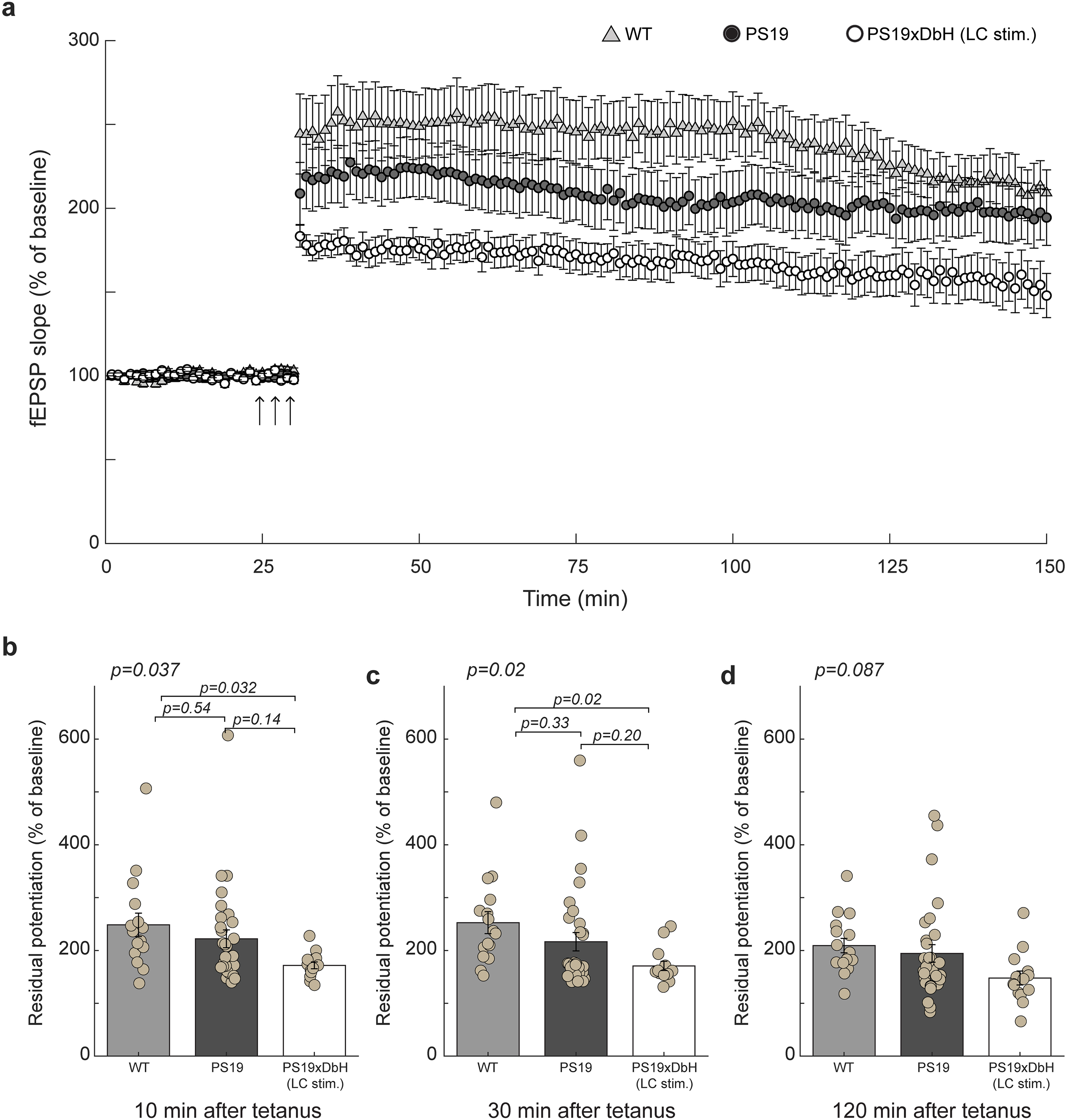
Hippocampal LTP of experimental mice. (a) Summary graph showing that long-term LC stimulation induced LTP impairment in PS19xDbH (LC stim.) mice. (b-d) Quantification of the residual potentiation at 10, 30 and 120 min after tetanus from LTP curves shown in a), respectively. (WT: n = 16 slices/7 animals; 4 males, 3 females. PS19: n = 32 slices/10 animals; 3 males, 7 females. PS19xDbH (LC stim.): n = 14 slices/6 animals; 3 males, 3 females). Statistical analyses in Figure 3 were performed using slices as the unit of analysis. A complementary analysis using individual animals as the biological replicate is provided in Supplemental Figure 1. At 10 min, one-way ANOVA: F(2, 59) = 3.48, p = 0.037; Tukey-Kramer: p = 0.032, PS19xDbH (LC stim.) versus WT. At 30 min, one-way ANOVA: F(2, 59) = 3.84, p = 0.02; Tukey-Kramer: p = 0.02, PS19xDbH (LC stim.) versus WT. At 120 min, one-way ANOVA: F(2, 59) = 2.54, p = 0.087.
Neurodegeneration and neuroinflammation increased in the hippocampus of PS19 mice undergoing chronic LC stimulation
Neuroinflammation is a key contributor to the pathogenesis of AD.38,39 Activated astrocytes and microglia can accelerate disease progression and promote neuronal loss and cognitive decline.40,41 To assess whether chronic LC stimulation increased inflammation levels in the hippocampus, we performed immunostaining for glial fibrillary acidic protein (GFAP), a marker of activated astrocytes, and Iba1, a marker of microglia (Figures 4(a) and 5(a)). PS19xDbH mice with chronic LC stimulation exhibited a significantly higher percentage of GFAP-positive area compared to both PS19 and WT controls (p = 0.021, and p = 0.045, respectively, post hoc Tukey-Kramer test; Figure 4(b)), indicating elevated astrocytic activation in the hippocampus. Compared with WT mice, PS19 mice showed a significant reduction in NeuN-positive cells (p = 0.049, post hoc Tukey–Kramer test; Figure 4(c)), indicating neuronal loss associated with tau pathology. Notably, PS19×DbH mice with chronic LC stimulation exhibited a further reduction in NeuN-positive cells compared with WT mice (p = 4.8 × 10⁻⁴, post hoc Tukey–Kramer test), and a trend toward decreased NeuN staining relative to PS19 mice (p = 0.075, post hoc Tukey–Kramer test), suggesting that prolonged LC activation may exacerbate hippocampal neuronal loss. Consistent with these results, Iba1-positive area was also significantly increased in PS19×DbH mice compared with PS19 and WT mice (p = 0.029 and p = 0.0498, respectively, post hoc Tukey–Kramer test; Figure 5(b)), suggesting enhanced microglial activation.
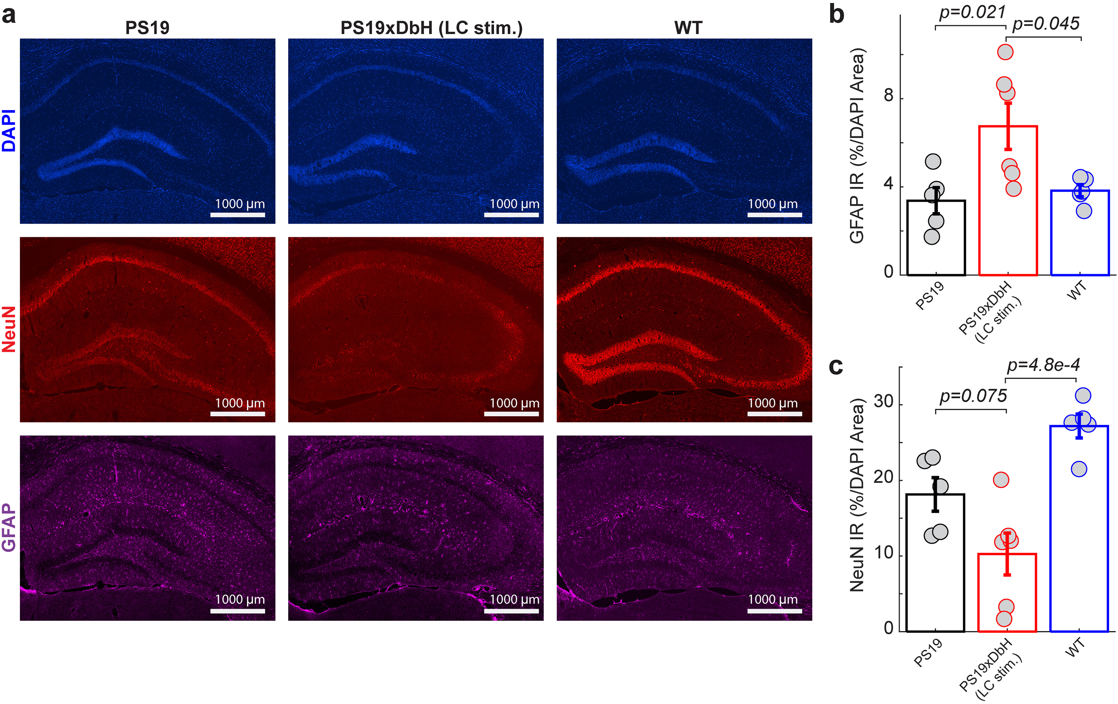
Chronic LC stimulation increased neurodegeneration and astrocyte activation in the hippocampus. (a) Example immunofluorescence images of DAPI, GFAP and NeuN signals in the hippocampus of PS19 mice (with and without LC stimulation) and WT mice. (b) Percentage of GFAP-DAPI signal overlap in the hippocampus of PS19 mice (with and without LC stimulation) and WT mice. One-way ANOVA: F(2,13) = 5.89, p = 0.015; Tukey-Kramer: p = 0.045, PS19xDbH (LC stim.) versus WT; p = 0.021 PS19xDbH (LC stim.) versus PS19. (c) Percentage of NeuN-DAPI signal overlap in the hippocampus of PS19 mice (with and without LC stimulation) and WT mice. One-way ANOVA: F(2,13) = 13.41, p = 6.9 × 10⁻⁴; Tukey-Kramer: p = 4.8 × 10⁻⁴, PS19xDbH (LC stim.) versus WT; p = 0.075, PS19xDbH (LC stim.) versus PS19. WT: n = 5; 3 males, 2 females. PS19: n = 5; 4 males, 1 female. PS19xDbH (LC stim.): n = 6; 3 males, 3 females.
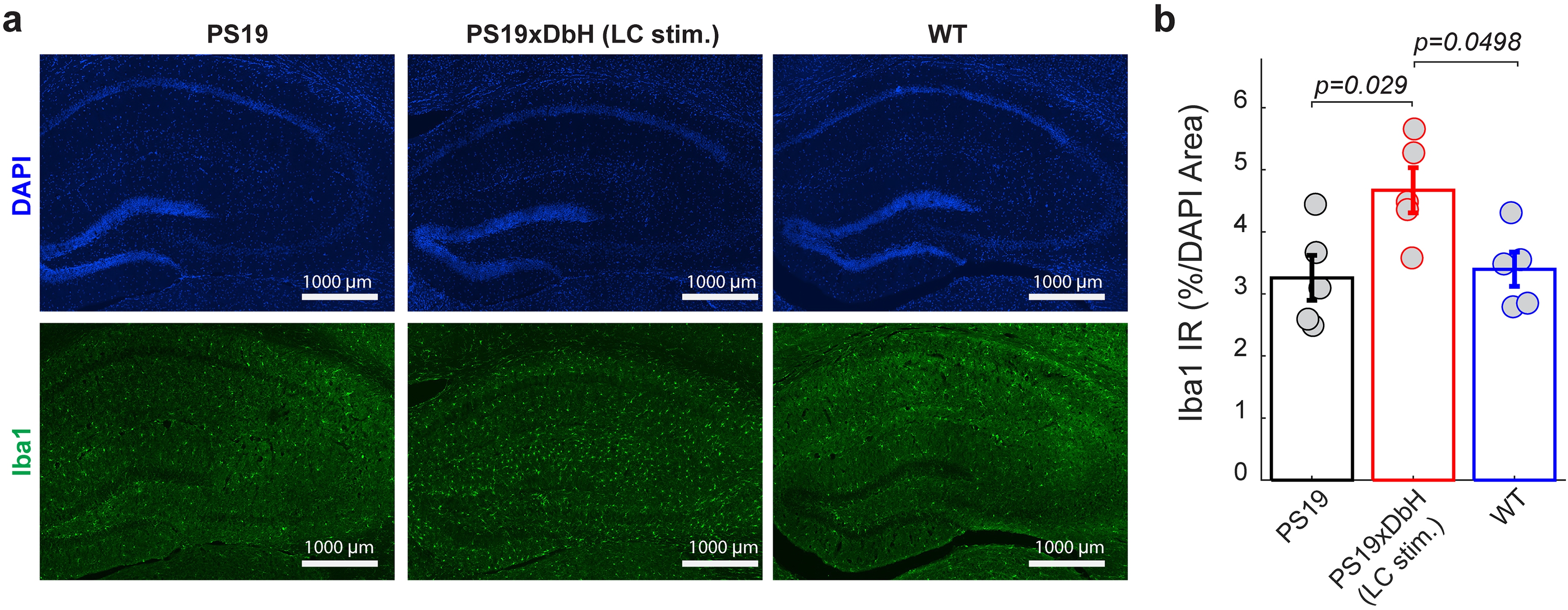
Chronic LC stimulation exacerbated microglia-mediated neuroinflammation in the hippocampus. (a) Example immunofluorescence images of DAPI and Iba1 signals in the hippocampus of PS19 mice (with or without LC stimulation) and WT mice. (b) Percentage of Iba1-DAPI signal overlap in the hippocampus of PS19 mice (with and without LC stimulation) and WT mice. One-way ANOVA among all: F(2,12) = 5.33, p = 0.022; Tukey-Kramer: p = 0.0498, PS19xDbH (LC stim.) versus WT; p = 0.029, PS19xDbH (LC stim.) versus PS19. WT: n = 5; 3 males, 2 females. PS19: n = 5; 4 males, 1 female. PS19xDbH (LC stim.): n = 5; 3 males, 2 females.
Chronic LC stimulation increased tau pathology in the LC
To assess tau pathology in the LC, we examined phosphorylated tau levels using AT8 immunostaining. AT8 recognizes tau phosphorylated at Ser202/Thr205, a well-established marker of pathological tau.42,43 Coronal brainstem sections containing the LC were imaged using confocal microscopy, and LC neurons were identified by their reactivity to DbH antibodies (Figure 6(a)). We then quantified the percentage of AT8 signal colocalized with DbH-positive neurons in a representative coronal slice for each animal. The analyzed section corresponded to the anatomical level containing the central LC region (AP −5.3 mm), where LC neuron density is highest according to the mouse brain atlas. LC-stimulated PS19xDbH mice exhibited significantly higher levels of phosphorylated tau in the LC compared to PS19 controls (p = 0.048, Student's t-test; Figure 6(b)). Representative images revealed stronger and more widespread AT8 immunoreactivity in the LC of stimulated mice. These results indicate that chronic LC stimulation aggravates tau pathology within LC neurons in PS19 mice.

Chronic LC stimulation increased tau pathology in the LC. (a) Example immunofluorescence images of DbH and phosphorylated tau (AT8) signals in the LC of PS19 mice with and without LC stimulation. (b) Percent of overlap of DbH and AT8 signals in the LC of PS19 mice with and without LC stimulation. Student's t-test, p = 0.048. PS19: n = 5; 4 males, 1 female. PS19xDbH (LC stim.): n = 6; 3 males, 3 females.
Chronic LC stimulation increased AEP expression in the LC
AEP has been shown to promote tau pathology by cleaving tau at residue N368 and facilitating its hyperphosphorylation. Prior studies have reported that the NE metabolite DOPEGAL can activate AEP, contributing to tau hyperphosphorylation and memory impairment.26,27 Therefore, the elevated tauopathy may result from an increase in AEP expression. To investigate whether chronic LC stimulation affects AEP activity, we quantified the colocalization of AEP with tyrosine hydroxylase (TH), a marker of LC neurons (Figure 7(a)). The percentage of AEP-positive signal overlapping with TH-positive cells was significantly higher in LC-stimulated PS19xDbH mice compared to both PS19 and WT controls (p = 0.036, and p = 0.044, respectively, post hoc Tukey-Kramer test; Figure 7(b)). Together, these results suggest that long-term LC stimulation upregulates AEP expression in LC neurons, potentially contributing to the exacerbation of tau pathology.
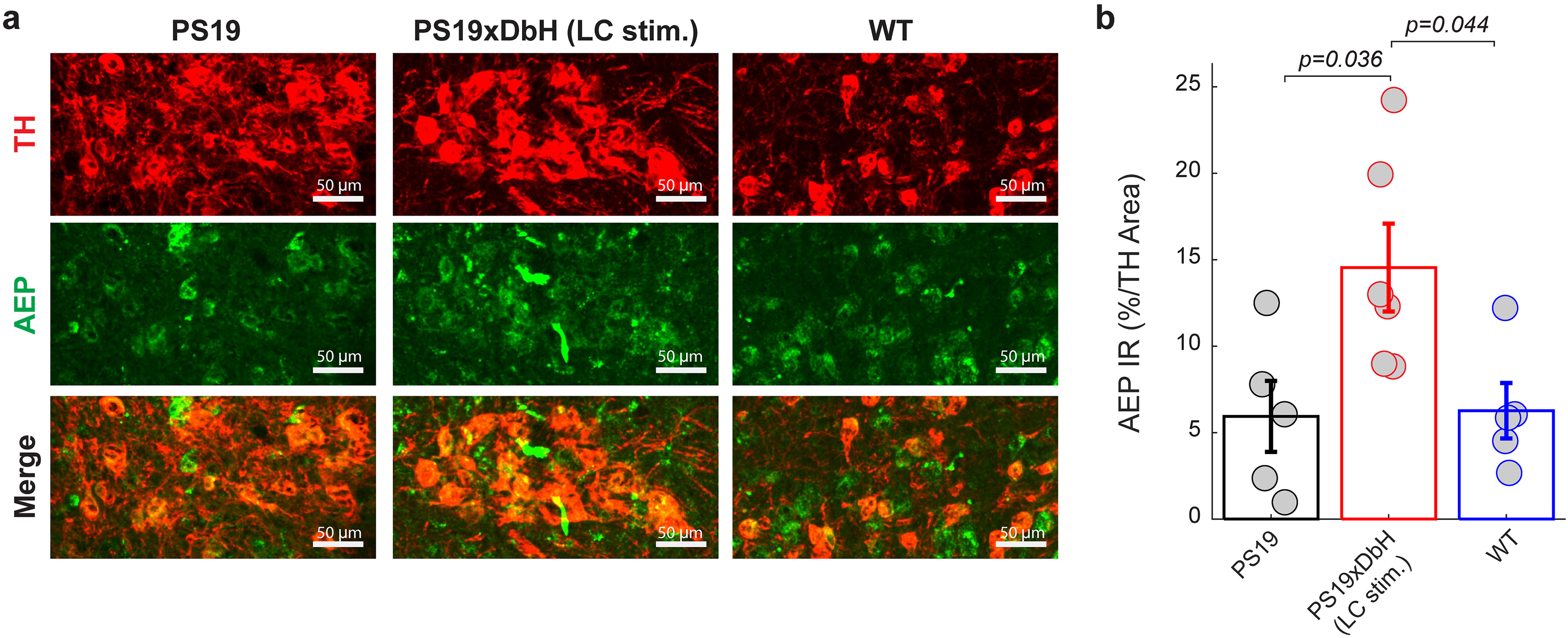
Chronic LC stimulation increased AEP expression in the LC. (a) Example immunofluorescence images of TH and AEP signals in the LC of PS19 mice (with and without LC stimulation) and WT mice. (b) Percentage of AEP-TH signal overlap in the LC of PS19 mice (with and without LC stimulation) and WT mice. One-way ANOVA: F(2,13) = 5.28, p = 0.021; Tukey-Kramer: p = 0.044, PS19xDbH (LC stim.) versus WT; p = 0.036, PS19xDbH (LC stim.) versus PS19. WT: n = 5; 3 males, 2 females. PS19: n = 5; 4 males, 1 female. PS19xDbH (LC stim.): n = 6; 3 males, 3 females.
Discussion
In this study, we investigated the long-term effects of chemogenetic activation of the LC on cognition and disease pathology in PS19 mice, a tauopathy model of AD. Our findings showed that long-term LC stimulation over a three-month period leads to worsening spatial and contextual memory performance, impaired hippocampal synaptic plasticity, and exacerbated tau pathology and neuroinflammation. These results suggest that overactivation of the LC-NE system may accelerate tau-related pathology in AD.
Previous studies have shown that short-term activation of the LC can enhance memory performance in a reversal learning task for a TgF344-AD rat model, which expresses both mutant APPsw and PS1ΔE9. 18 Although this rat model eventually develops tau pathology at older ages, it is primarily an amyloid-driven model. In this context, transient increases in NE via CNO-mediated activation of DREADD receptors expressed in LC neurons before each behavioral session may have exerted protective effects against Aβ-related toxicity, likely by inhibiting Aβ aggregation and enhancing TrkB signaling.44,45 On the other hand, long-term elevation of NE has been associated with detrimental effects in the context of tauopathy. For example, chronic treatment with the NE reuptake inhibitor reboxetine led to worsened memory deficits and neurodegeneration in tauopathy mouse models. 24 These findings align with our results, in which chronic LC stimulation in PS19 mice led to a significant decline in spatial working memory and impaired contextual fear memory. Together, these findings highlight a context-dependent role of noradrenergic signaling in AD-related pathology. Rather than being uniformly protective or detrimental, LC-NE activation may produce different outcomes depending on disease context. In amyloid-driven models, increased NE signaling may improve cognitive and synaptic function through enhanced TrkB-related neurotrophic signaling.44,46 It has also been reported that microglial Aβ phagocytosis is impaired in NE-depleted APP-transgenic mice, presumably due to reduced NE signaling, suggesting that NE may additionally counteract Aβ-related toxicity by promoting Aβ clearance via modulation of microglial function. 47 In contrast, in tauopathy, sustained LC activation may increase NE turnover and catecholamine metabolism, potentially engaging DOPEGAL/AEP-associated pathways and exacerbating tau pathology. Thus, the consequences of LC-NE activation may differ between amyloid- and tau-dominant disease contexts, emphasizing the need to tailor noradrenergic interventions to the underlying pathological state.
When interpreting the behavioral findings, it is also important to consider that chronic LC activation may alter arousal, attention, stress responsivity, or exploratory drive, which could contribute to performance deficits in RAWM and fear conditioning. The increased open-field locomotor activity on day 1 supports this possibility, although the visible platform, sensory threshold, and anxiety-related open-field results argue against simple visual-motor, sensory, or anxiety-related explanations.
The cognitive deficits observed in PS19 mice subjected to long-term LC activation were further supported by electrophysiological evidence of impaired synaptic plasticity. LTP in the hippocampus, a well-established cellular correlate of learning and memory, was significantly reduced in PS19 mice that received long-term LC stimulation as compared to their WT littermates (Figure 3(b), (c)). In the slice-level analysis, this reduction was significant at 10 and 30 min after tetanus but did not reach significance at 120 min, suggesting a stronger effect on short-term potentiation (first 10 min) and early LTP (∼first 1 h).48,49 However, additional animal-level analysis showed a significant difference at 30 min and a borderline difference at 120 min, while the 10-min difference no longer reached significance. Therefore, although our data support an overall reduction in hippocampal LTP following chronic LC stimulation, the precise temporal profile of this impairment should be interpreted cautiously. Chronic LC stimulation may have an even more deleterious impact in PS19 mice, which express the human MAPT P301S tau mutation associated with familial frontotemporal dementia. 50 Thus, our findings may reflect maladaptive NE signaling in the presence of tau pathology, which disrupts mechanisms underlying synaptic potentiation.
To investigate potential mechanisms underlying these functional impairments, we examined tau phosphorylation in the LC using AT8 immunostaining. We observed a significant increase in pTau levels in the LC neurons of stimulated PS19xDbH mice compared to PS19 controls. Since the LC is the earliest region to exhibit pTau accumulation in both humans and rodent models of AD,3,18 this result supports the notion that LC overactivity may initiate or exacerbate local tau pathology. Our data provide in vivo evidence that prolonged LC stimulation alone is sufficient to elevate pTau in this region, without requiring additional factors such as Aβ accumulation.51–54
To further investigate the potential mechanisms underlying tau pathology, we examined the expression of AEP, an enzyme that cleaves tau at residue N255/N368 and facilitates its aggregation and phosphorylation. 25 AEP has been reported to be activated by DOPEGAL, a reactive metabolite produced during NE degradation via monoamine oxidase.26,27 In our study, AEP expression was significantly elevated in LC neurons following chronic stimulation, supporting its potential involvement in LC stimulation-associated tau pathology. Although NE levels, DOPEGAL production, and AEP enzymatic activity were not directly measured in the present study, the observed increase in AEP expression is consistent with previous reports linking enhanced NE metabolism to AEP activation. Therefore, our findings support a plausible mechanistic framework in which excessive LC activity and altered NE metabolism may contribute to AEP-associated tau pathology. Further work is needed to validate the mechanistic link between LC stimulation, NE metabolism, and AEP activation, which ultimately contributes to synaptic dysfunction and cognitive decline. 26
Beyond the LC, we also examined the hippocampus for signs of neuroinflammation and neurodegeneration. We found that astrocyte activation, assessed via GFAP staining, was significantly increased in the hippocampus of LC-stimulated PS19 mice, indicating a heightened inflammatory response. Although NE is generally anti-inflammatory,47,55,56 previous studies have shown that chronic stress or NE elevation can trigger glial activation and cytokine release, 57 which in turn contribute to synaptic dysfunction and neuronal vulnerability.58,59 In addition, NeuN staining in this study revealed a significant reduction in neuronal density in the hippocampus, indicating that prolonged LC activation not only accelerates glial activation but also drives overt neuronal loss. Although future work is necessary to understand the extent to which the observed apoptosis of hippocampal neurons is due to aggregated tau forms or elevated cleaved caspase3 activity, our findings are consistent with previous reports linking sustained NE elevation to neuronal vulnerability in AD models. 24
Taken together, our findings reveal a complex role for the LC-NE system in AD. While the LC is essential for regulating various perceptual and cognitive functions, excessive or prolonged LC activation may have harmful effects in the context of tauopathy. These results highlight the importance of considering both the timing and duration of neuromodulatory interventions. Although targeting the LC remains an appealing strategy for AD treatment, our data suggest that overactivation of the LC during early disease stages may aggravate rather than improve pathology.
Several limitations of this study should be acknowledged. First, our study focused on one tauopathy mouse model (PS19) and may not generalize to other mouse models used in AD studies, particularly those with Aβ pathology. Second, we used a single dose and duration for chemogenetic LC stimulation. Daily chemogenetic activation likely produces a sustained, tonic activation pattern that differs from the endogenous firing dynamics (mixed tonic and phasic firing) of LC neurons. Physiological studies distinguish phasic LC firing, which is associated with adaptive responses and improved cognitive performance, from tonic firing. Prolonged tonic activity has often been linked to stress-like states and impaired learning and may also promote neuroinflammatory responses under chronic conditions. Thus, the chronic tonic activation induced by our stimulation paradigm may have contributed to the detrimental outcomes observed here. In addition, we did not directly validate LC activation throughout the three-month stimulation period, and prolonged DREADD activation may lead to receptor desensitization or altered neuronal responsiveness over time. Third, while our results point to AEP and pTau accumulation in LC neurons as potential factors, additional studies are needed to directly measure NE and DOPEGAL levels and to confirm their causality in this pathway. Fourth, some group sizes were relatively small, including in the fear conditioning experiment, the vehicle-treated PS19xDbH-Cre cohort, and several histological analyses. This may reduce statistical power and make borderline significant findings more difficult to interpret and should be considered when interpreting these results. Fifth, histological quantification was performed using a single anatomically matched coronal section per animal, which, although ensuring consistency across animals, may not fully capture regional variability within the hippocampus and the LC, and reproducibility across adjacent sections was not directly quantified. Sixth, although our behavioral experiments included an AAV-injected PS19xDbH-Cre vehicle group, WT and PS19 controls received saline injections rather than a matched control virus. Therefore, possible effects related to viral transduction, transgene expression, or local inflammatory responses associated with AAV delivery cannot be fully excluded. Finally, variability in viral expression of DREADDs across animals could lead to differences in the degree of LC activation following ligand administration, which may contribute to variability in the observed effects.
In conclusion, our study provides new evidence that LC overactivation worsens memory deficits, impairs hippocampal LTP, and promotes tau pathology in a tau-overexpressing AD model. These findings suggest that while the LC plays a central role in numerous brain functions, the consequences of its activation depend on both disease context and stimulation dose. Prolonged NE elevation in a tauopathy model may worsen pathology, whereas its effect in amyloid-related models may yield different outcomes (unpublished data). Future studies will be important to clarify how LC activity varies across disease stages and pathologies, and to develop treatment strategies that fine-tune LC activity to harness the beneficial effects of NE while avoiding downstream toxicity.
Supplemental Material
sj-docx-1-alz-10.1177_13872877261468577 - Supplemental material for Long-term locus coeruleus stimulation exacerbates tau pathology in PS19 mice
Supplemental material, sj-docx-1-alz-10.1177_13872877261468577 for Long-term locus coeruleus stimulation exacerbates tau pathology in PS19 mice by Yuhan Nong, Steven Wellman, Hong Zhang, Yuxiang (Andy) Liu, Elentina K. Argyrousi, Ottavio Arancio and Qi Wang in Journal of Alzheimer's Disease
Footnotes
Acknowledgements
The authors would like to thank the Zuckerman Institute Cellular Imaging Facility for its technical support with confocal imaging.

Ethical considerations
All animal experiments were approved by the Columbia University Institutional Animal Care and Use Committee (IACUC).
Consent to participate
Not applicable
Consent for publication
Not applicable
Author contribution(s)
Funding
The authors disclosed receipt of the following financial support for the research, authorship, and/or publication of this article: This work was supported by National Institutes of Health (NIH) grant numbers R01AG075114 and R01NS119813.
Declaration of conflicting interests
The authors declared the following potential conflicts of interest with respect to the research, authorship, and/or publication of this article: Dr. Qi Wang is the co-founder of Sharper Sense. Dr. Qi Wang is an Editorial Board Member of this journal but was not involved in the peer-review process of this article nor had access to any information regarding its peer-review.
Data availability statement
All data will be available from the corresponding author upon request.
Supplemental material
Supplemental material for this article is available online.
References
Supplementary Material
Please find the following supplemental material available below.
For Open Access articles published under a Creative Commons License, all supplemental material carries the same license as the article it is associated with.
For non-Open Access articles published, all supplemental material carries a non-exclusive license, and permission requests for re-use of supplemental material or any part of supplemental material shall be sent directly to the copyright owner as specified in the copyright notice associated with the article.
