



Abstract
Purpose:
Electrified wire in situ fenestration (EWISF) represents an effective off-the-shelf option in time-sensitive endovascular procedures. This article highlights the importance of radiofrequency energy delivery duration for successful catheter crossing with subsequent ballooning and bridging stent placement.
Case Report:
An 89-year-old man with symptomatic pararenal abdominal aortic aneurysm was transferred to our endovascular center. Given the patient’s advanced age and the urgent clinical scenario, an endovascular approach was preferred. Pre-stenting of the stenotic left renal artery (LRA) was followed by intentional coverage of both renal arteries using a tubular aortic endograft with suprarenal fixation (Endurant). The right renal artery was revascularized using a bare metal stent (Visi-Pro) via the parallel technique, whereas the LRA was targeted with EWISF. Intraoperatively, catheter crossing after initial EWISF proved highly challenging. A repeat EWISF procedure with prolonged radiofrequency application was required to accomplish revascularization. Distal extension with a bifurcated endograft, appropriately sized limbs, and subsequent implantation of a physician-modified thoracic endograft, to manage insufficient overlap between the previously implanted endografts, completed the operation.
Conclusion:
Electrified wire in situ fenestration is effective for endograft modification; prolonged energy delivery may be required to achieve catheter passage.
Clinical Impact
The electrified wire technique has emerged as a valuable alternative for in situ fenestration in urgent cases, enabling target vessel revascularization at low cost using readily available equipment. In a patient with a symptomatic pararenal aneurysm undergoing urgent endovascular repair, electrified wire in situ fenestration (EWISF) proved challenging. After intentional coverage of the renal artery ostium with a tubular aortic endograft, the initial EWISF attempt did not allow catheter passage through the endograft. A second attempt with prolonged electrical energy application ultimately enabled successful revascularization of the renal artery. Application duration may affect EWISF success, and acknowledgment of this parameter seems essential for enhanced technique reproducibility.
Keywords
Introduction
In situ fenestration (ISF) represents a reliable technique in complex endovascular aortic repair, particularly in urgent settings and challenging anatomies.1,2 Over the past decades, several reports have addressed the technical aspects and clinical outcomes of the 3 pivotal ISF approaches—laser fenestration, needle puncture, and the electrified wire technique—highlighting that each method carries inherent limitations.1,2 High cost and instrumental dependency mainly limit the laser technique, whereas needle puncture techniques are hampered by the high risk for graft tearing. 1
Originally developed for trans-septal puncture in interventional cardiology, electrified wire in situ fenestration (EWISF) has gained traction in the endovascular field as an affordable, precise, and widely accessible alternative. 3 An advantage of this technique is its ease of use and availability, requiring a standard surgical electrocautery device and an appropriate guidewire.2,3 Most published experience so far has focused on its use in the subclavian artery, transcatheter aortic valve replacement and puncture, as well as fenestration and septotomy of dissection membranes.2,3 In contrast, its application to visceral vessels remains limited, due to greater technical challenges. 4
The aim of this case report is to highlight the importance of energy application duration during EWISF, which may potentially increase its technical feasibility and applicability.
Case Presentation
An 89-year-old man with a known 54-mm pararenal abdominal aortic aneurysm, first diagnosed 3 months earlier, presented with persistent abdominal pain attributed to the aortic aneurysm. A new computed tomographic angiography (CTA) showed an increased abdominal aortic diameter of 57 mm with a severe left renal artery (LRA) stenosis (Figure 1A-D) and dilated iliac arteries, up to 23 mm.

Panel A: Three-dimensional aortic model based on the preoperative computed tomographic angiography (CTA) demonstrating a pararenal abdominal aortic aneurysm (PRAAA) along with iliac artery aneurysms. Panel B: Maximum aortic diameter of 57 mm. Panel C: Three-dimensional model of the renovisceral segment demonstrating high-grade stenosis of the left renal artery (LRA). Panel D: computed tomography (CT) in coronal view showing severe LRA stenosis. Panel E: Intraoperative control angiography confirming severe LRA stenosis. Panel F: Intraoperative angiography demonstrating LRA patency after pre-stenting. Panel G: Intraoperative image demonstrating main endograft and chimney stent for right renal artery.
Given the urgent scenario and the patient’s stable cardiopulmonary status, an endovascular repair was scheduled for the following days. Due to the short, conical proximal landing zone, with the right renal artery (RRA) located 18.5 mm proximal to the LRA and presenting a short main trunk (Figure 1C and D), a temporary coverage of both renal arteries was planned using an off-the-shelf infrarenal stent graft with suprarenal fixation, intended to land just below the superior mesenteric artery (SMA) followed by a parallel graft for the RRA and EWISF for the LRA.
An informed consent was obtained after explanation, including the off-label and physician-modified aspects of the procedure, as well as the urgent setting.
Procedure
The patient was operated in a hybrid operating room under general anesthesia using ultrasound-guided bilateral common femoral artery (CFA) access and systemic heparinization (target ACT 250-300 s).
Initial angiography confirmed a high-grade LRA stenosis (Figure 1E). A 12F Oscor Destiny (Oscor Inc., Palm Harbor, Florida) steerable sheath within a 16 F Gore DrySeal Flex Introducer Sheath (W. L. Gore & Associates, Flagstaff, Arizona) was used to catheterize the LRA. After pre-dilatation with 4-mm percutaneous transluminal angioplasty (PTA) balloon, a balloon-expandable covered stent (Advanta V12; Getinge, Atrium Medical Corporation, Hudson, New Hampshire) was implanted, marking the vessel for later EWISF (Figure 1F).
The RRA was catheterized via the same steerable sheath, followed by delivery of a balloon-expandable bare metal stent (Visi-Pro Peripheral Balloon-Expandable Stent System; Medtronic, Minneapolis, Minnesota). After that, a tubular stent graft with suprarenal fixation (ETTF 36/36/70; Medtronic) was introduced through the right CFA access, deployed just below the SMA and the previously placed stent in the RRA was deployed, restoring right renal perfusion (Figure 1G).
For EWISF, the steerable sheath was reoriented toward the previously stented LRA. A 0.014″ Astato XS20 wire (Asahi Intecc, Aichi, Japan) was stripped off its polytetrafluoroethylene cover at a proximal segment and advanced through a 5F Berenstein catheter. 2 An electrosurgical generator was connected to the distal external end of the wire. After confirming correct positioning in double perpendicular projections, radiofrequency energy was delivered at 40 W in cutting mode (Figure 2A), achieving graft perforation under continuous flushing of the Berenstein catheter with 5% glucose solution.
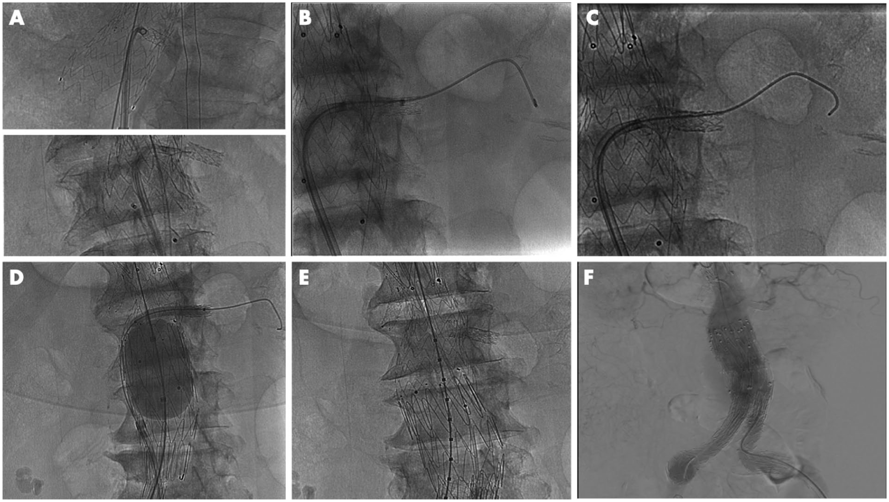
Panel A: Double perpendicular intraoperative projection checks before the in situ fenestration attempts to target the precise LRA ostium spot. Panel B: LRA cannulation with 7F sheath, demonstrating an adequately dilated fenestration. Panel C: Intraoperative image of the implanted balloon-expandable covered stent bridging the newly created fenestration to the LRA. Panel D: Intraoperative image demonstrating the LRA ballooning was performed to secure patency at the bridging point. Panel E: Intraoperative image demonstrating the inadequate overlap between the proximal tubular and distal bifurcated endograft. Panel F: Final angiography showing target vessel patency.
Despite a quick and successful endograft penetration and wire advancement into the LRA, subsequent advancement of the Berenstein catheter was not possible. Several catheters failed to cross the graft perforation site, including a 5F Quick-Cross support catheter (Spectranetics, Colorado Springs, Colorado) and PTA balloon catheters for 0.014″ wire (3 × 40 mm Amphirion; Medtronic and 2 × 20 mm Saber; Cordis, Miami Lakes, Florida). An additional 0.014″ wire was introduced through the steerable sheath, establishing a through-and-through wire to provide additional support, but crossing remained unsuccessful.
Ultimately, the Astato wire positioned in the LRA was almost completely withdrawn, and its tip was redirected toward the previously obtained fenestration. A prolonged application of radiofrequency (approximately 2 seconds at 40 W) was then delivered, while maintaining stable wire positioning at the level of the graft. Following this maneuver, successful 2-mm PTA balloon catheter passage through the fenestration was achieved, followed by PTA with residual waist resolved by inflation at 12 bar.
Using an exchange 0.035″ catheter, a Rosen Wire was then placed and over this, a 7F sheath was delivered through the fenestration (Figure 2B). Finally, the bridging stent was deployed (BeFlared 6x22 Balloon-Expandable Covered Stent; Bentley Innomed, Germany; Figure 2C). The procedure was completed by distally extending the graft with a bifurcated endograft (TFFB-36-95-ZT; Cook Medical LLC, Bloomington, Indiana), physician-modified by removing its proximal bare-stent and introduced through the right CFA. Simultaneous balloon molding of the bridging stent and graft overlap was performed (Figure 2D).
Distal extensions were added (ZSLE-24-74-ZT on the right and ZSLE-24-90-ZT on the left).
Intraoperatively, a type III endoleak was detected between the 2 aortic graft components, likely due to insufficient overlap (Figure 3E). This was corrected by relining with an additional physician-modified tube graft (Zenith TX2, ZTEG-2P-38-127-PF; Cook Medical), shortened by 2 stents. Final angiography confirmed adequate overlap, patency, and no residual endoleak (Figure 2F).
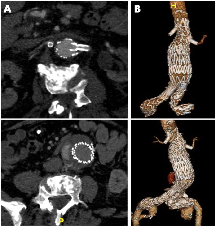
Panel A: Postoperative CTA image showing LRA patency. Panel B: Three-dimensional aortic model demonstrating successful aneurysm exclusion and target vessel patency. Panel C: Postoperative delayed-phase CTA image showing type II endoleak from a lumbar artery. Panel D: Three-dimensional aortic model demonstrating small type II endoleak and target vessel patency.
Postoperative Course
Postoperatively, the patient was monitored for 12 hours in the intensive care unit remaining hemodynamically stable. After transfer to a regular ward the following day, the postoperative CTA confirmed LRA patency and correct exclusion of the pararenal aneurysm (Figure 3A and B). Six-month follow-up CTA revealed a small type II endoleak from a lumbar artery without aneurysm diameter increase and sustained renal artery patency (Figure 3C and D).
Discussion
In the last decade, physician-modified endografts, ISF, and the use of parallel grafts have substantially expanded the spectrum of endovascular repair options, particularly in urgent and complex aortic scenarios.5-7 These techniques provide clinicians with flexible, off-the-shelf solutions that can be tailored to challenging anatomies or urgent setting when custom-made devices are not feasible or available in time.5-7 Current guidelines increasingly recognize their role, especially in cases when patient anatomy, comorbidities, or procedural timing limits conventional approaches. 8
In the present case, considering the advanced age and renal artery morphology of the patient, an endovascular multimodality approach combining EWISF and parallel grafts was selected.2,9 Severe LRA stenosis and RRA early bifurcation made traditional fenestrated or branched designs impractical in the urgent setting. Left renal artery pre-stenting and targeting through a fenestration could offer some flexibility regarding the choice of preferred strategy, but the RRA early bifurcation urged for a tailored solution to preserve both sub-branches, avoiding parenchymal loss. Although the use of balloon-expandable bare metal stent parallel graft has been limited in available literature and with controversial outcomes, it enabled complete renal preservation in this case.9-11 For the pre-stenting of the LRA, selection of the Advanta V12 instead of a bare metal balloon-expandable stent served 2 main purposes: its covered configuration, along with stable wire positioning at the level of the graft during radiofrequency application, enhanced native vessel protection against potential injury, whereas the fabric covering of both the inner and outer surfaces of the stent provided a smooth interface, facilitating subsequent catheter and wire manipulation within the stent lumen. 11
Electrified wire in situ fenestration combined with a parallel graft was preferred over on-table graft modification to avoid the technical challenges and potential risks due to the high loading force required to re-sheath a 36-mm graft into a 20F delivery system. The choice of EWISF over the more extensively studied laser-assisted alternative, although primarily driven by device availability in our case, is further supported by its use of readily available and cost-effective equipment, making it particularly attractive in urgent settings. 12 Regarding off-the-shelf branched endograft alternatives, currently available options require extensive aortic coverage that may increase the risk of spinal cord ischemia in elderly patients and urgent settings. 13 Thus, the adopted approach represented a safe and pragmatic solution in this specific setting.
The use of an Endurant graft was deliberate, given its strong scaffolding properties, short stent configuration, and subsequent suitability for a conically shaped aorta. 14 Its nitinol framework provides excellent conformability in tortuous anatomies maintaining ISF integrity and reducing the likelihood of fabric tearing, although increased resistance and recoil may complicate fenestration creation. 15 Emerging evidence suggests that cutting balloon usage may improve fenestration dimensions of Endurant grafts. 16 Moreover, the use of an Endurant endograft was based on available evidence supporting its established clinical acceptance for chimney techniques. 17 However, the lack of an appropriately sized bifurcated Endurant device necessitated the use of an aortic cuff, resulting in limited distal overlap and potential implications for long-term durability.
A critical technical observation from this procedure was the importance of sustained and controlled radiofrequency exposure during EWISF. Prolonged, controlled energy delivery was required to create a fenestration large enough for catheter passage while avoiding excessive graft injury. 15 Continuous 5% glucose solution injection during radiofrequency application and wire coverage by respective catheter was performed to protect from blood coagulation. 2 Factors potentially contributing to initial failure included possible suboptimal positioning near a stent strut (even though the Astato wire was positioned after double perpendicular projection control) and low Wattage of the applied current, especially when compared with previous evidence.2,15 Usage of a larger, 0.018″ has been previously reported and may have contributed to larger fenestration size; however, the need for larger compatible catheters could offset this advantage, rendering the overall benefit uncertain. 15
Although optimal energy settings would ideally be evaluated in vitro, variability in patient-specific impedance limits standardization. To minimize the risk of excessive mechanical damage, our approach is based on initial energy application at 40 W in pure cut mode with stepwise escalation as needed. In addition, a low-profile angioplasty catheter (compatible with a 0.014″ guidewire) may be used to facilitate initial crossing of small, newly created fenestrations, enabling progressive dilation, beyond its role in providing additional support as part of a coaxial microcatheter system. 18
Conclusion
Electrified wire in situ fenestration is a feasible and cost-effective option for endovascular repair, especially when dedicated devices are unavailable. In this case, its combination with parallel grafts provided an effective, renal-preserving solution tailored to complex anatomy. Adequate duration of energy delivery may be important for achieving effective fenestration and catheter passage.
Footnotes

Ethical Considerations
This study complied with the Declaration of Helsinki and no approval by the ethics committee was required according to the current state law. A written consent has been assigned by the patient, confirming the agreement to publication.
Funding
The authors received no financial support for the research, authorship, and/or publication of this article.
Declaration of Conflicting Interests
The authors declared the following potential conflicts of interest with respect to the research, authorship, and/or publication of this article: Tilo Kölbel is a consultant for Cook Medical and Getinge, and proctor for and has intellectual property with Cook Medical, receiving royalties, speaking fees, and research, travel, and educational grants. All authors declare no support from any organization for the submitted work; no financial relationships with any organizations that might have an interest in the submitted work; no other relationships or activities that could appear to have influenced the submitted work.