
Abstract
The bone defect repairing is still a challenge in orthopedics. As the gene engineering bones have been used in the bone repairing clinic, the scaffold construction is a critical fact to be considered. This study aims to construct optimal scaffolds using adipose tissue in the bone repair together with the gene engineering osteocytes. Rat adipose stem cells (ASC) were prepared; the cells were transduced with the OCT-4 gene carrying lentiviral vectors (OCT-4-Lv). Artificial bone defects were created in the rat femoral bone. The bone defects were filled up with adipose scaffolds and shaped by using surrounding muscles and supported with orthopedic splints. ASCs with or without transducing the OCT-4-Lv were injected into the adipose scaffolds. The rats were sacrificed 12 weeks after the surgery. After receiving the OCT-4-Lv, the expressions of OCT-4, RUNX2 and osteocalcin were detected in the ASCs. X-ray examination showed that rats received the OCT-4-Lv transduced ASCs together with the adipose pad had new bone formation in the defect area; none of the control rats showed any new bone formation in situ. The results were supported by histological assessment. Using adipose scaffold and OCT-4-modified ASC transplantation can repair bone defects.
Introduction
The segmental bone defects can be resulted from non-union fractures, surgical or traumatic injuries, bone resection for infection or tumors or femoral head necrosis. The repair of bone defects is a challenge in orthopedic clinic because the capacity of self-regeneration of bone is limited; other bone repairing materials have to be utilized. Autologous bone, allograft bone, bone xenograft or artificial bone can be used in the segmental bone defect repair. The disadvantage of using those materials is that the graft may cause immune rejection or need secondary surgery to remove the artificial materials. The own materials or allograft materials are ideal ones, but the sources of these materials are limited. 1
The bone-forming cells or osteoblasts are the most important cells in the process of bone repair. The results of bone repair are dependent on the number and quality of the bone-forming cells. 2 Stem cells can differentiate into bone-forming cells under certain microenvironment. 3 Adipose tissue is one of the sources of adult stem cells because it is easy accessible by liposuction or resection, and contains a large number of stem cells. 4 In addition, the bone marrow is also another source of stem cells. 5
The gene engineering approach has been employed to expand the osteoblast population. Using stem cells as the precursors, several genes have been transplanted into stem cells to drive these cells to become the osteoblasts. The genes include BMP2, 4, or 6, Wnt4, Shh, Osterix or Runx2. 6 The embryonic transcription factor Oct-4 is one of the well-characterized transacting elements, and is sufficient to generate pluripotent stem cells independently. 7 Oct-4 can induce the expression of STRO-1, CD146, CD29 and CD44; the cells are able to advance into the osteogenic cells culturing in osteoblast medium. 8
A scaffold is required in the stem cell-based tissue engineering used in bone repair 9 in order to form a desired shape of the new bone. However, the materials of a scaffold currently used in the clinic are usually foreign substances; these materials have the potential to induce an immune response to be rejected by the recipients. Thus, using a self-tissue serving as the scaffold can be one of the choices to avoid the rejection. In this study, we developed gene engineering osteoblasts by transducing the Oct-4 gene to adipose-derived stem cells (ASCs). In a rat model, the ASCs were transplanted into the bone defects where an adipose pad was used as a scaffold. New bone was formed in situ as we observed 12 weeks after the transplantation.
Materials and methods
Reagents
Anti-rat Oct-4 antibody was purchased from Acris Antibodies, Inc. (Shanghai, China). Anti-rat RUNX2 was purchased from Aviva Systems Biology (Beijing, China). Anti-rat osteocalcin antibody was purchased from Life Span Bioscience, Inc. (Seattle, WA). An osteogenic medium kit was purchased from Millipore China (Shanghai, China).
Preparation of ASCs
Adult Sprague Dawley rats were purchased from the experimental animal facility of our university. The experimental procedures were approved by the University Animal Care Committee. The ASCs were isolated from the abdominal subcutaneous fat of the same rats referring to published procedures. 10 Briefly, the harvested tissue was cut into small pieces (less than 3 mm3), treated with 0.1% collagenase in, and placed on a shaker at 37℃ for 20 min. The fraction of mononuclear cells was harvested and plated in tissue-culture dishes at a density of 5 × 10⁶ cells in the osteogenic medium in an atmosphere of 5% CO2/95% air at 37℃. The medium was changed every three days thereafter.
Constructing Oct-4 expression vectors
Recombinant lentiviruses (Lv) were constructed using the Lv system (Invitrogen, Shanghai, China). Production and purification of the recombinant Lv were performed as reported. Briefly, the cDNA of rat Oct-4 (Genbank no: NM_001009178.2) was subcloned into pET-32 a (+) to yield Lv-Oct-4. The resultant plasmid was transformed into Escherichia coli strain BL-21 based on the designed restriction enzyme sites. The recombinant Lv plasmid was selected on purimycin and confirmed by restriction endonuclease digestion. The recombinant Lv plasmids were transduced into HEK293 cells where they were packaged into virus particles (Lv-Oct-4). Viral titers were estimated by optical density and standard plaque assay. 1 × 10¹⁰ particles/mL Lv were prepared.
Transduction of ASCs with Lv-Oct-4
ASCs were plated at a density of 106 cells/mL. Transductions of Lv-Oct-4 at multiplicity of infection (MOI) of 25 were carried out. Polybrene was added to the culture at a concentration of 8 µg/mL. Twenty-four hours after the culture, the medium was replaced with fresh Dulbecco's modified Eagle medium with 10% fetal bovine serum.
Quantitative real-time RT-PCR
The total RNA was extracted from the ASCs using an RNeasy Mini kit. The RNA was reverse transcribed using the iScriptTMcDNA Synthesis Kit. The cDNA was then subjected to quantitative reverse transcriptase polymerase chain reaction (qRT-PCR) with SYBR Green reagent kit. The primer sets included: OCT-4: forward, gagatttgcaaagcggagac; reverse, cggttacagaaccacacacg; (NCBI: AB182322). RUNX2: forward, tctggccttccactctcagt; reverse, gactggcggggtgtaagtaa; (NCBI: AY598934). Osteocalcin (Osc): forward, gccgagaaatgttggagaaa; reverse, ctccttaatctggccaacca; (NCBI: AL446200). Expression levels were normalized to β-actin amplification levels in each sample.
Western blotting
The total proteins were extracted from the ASCs. A total of 80 µg denatured proteins was separated in 12.5% sodium dodecyl sulfate polyacrylamide gel electrophoresis gel and transferred onto nitrocellulose membrane. The membranes were then incubated with the antibodies (0.5–1 µg/mL) of OCT-4, RUNX2 and Osc, respectively, overnight at 4℃. Reactions were developed using the Pierce ECL chemiluminescence substrate kit. Results were recorded with X-ray film. The immune blots were semi-quantified by the densitometry. The intensity of the specific band was normalized to the intensity of β-actin immune blots.
Femoral bone defect model and bone defect repairing
Twenty adult Sprague Dawley (body weight 0.3 kg) were divided into four groups. Rats were anesthetized with an intramuscular injection of 3% sodium pentobarbital (1.5 mL/kg). One femoral bone was clipped and a 10 mm segmental defect was made in the femoral bone for each rat. A piece of abdominal wall adipose tissue was cut to fill up the bone defects. The surrounding muscle were sutured back the place to shape the adipose scaffold. A total of 107 ASCs (with or without transducing with OCT-4-Lv) in 0.5 mL transducing reagents were injected into four spots of the transplanted adipose tissue with 0.125 mL cells each spot. The surgical limb was fixed with the orthopedic splint to support the bone repair area and to facilitate the well-being of the experimental rats. The rats were sacrificed 12 weeks after the surgery and subjected to the following assessments.
X-ray examination
Rats were anesthetized with 3% sodium pentobarbital (1.5 mL/kg). Radiographs were taken from the operated limbs of each rat at week 0 and 12, respectively.
Histology
The osteotomy regions were excised and fixed in 4% paraformaldehyde, and then were decalcified, embedded in paraffin; sections were cut and stained with hematoxylin and eosin. The sections were observed under a light microscope.
Biomechanical test
The femoral bones were dissected and fixed with clamps. With a Universal Material Testing Machine (Zwick/Roell Z020, Germany), the maximal anti-bending strength of the femoral bone was recorded. The test motion speed was 2 mm/min. The normal femoral bones in the opposite legs were used as normal controls. The strain curves (N mm−1) were analyzed for the elastic linear deformation zone with exclusion of the plastic deformation zone. The stiffness (Young’s modulus) was calculated from the slope of the linear elastic deformation curve.
Statistics
The data were expressed as mean ± SD. Differences between groups were determined by Student’s t-test. P < 0.05 was considered significant.
Results
ASCs express OCT-4, RUNX2 and osteocalcin (Osc) after transduction of OCT-4-Lv
After the transduction of OCT-4-Lv, we evaluated the expression of OCT-4, RUNX2 and Osc in ASCs at week 1, 2, 3 and 4, respectively. As shown by qRT-PCR and Western blotting, the expression of OCT-4, RUNX2 and Osc in ASCs was increased in a time-dependent manner (Figure 1).
Induction of expression of Oct-4, RUNX2 and Osc in ASCs. ASCs were prepared and transduced with Oct-4-Lv as described in the text. The ASCs were collected at indicated time points and analyzed by qRT-PCR and Western blotting. (a) The bars indicate the mRNA levels of Oct-4, RUNX2 and Osc in ASCs. (b) The immune blots show the blots of Oct-4, RUNX2 and Osc in ASCs. The bars below the immune blots show the integrated density of the immune blots that were determined by densitometry. Con: naïve ASCs were cultured in the osteognic medium using as controls. The data represent six experiments. ASCs: adipose-derived stem cells; qRT-PCR: quantitative reverse transcriptase polymerase chain reaction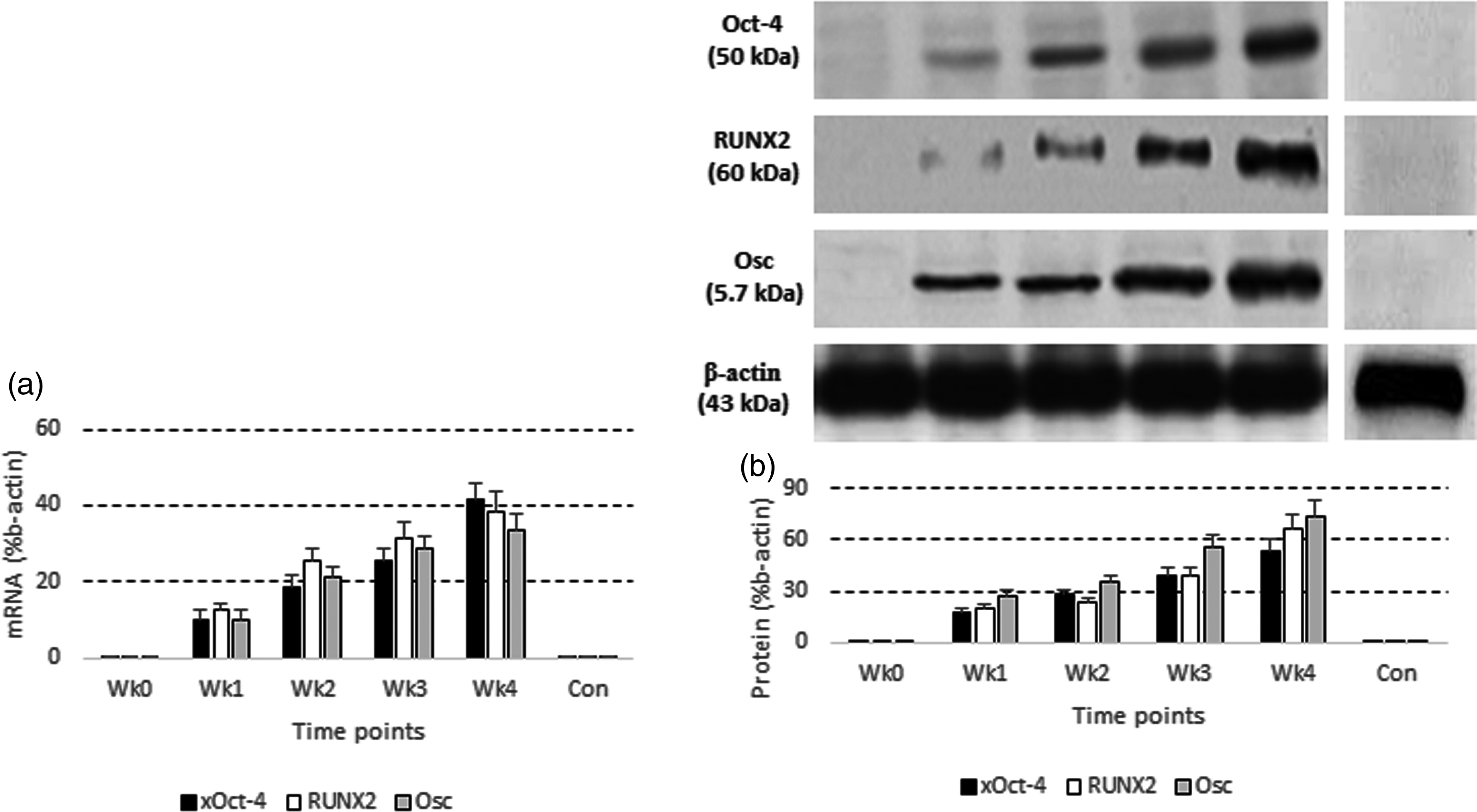
OCT-4-Lv-transduced ASCs facilitate bone repair
The femoral bone defects in the grouped rats were filled with a fat pad. The OCT-4-Lv-ASCs were injected into the fat pad. One group was injected with the OCT-4-Lv-transduced ASCs; another group was injected with naive ASCs using as a control group. Another control group was treated with OCT-4-Lv-transduced ASCs on site without the adipose pad. The femoral bone of the experimental rats was examined with X-ray in week 2, 6 and 12, respectively. The results showed that the bone defects were filled with new bone-like materials in rats treated with OCT-4-Lv-ASCs while none of the rats in the control group showed the new bone generation in the bone defects (Figure 2).
Oct-4-Lv-transduced ASCs facilitate bone repair. Rat femur bone defects (1 cm) were filled with fat pads with Oct-4-Lv-transduced ASCs or with naïve ASCs transplanting. Control group was not used either adipose pad or ASCs, or using Oct-4-Lv-ASC alone. Each group consisted of five rats. ASCs: adipose-derived stem cells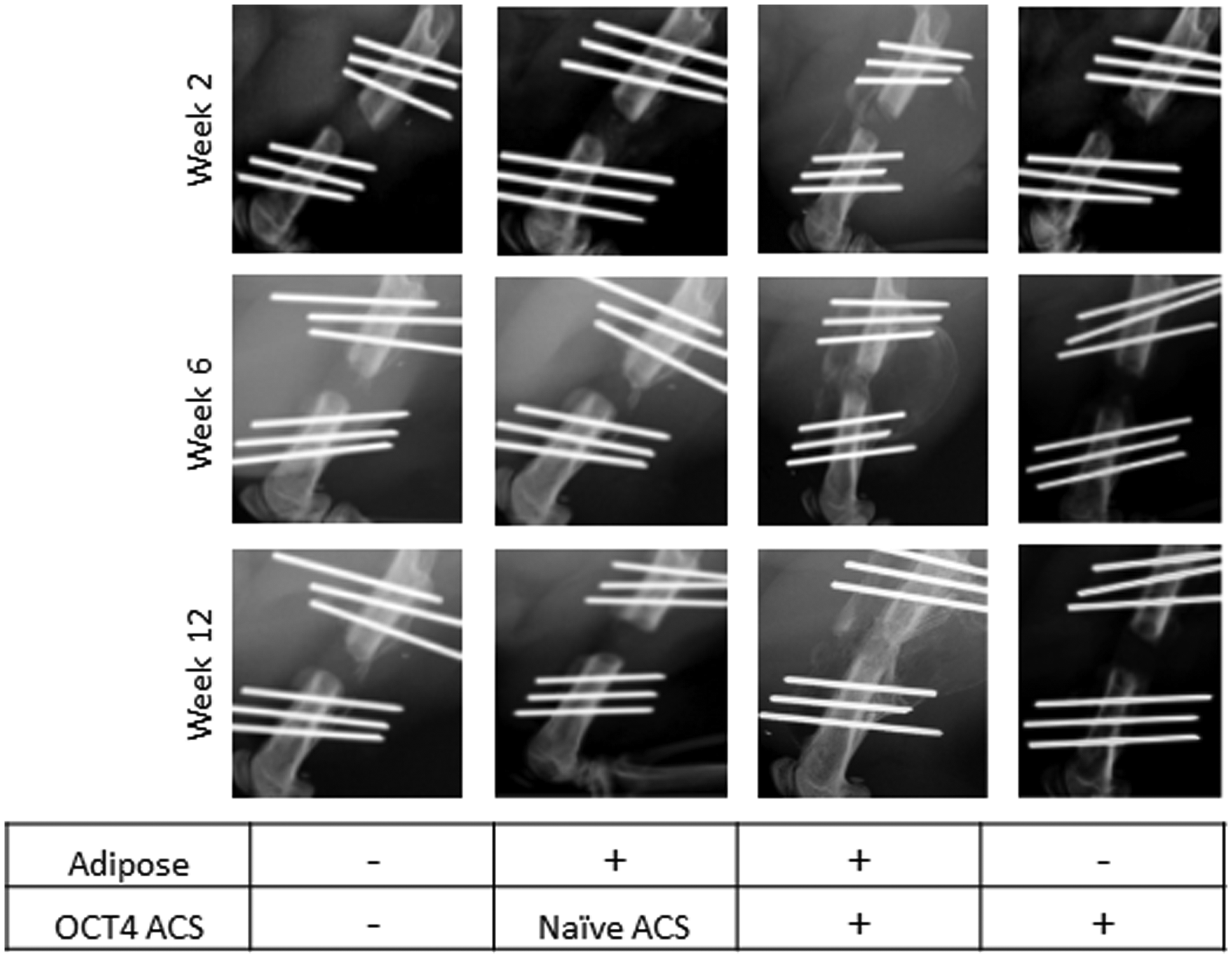
Histology shows the new bone formation in the bone defects of rats
We then collected the new bone from the rats and processed for histology observation. As shown in the histology images, bone like materials were observed in the group treated with OCT-4-Lv-transformed ASCs, which was not observed in the control groups; the bone defects were filled up in 5 out of 5 rats with fat like materials (Figure 3).
Histology of the rats femurs. The ASC transplantation part in the femoral bone defects was processed for histology observation. (a and b) The representative images show the new bone tissue (the dense red area) in the Oct-4-Lv transduced group (a). No new bone was observed in the naïve cell control group (b) or adipose +nASC group (c). Magnification: ×100. Samples from individual rat were processed separately. ASC: adipose-derived stem cell. (A color version of this figure is available in the online journal)
Biomechanical test
At 12 weeks post-surgery, the maximal anti-bending strength of the femoral bone was 168.68 ± 14.29 N in Oct-4-Lv group and 16.23 ± 3.55 N in the control group (used naïve ASCs; P < 0.01).
Discussion
To repair the bone defects, using the bone-forming cells is a good choice. Two requirements are important in bone repair; one is the development of bone-forming cells; another is to choose a suitable scaffold. In this study, we transduced ASCs with the OCT-4 gene via genetic engineering approach. The cells showed the properties of osteoblasts manifesting expressing high levels of RUNX2 and Osc. In addition, we used an adipose pad to form a scaffold in bone defects. The results showed that the adipose pad met the requirement of the scaffold, which supports the transplanted ASCs to form new bones in a designed shape in the bone defect sites; the new bone was generated within 12 weeks.
Several molecules have been recognized to expand stem cells to osteoblasts to be used in bone repair. Chen et al. reported that nanospheres of recombinant human bone morphogenetic (BMP)-2 and polylactic acid have excellent biological activity, and can promote proliferation, differentiation and mineralization of osteoblasts. 11 Other types of BMP, such as BMP-4 and BMP-7, are also involved in the development of osteogenesis. 12 OCT-4 is a transcription factor in the center of transcription network, cooperates with high mobility group (HMG) box transcription factor Sox2 and c-Myc to synergistically upregulate “stemness” genes. 13 Using genetic engineering approach, ASCs were transduced with the OCT-4 gene, which resulted in the expression of RUNX2 and Osc. Both RUNX2 and Osc are the signature molecules in the osteogenesis. RUNX2 is suggested as the activated osteoblast determinant.14,15 Osc is associated with osteogenesis. 16 Our data are in-line with previous reports by showing that the expression of OCT-4 in ASCs can expand the cells to osteoblasts.
To use bone-forming cells to repair bone defects, scaffolds are necessary. A number of different types of scaffolds have been used in the clinic and animal model studies for bone defect repairing. 17 Using biocompatible materials to repair bone defects is feasible and has its advantage, but has the potential to be rejected by the recipients. Using self-material in bone repair can avoid immune rejection; but self-materials are limited. The present study sheds new light on this area. We used self-adipose pad in the bone defect repairing. The results are very encouraging and have the potential to be used in the human bone repair.
Author contribution
WL, JF, FC, WY and JS performed the experiments, analyzed data and reviewed the manuscript. ZB designed the projects, supervised the experiments carried out and wrote the manuscript.
Footnotes
ACKNOWLEDGMENT
The study was supported by the funding from China National Natural Science Foundation (No. 81171692).