
Abstract
Epigenetic changes have been recently recognized as important in human cancers. Enhancer of zeste homologue 2 gene (EZH2) has been shown the overexpression in various human cancers, consistent with a straightforward role of EZH2 as an oncogene, but its function in carcinogenesis is partly contradictory. The role of EZH2 in development of human colorectal cancer (CRC) has not yet been clarified. In the present study, we observed up-regulation of EZH2 expression in tumor tissues from CRC patients. The expression of EZH2 in CRC cell lines is consistent with the trend in cancer tissues using reverse transcription polymerase chain reaction (RT-PCR). We showed that TNM stage and lymph node metastasis in CRC patients are significantly correlated with EZH2 expression levels. EZH2 level of transcription and protein is inhibited by small interfering RNA (siRNA). More importantly, EZH2-siRNA inhibits the proliferation and migration of SW620 cells while promoting their apoptosis, and inducing G0/G1 cell cycle arrest of SW620 cells. Collectively, our results suggest that upregulated EZH2 expression may contribute to the progression of the patients with CRC. A comprehensive study of epigenetic mechanisms and the relevance of EZH2 in CRC is important for fully understanding this disease and as a basis for developing new treatment options in patients with CRC.
Introduction
Colorectal cancer (CRC) is the third most common cause of cancer-related death in women and the fourth leading cause of cancer mortality in men. 1 Meanwhile, there is a swift growth in CRC morbidity in developing countries such as China where lifestyle has significantly changed. 2 Although the therapies of CRC such as chemotherapy and radiation have made great progress, it is regrettably still hard to cure advanced CRC, which has a poor five-year overall survival rate and a 40% overall mortality rate.3,4 Hence, identifying the molecular mechanisms during the development of CRC is important for fully understanding this disease and as a basis for developing new treatment options in patients with CRC.
It has been demonstrated that epigenetic changes including histone modifications are important in many human cancers. 5 The enhancer of zeste homologue 2 gene (EZH2) is a core component of the poly-comb repressive complex 2 (PRC2), which modifies transcription at the epigenetic level by affecting both histone and DNA methylation. 6 Recent studies have shown the overexpression of this epigenetic molecule, EZH2, in various human cancers such as breast cancer, prostate cancer, pancreatic cancer, renal cell carcinoma, bladder cancer, and lung cancer.7–12 Meanwhile, experimental evidence from a series of studies demonstrated that EZH2 can contribute to the carcinogenesis by stimulating cell proliferation, blocking apoptosis, activating tumor angiogenesis, and promoting cell invasion.13–16 Moreover, EZH2 also has an important pro-metastatic role in development of cancer by silencing of tumor and metastasis suppressor genes.17,18 However, there is also study suggesting that EZH2 acts as an effect of antitumor in certain cancers. 19 In this context, there is considerable controversy, over the function of EZH2 in CRC despite of the high biomedical significance of this cancer. 20
However, the role of EZH2 in CRC and the effects of targeting EZH2 on the biological behavior of CRC are not clear and required elucidation. To address these issues, in this study, we firstly showed the expression level of EZH2 in human CRC tissues and various CRC cell lines. Next, we evaluated the effects of transfection of SW620 cells with EZH2-small interfering RNA (EZH2-siRNA) on the expression of EZH2 mRNA and protein level. Finally, we investigated the contribution of inhibition of EZH2 expression to the biological behavior of CRC including the proliferation, apoptosis, and migration by RNA interference.
Materials and methods
Patients and specimens
Paraffin-embedded CRC tissue samples were collected at the First Affiliated Hospital of Soochow University from 2011 to 2013. There were also 42 paired non-tumor tissue samples (used as controls). These patients had not received any preoperative treatment. Tumors were staged according to the American Joint Committee on Cancer pathologic tumor-lymph node-metastasis classification. The study protocol was approved by the Ethics Committee at the First Affiliated Hospital of Soochow University.
Cell culture
All human CRC cell lines were purchased from the Chinese Academy of Sciences (Shanghai, China). These cell lines were routinely maintained as described previously 21 and used in the next experiments.
Immunohistochemical (IHC) staining and evaluation of the results
Sections were subjected to routine IHC staining as described previously.21,22 Two pathologists who were unaware of these patients’ outcome operated IHC staining independently. Evaluation of IHC results was as described previously. 21
Transfection of SW620 cells with EZH2-siRNA
EZH2-siRNA sequences were designed and synthesized by Genepharma (Shanghai, China), which included one 25-nucleotide stealth RNAi targeting EZH2, and a fluorescently labeled siRNA oligos segment which was used to detect the transfection efficacy by flow-assisted cell sorting (FACS). There are three groups in this section: (a) EZH2-siRNA1, (b) scrambled-siRNA, and (c) non-siRNA. The sequence of EZH2-siRNA was 5'-AAGACTCTGAATGCAGTTGCT-3'. In brief, SW620 cells were transfected using Lipofectamine 2000 (Invitrogen) on the basis of the manufacturer’s protocol. SW620 cells were exposed to siRNA in DMEM, and cells were continued to be incubated for two days. In the preliminary experiment, the maximal transfection efficacy was obtained when the ratio of Lipofectamine 2000 to siRNA was 4 mL:4 mL.
Reverse transcription polymerase chain reaction (RT-PCR) analysis
The mRNA expression of EZH2 in CRC cell lines and SW620 cells after EZH2-siRNA transfection was routinely quantified by RT-PCR as described previously. 21 The primer sequences of EZH2 were 50-AGGAGGACGAGGTA GATGCTTG-30 (forward) and 50-CATTGTTCCCTTG GTCGTAGTT-30 (reverse); and the primer sequences of b-actin were 50-AACTCCATCATGAAGGGTTGTGA-30 (forward) and 50-ACTCCTGCTTGCTGATCCAC-30 (reverse).
Western blot analysis
Following a 72 h transfection, protein was extracted from SW620 cells performed as previously described and then subjected to sodium dodecyl sulfate-polyacrylamide gel electrophoresis (SDS-PAGE). 22 In brief, protein concentrations were transferred onto polyvinylidene difluoride (PVDF) membrane (Merck KGaA, Darmstadt, Germany), then membranes were blocked and incubated with rabbit anti-human EZH2 (1 : 1000) antibody (1 : 1000) at 4°C overnight.
The membranes underwent hybridization with a goat anti-rabbit IgG secondary antibody (1:1000) at 37C for 1 h after three washes with TBS-T solution. Finally, we use an ECL chemiluminescence kit (Merck KGaA, Darmstadt, Germany) to detect EZH2 level.
MTT assay
SW620 cells were subjected to routine MTT assay as described previously. 24 The reaction product was quantified by measuring the optical density (OD) using test wave length for 490 nm at room temperature.
Flow cytometry analysis
Transfection efficiency was estimated with the use of fluorescein phosphoramidite (FAM)-antisense oligodeoxynucleotides by FACS, as previously described. 20 Cells were transfected with the mixture of Lipofectamine™ 2000 and FAM-NC-siRNA according to preset mixing ratio. In addition, SW620 cells were analyzed for cell cycle by FACS as described previously. 24
Cell migration assay
Cell migration assay was performed by wound-healing method. Cells (1 × 10⁵) of each group were plated in six-well plates and grown to confluence. The wound-healing method was carried out as described previously. 25 In brief, SW620 cells were wounded by scratching with a sterile pipette tip lengthwise along the chamber and were washed with PBS and cultured at 37C for 1 day. After cell wounding for 0 h and 24 h, images were captured promptly, and OpenLab software was used to detect the wound width (mm). Wound healing rate = (0 h scratch width–24 h scratch width)/0 h scratches width × 100%.
Statistical analysis
Differences were evaluated using Statistical Package for Social Science software (SPSS, Version 17.0, Chicago, IL, USA). All measurement data are presented as meanstandard deviation (SD). Statistical significance was evaluated by the Student’s t-test, and F test was used for correlation analyses. Values of P < 0.05 were considered to be statistically significant.
Results
Up-regulated expression of EZH2 in CRC tissues and CRC cell lines
To obtain insights into the role of EZH2 in colorectal tumorigenesis, we firstly investigate the expression of EZH2 in primary CRC tissues by IHC staining. In Figure 1(a), we observed that EZH2 staining was mainly presented in the nuclei of cells in tumor tissues, while there is almost no EZH2 staining observed in the paired nontumor tissues. Among 42 CRC patients, a higher percentage (78.6%) of tumor tissues expressed EZH2. However, expression of EZH2 was absent or weakly present in the paired non-tumor tissues (P < 0.01, Table 1). This indicated that EZH2 might facilitate the progression in CRC.
EZH2 Expression in CRC tissues and cell lines. A: Expression of EZH2 in tumor tissues and the paired non-tumor tissues was detected by IHC. Negative expression of EZH2 in the paired non-tumor tissues (a and b); EZH2 was up-regulated expressed in tumor tissues (c and d). Original magnification × 200 (a and c), × 400 (b and d). B: Relative expression of EZH2 mRNA in four CRC cell lines (HCT116, LoVo, SW480, and SW620) detected by RT-PCR. Results are given as average value of the gray in three target genes and internal controls from three independent experiments. (A color version of this figure is available in the online journal.) Immunohistochemical staining of EZH2 in CRC tissues CRC: colorectal cancer; –: negative; +: weakly positive; ++: positive; +++: strongly positive.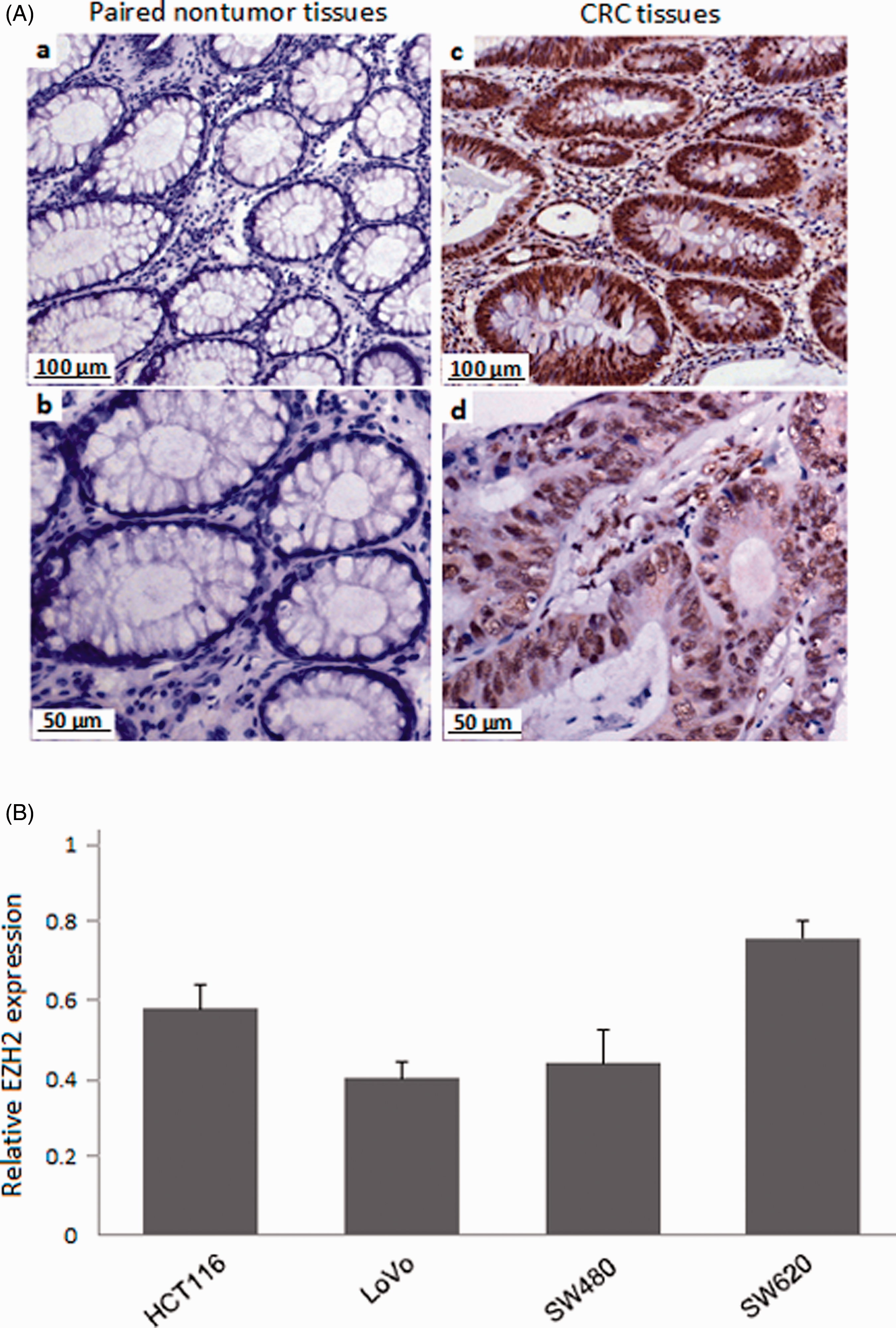

Based on the EZH2 expression in tumor tissues, we are interested to explore the biology of EZH2 in human CRC. In order to investigate the expression of EZH2 in CRC cells, we detected a panel of CRC cell lines (HCT116, LoVo, SW480, and SW620) by RT-PCR. As shown in Figure 1(b), there were relatively high expression in these cell lines and SW620 cells showed the highest elevation among them, which were used for further studies (Figure 1(b)).
Correlations between expression of EZH2 in CRC tissues and clinicopathological features
Correlation of EZH2 expression with clinicopathological features in CRC
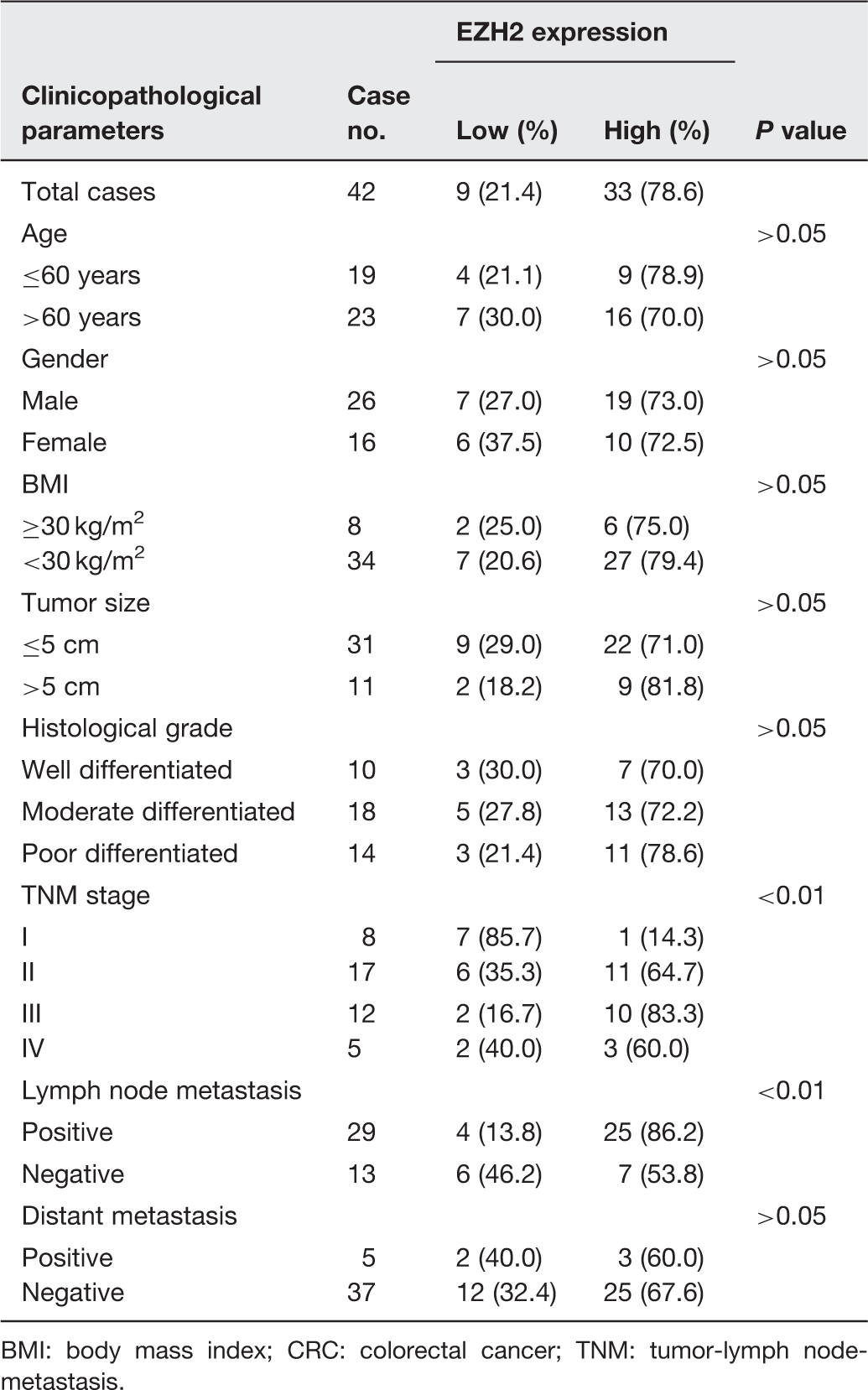
BMI: body mass index; CRC: colorectal cancer; TNM: tumor-lymph node-metastasis.
Inhibited expression of EZH2 mRNA and protein after transfection of SW620 cells with EZH2-siRNA
In order to assess the efficacy of EZH2 siRNA in downregulation of expression of EZH2, we investigated the EZH2 expression in transfected cells at level of transcript and protein levels, respectively. As shown in Figure 2(b), we observed that mRNA expression of EZH2 was significantly higher in the control groups than in the EZH2-siRNA group (P < 0.05, respectively). Likewise in Figure 2(b), EZH2 expression of protein level was remarkably higher in the control groups than in the EZH2-siRNA group (P < 0.05, respectively).
EZH2 expression level in EZH2-siRNA transfected SW620 cells. A: EZH2 mRNA expression of EZH2-siRNA transfected SW620 cells detected by RT-PCR. Forty-eight hours later of EZH2-siRNA transfection, RT-PCR was performed to detect EZH2 mRNA expression. B: EZH2 protein expression of EZH2-siRNA transfected SW620 cells detected by Western blot. Forty-eight hours later of EZH2-siRNA transfection of SW620 cells, protein expression of EZH2 was determined by western blot assay. a: EZH2-siRNA group; b: scrambled-siRNA group; c: non-siRNA group; d to f: represented corresponding internal reference. *, compared with scrambled-siRNA group and non-siRNA group, P < 0.05. The results shown were representative of three independent experiments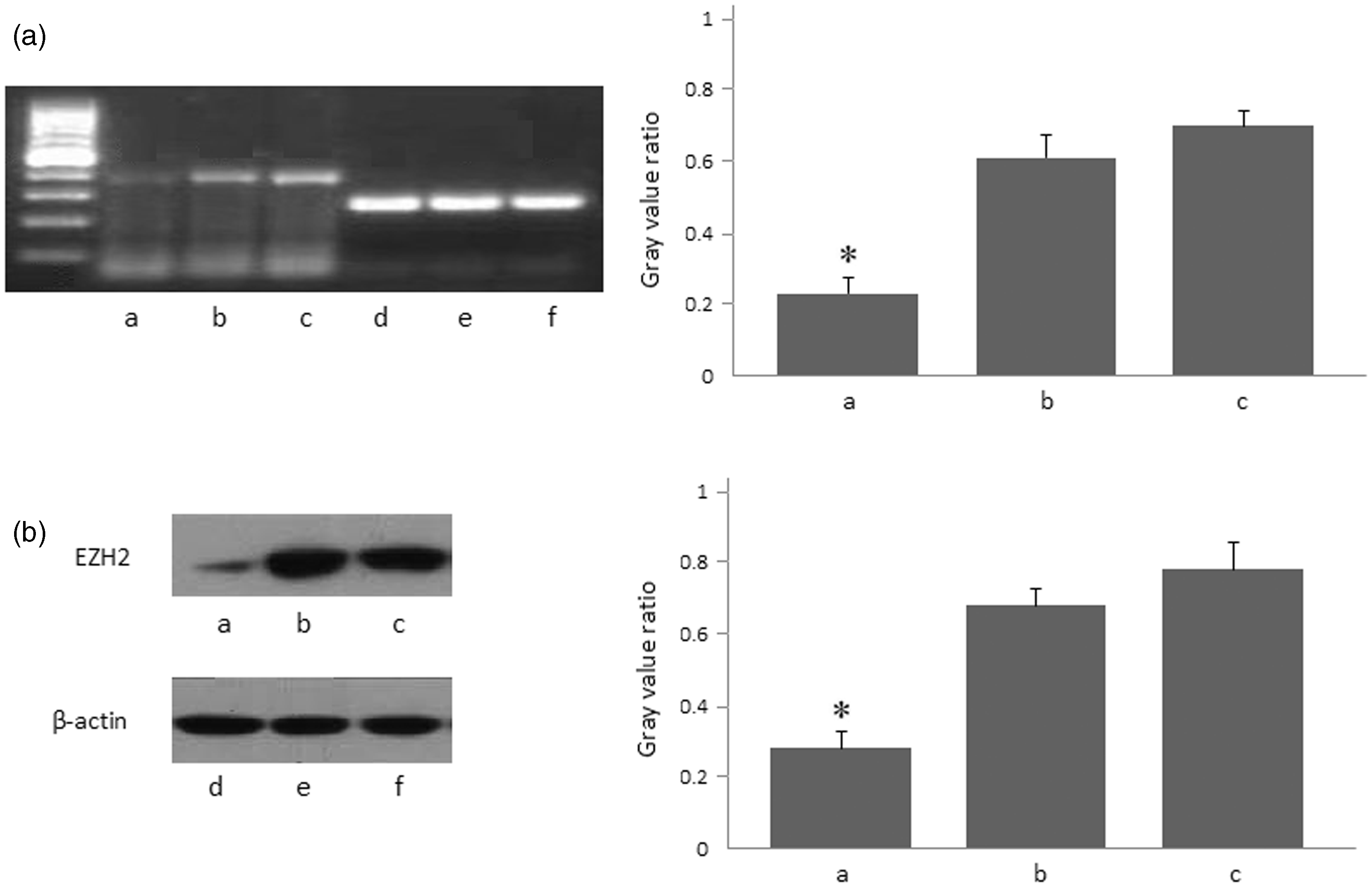
Influence of EZH2-siRNA transfection on the growth of SW620 cells
We sought to investigate its influence on cell proliferation in vitro. The results showed that the number of viable cells in the EZH2-siRNA group was markedly decreased compared to the other two groups 24, 48, and 72 h after the treatment, respectively (P < 0.05, Figure 3). The results demonstrated that RNA interference mediated specific down-regulation of EZH2 induced strong inhibition of CRC cells growth.
Effect of SW620 cells growth after transfection of EZH2-siRNA by MTT assay. Curves of SW620 cells growth after transfection of EZH2-siRNA for 24, 48, and 72 h by MTT assay. OD: optical density. *, compared with scrambled-siRNA group and non-siRNA group, P < 0.05. The results shown were representative of three independent experiments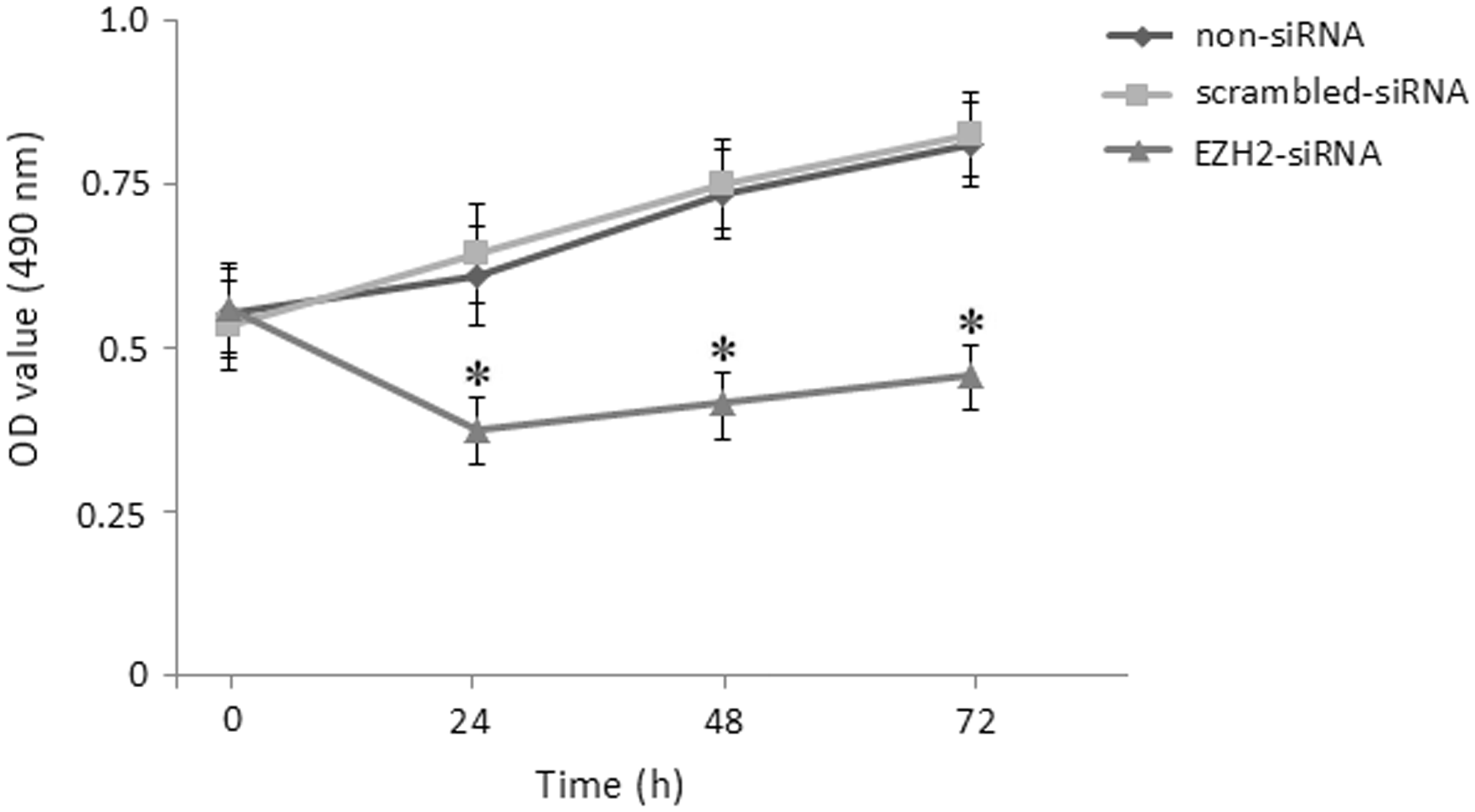
Influence of EZH2-siRNA transfection on the apoptosis and cell cycle of SW620 cells
To determine whether down-regulation of EZH2 has influence on the apoptosis and cell cycle distribution of SW620 cells, we further carried out the experiments by PI staining. The results showed that the rate of apoptosis in the EZH2-siRNA group (17.93.55)% was remarkably higher than that of the scrambled-siRNA (7.51.58)% and non-siRNA groups (5.9 ± 1.29)% (P < 0.05, respectively, Figure 4(a)). In addition, we also found that the cell cycle analysis in the EZH2-siRNA group showed increased G0/G1 phase cells (59.25 ± 4.95)% and decreased percentage of S (11.28 ± 2.78)% and G2/M (26.95 ± 4.50)% phase cells although there are no significant differences between the two control groups (Figure 4(b)).
Influence of apoptosis and cell cycle in SW620 cells after EZH2-siRNA transfection. (a) Influence of apoptotic rate in SW620 cells 48 h after EZH2-siRNA transfection determined by FACS. *, compared with scrambled-siRNA group and non-siRNA group, P < 0.05. (b) Influence of cell cycle in SW620 cells after EZH2-siRNA transfection. Forty-eight hours after transfection, cell cycle was determined by FACS. The results shown were representative of three independent experiments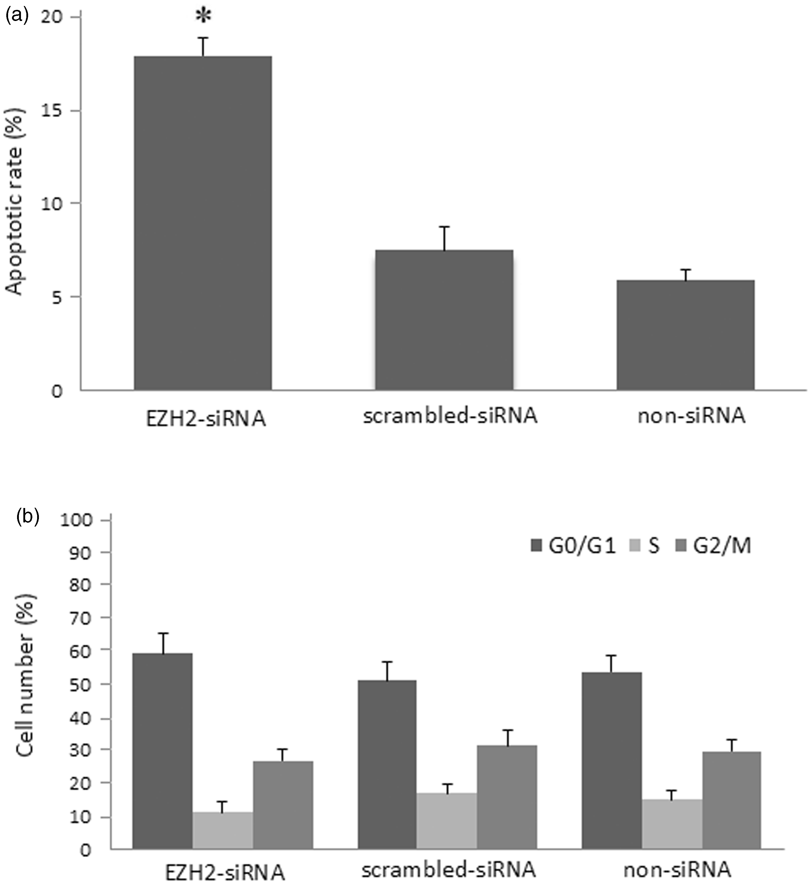
Influence of EZH2-siRNA transfection on the migration of SW620 cells
In Figure 5(a), the result showed that the wound healing rate of EZH2-siRNA transfected group was (31.66 ± 4.27)%, which was remarkably lower than that of scrambled siRNA (71.55 ± 6.25)% and non-siRNA group (73.28 ± 5.11)% (P < 0.05, Figure 5(b)). The results suggested that SW620 cells after transfection with EZH2-siRNA were sufficient to restore the migratory capability.
Influence of migration in SW620 cells after EZH2-siRNA transfection. (a) The SW620 cells were subjected to the migration assay 24 h after transfection of EZH2-siRNA. (b) The histograms represent the quantification of cells having wounding remaining width. *, compared with scrambled-siRNA group and non-siRNA group, P < 0.05. The results shown were representative of three independent experiments. (A color version of this figure is available in the online journal)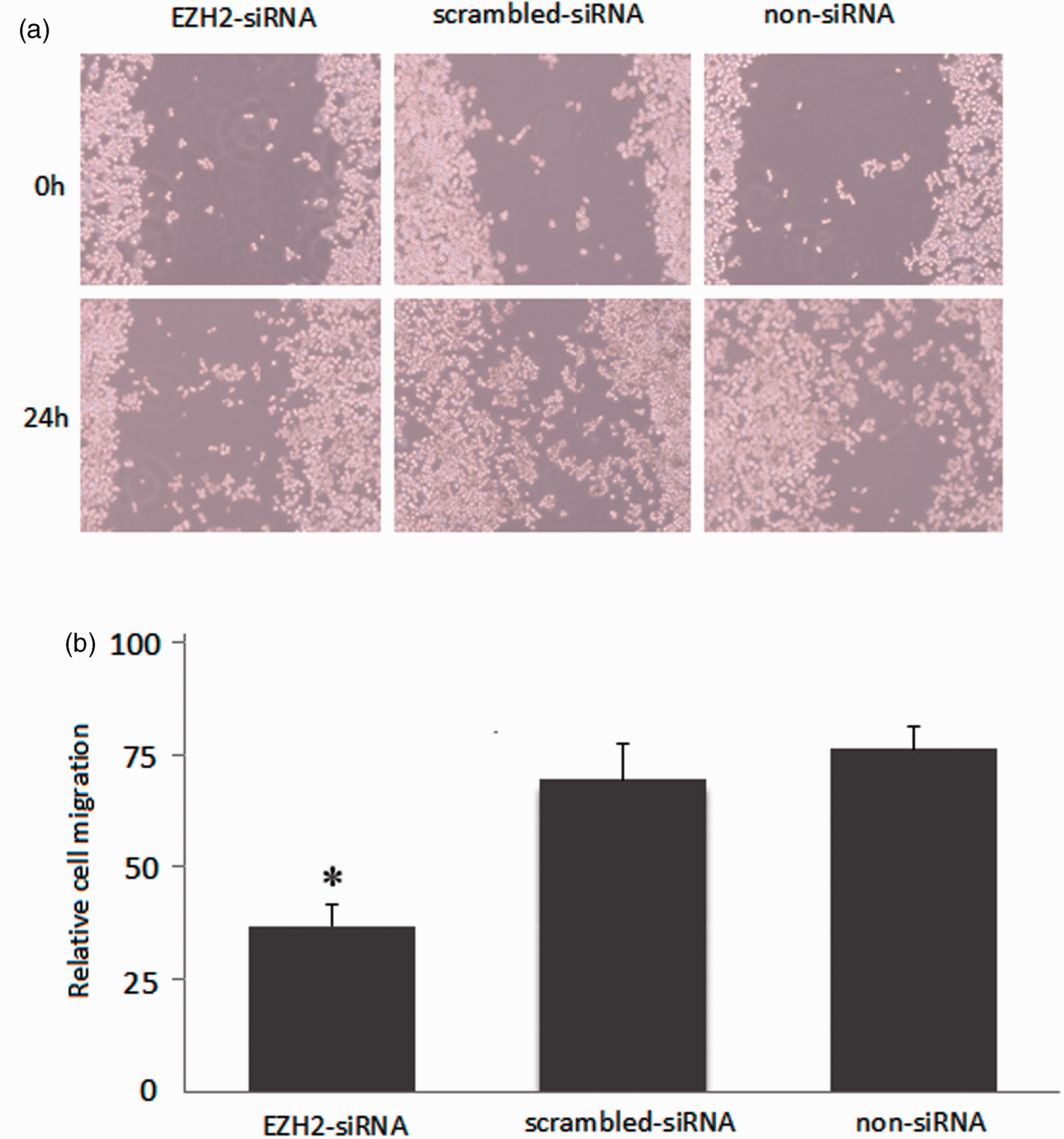
Discussion
In the present study, we have demonstrated up-regulated expression of EZH2 in CRC tissues compared with the paired non-tumor tissues, which was related with the clinicopathological significance of the patients. The expression of EZH2 in CRC cell lines was consistent with the trend in cancer tissues. We also observed that the EZH2 level of transcription and protein was remarkably reduced by RNA interference. More importantly, we explored the role of EZH2 in the biological behavior of CRC through silencing EZH2. Collectively, these data indicate a significant role for targeting EZH2 in the influences of the development and progression in CRC.
As a transcriptional repressor, EZH2 plays a critical role in the control of cell proliferation, determination of stem cell fate, and carcinogenesis.26,27 Recently, overexpressions of EZH2 in a variety of malignancies were observed in several studies.7–12 Moreover, up-regulated expression of EZH2 in cancers is related with poor prognosis or increased cancer risk from several epithelial cell derived tumors, consistent with a straightforward role of EZH2 as an oncogene.28–30 Intriguingly, Simon et al. 27 found that deregulation of EZH2 and associated genes is sufficient to induce aggressive T-acute lymphoblastic leukemia (T-ALL) in mouse and human indicating EZH2 as a tumor suppressor in T-ALL. Mutations of PRC2 and its marker, H3K27me3, were discovered recently, and they were found to have pro-oncogenic effects. 32 Moreover, no direct results were shown to address whether the abnormalities of EZH2 in CRC have a potential role in the pathogenesis of CRC.
The expression of EZH2 in CRC remains a critical first step in furthering the understanding and characterization of this molecule in CRC patients. Herein, we observed an increase in EZH2 expression in 42 CRC tumor tissues and several CRC cell lines. Moreover, up-regulated expression levels of EZH2 were remarkably associated with TNM stage and lymph node metastasis. Likewise, in prostate cancer, EZH2 overexpression is associated with aggressive and metastatic disease as well as a poor clinical outcome.8,20 A remarkable relevance has been described between PRC2 occupancy and the aberrant methylation of CpG islands at promoters seen in some cancers, although the precise contexts in which this occurs is still unclear. Therefore, our results impelled us to comprehensively study the potential function of EZH2 in the progression of CRC.
To determine the influence of EZH2 on the biological behavior of CRC, we employed the RNA interference technique was used to down-regulate EZH2 expression in SW620 cells. In the present study, our results showed that silencing EZH2 can markedly inhibit the cell proliferation in SW620 cells. Apoptosis constitutes a system for the removal of aged or damaged cells, which plays a specific role in the process of carcinogenesis. 33 Notably, FACS analysis found that the cell apoptotic rate in EZH2 inhibitor group was significantly higher than the control groups. Similarly, the data of cell cycle also revealed that the cells of S and G2/M phase in the EZH2 inhibitor group were lower than control groups, while cells of G0/G1 phase were also increased. Migration of tumor cells is one of the most important prerequisites of tumor progression and metastasis. Consistent with our hypothesis, the migration of tumor cells was inhibited in varying degrees after silencing EZH2. Moreover, it would be of great interest to reveal the mechanisms of migratory capability of the tumor cells. The therapeutic modulation of epigenetic marks has led to the improvements in treatments of some cancers, which has been proposed as a therapeutic goal and our study also provides the evidence for this concept.34,35 Targeting of down-regulated epigenetic proteins has been an attractive strategy in cancer treatment, and eradicating tumor-initiating HCC cells in nude mice and inducing apoptosis in breast cancer cells have been observed in previous studies.36,37
There are still several limitations in our present study. First, other CRC cell lines should be also investigated to support the relevance of EZH2 in CRC. Second, the regulation effect of silencing EZH2 in vivo should be further studied. Though the precise molecular mechanisms of EZH2 on apoptosis and cell death in CRC have not been clearly clarified, to the best of our knowledge, the regulation effects of malignant phenotypes by silencing EZH2 expression in CRC have not been reported to date. Finally, future studies are warranted to further elucidate the comprehensive mechanisms for the influence of EZH2 in the development of CRC, and we thought that it is very important for the forming of targeted treatment strategy in patients with CRC. It has been increasingly recognized that dysregulation of autophagy pathway is related to different types of tumor progression. 38 Autophagy has also been reported to serve a particular effect in CRC cells as autophagy can improve the aggressiveness of CRC cells and their ability to adapt to apoptotic stimuli. 39 Meanwhile, our results have shown that Ambra1, a new autophagy-related protein took a pro-survival role of the balance between autophagy and apoptosis in CRC cells in our previous study. 40 In this regard, a crosstalk between apoptosis and autophagy on interfering EZH2 could be further investigated. On the other hand, emerging evidence including our previous study demonstrated that miRNAs play significant roles in cancer progression by regulating expression of their downstream target genes including EZH2.41–43 We thought that will be interesting to explore the interrelationship between EZH2 and its target miRNA and biological relevance in the process of CRC.
In conclusion, our findings indicated that epigenetic molecule EZH2 may not only play a significant role in the progression of CRC but also serve as a potential biomarker for diagnosis and novel prognostic indicator in CRC. Moreover, inhibition of EZH2 expression has a significant influence on the proliferation, apoptosis, and migration of CRC cells. These results suggest that elevated EZH2 level might contribute to the development and progression of CRC. A comprehensive study of epigenetic mechanisms and the relevance of EZH2 in CRC are important for fully understanding this disease and as a basis for developing new treatment options in patients with CRC.
Footnotes
Author contributions
SBH and HZ designed the research; SBH, HZ, JZ, TH, DWW, and WG performed the research; SBH, GQZ, LG, YZ, XFX, LFZ, MF, SQH, XDY, XGZ, LW, and DCL contributed to the reagents/analytic tools; SBH, HZ, JZ, TH, and XGZ analyzed the data; SBH wrote the manuscript. All authors read and approved the final manuscript. SBH, HZ, JZ, and XGZ contributed equally to this work.
ACKNOWLEDGEMENTS
This work was supported by China Postdoctoral Science Foundation (2013M540374), Shanghai Postdoctoral Scientific Program of China (13R21415200), Project of Nature Science Foundation of China (81201905, 81302145), Nature Science Research Grants in University of Jiangsu Province of China (12KJB320009), Science and Technology Research Project of in Science and Technology Bureau of Suzhou City of China (SYS201220, SYS201330), Post-Graduate Scientific Research Innovation Project of Education Department of Jiangsu Province of China (CXZZ12_0842), and sponsored by Government Overseas Scholarship from Department of Education of Jiangsu Province.