
Abstract
We previously reported that cyclin D1 silencing interferes with RAD51 accumulation and increases the sensitivity of BRCA1 wild-type ovarian cancer cells to olaparib. However, the mechanisms associated with cyclin D1 overexpression in ovarian cancer are not fully understood. TargetScan predicted the potential binding sites for microRNA-20b (miR-20b) and the 3′-untranslated region of cyclin D1 mRNA; thus, we used luciferase reporter assay to verify those binding sites. The Kaplan-Meier method and log-rank test were used to examine the relationship between miR-20b and progression-free survival of ovarian cancer patients in The Cancer Genome Atlas (n = 367) dataset. In vitro experiments were performed to evaluate the effects of miR-20b on cyclin D1 expression, cell cycle and response to olaparib. A peritoneal cavity metastasis model of ovarian cancer was established to determine the effect of miR-20b on the sensitivity of olaparib. Immunohistochemistry was performed to evaluate molecular mechanisms. In this work, we demonstrated that miR-20b down-regulates cyclin D1, increases the sensitivity of ovarian cancer cells to olaparib, reduces the expression of RAD51, and induces cell cycle arrest in G0/G1 phase. Ovarian cancer patients with higher expression of miR-20b had significantly longer progression-free survival. These results indicate that miR-20b may be a potential clinical indicator for the sensitivity of ovarian cancer to olaparib and the survival of ovarian cancer patients. Our findings suggest that miR-20b may have therapeutic value in combination with olaparib treatment for ovarian cancer.
Impact statement
Although PARP inhibitors have shown significant clinical efficacy in the treatment of ovarian cancer, they are effective only in tumors with homologous recombination (HR) repair defect. Our previous study reported that cyclin D1 silencing interfered with RAD51 accumulation, enhanced the therapeutic efficacy of olaparib against BRCA1 wild-type ovarian cancer. Here, we found that miR-20b targets the cyclin D1 gene and induces G0/G1 cell cycle arrest in ovarian cancer cells. MiR-20b also interfered with RAD51 expression, thereby affecting HR efficacy (indicated by decreased γ-H2AX expression) and consequently sensitizing ovarian cancer cells to olaparib. Analysis of TCGA dataset showed that increased miR-20b expression was significantly associated with longer progression-free survival of ovarian cancer patients. This study suggests the possibility to predict the prognoses of patients with ovarian cancer using miRNAs and indicates that PARP inhibition may be useful to treat BRCA wild-type ovarian cancer when miRNA-based drug delivery systems are developed.
Introduction
Epithelial ovarian cancer has the highest mortality rate among all gynecological tumors. Although the initial response to treatment (surgery plus platinum-based chemotherapy) is usually effective, drug resistance is still a major obstacle to successful treatment. 1 Recently, the development of molecular targeted therapy based on DNA damage repair has opened a new chapter in ovarian cancer therapy. The FDA-approved poly (ADP-ribose) polymerase inhibitors (PARPi) are representative agents for DNA repair-targeted treatment and are currently used as monotherapy for advanced ovarian cancer with germline BRCA1/2 mutations. 2 Although PARPis are highly effective, they can only treat tumors with homologous recombination (HR) defects. A large number of tumors with de novo or acquired HR proficiency do not respond to PARPi, which narrows the use of these drugs.3–5 Therefore, the development of strategies to block HR repair may enhance the anti-tumor effect of PARPis and expand their application in the treatment of ovarian cancer.6–10 Our previous work demonstrated that silencing of cyclin D1 enhanced the therapeutic efficacy of the PARPi olaparib against BRCA1 wild-type ovarian cancer cells via interference with the accumulation of RAD51 and arresting the cell cycle in G0/G1 phase. In addition, overexpression of cyclin D1 reduced the sensitivity of ovarian cancer cells to olaparib. 11 However, the mechanisms associated with cyclin D1 overexpression in ovarian cancer have not been fully studied.
MicroRNAs (miRNAs) repress the expression of target genes by binding to the 3′-untranslated regions (UTR) of the messenger RNA (mRNA). Multiple studies have shown that miRNAs are highly dysregulated in cancer, indicating that miRNAs may be biomarkers for predicting treatment outcome or serve as therapeutic tools.12,13 We speculated that cyclin D1 expression in ovarian cancer may be regulated by miRNA.
Materials and methods
Prediction for miRNAs that targeting cyclin D1 in silico
TargetScan was introduced to predict the possible miRNAs that regulate CCND1 gene (cyclin D1 is encoded by CCND1gene).
Luciferase reporter assay
The 3′-UTR of CCND1 includes two predicted binding sites. Both the wild-type 3′-UTRs and the mutant 3′-UTRs (synthesized by General Biosystems (Anhui) Co., Ltd) were cloned into the pGL3-control plasmid (Promega) at XbaI/XbaI sites directly downstream from the luciferase coding sequence. For the luciferase reporter assay, cells were seeded in 96-well plates 24 h before transfection at a density of 5 × 10³ per well. Then, cancer cells were co-transfected with pRL-TK plasmid and miR-20b mimic or control mimic plus the parent luciferase expression vector, wild-type or mutant CCND1 3′-UTR plasmids using Lipofectamine 2000 (Thermo Fisher). Forty-eight hours later, the cells were subjected to lysis, and the luciferase activity was tested via the dual-luciferase assay reporter system (Promega).
Cell culture and reagents
The A2780 and SKOV3 ovarian cells and ES2 cells (BRCA1 wildtype) 14 were from Prof. Yuquan Wei. The cells were cultured according to the previous report.1,15 Olaparib was purchased from Sigma-Aldrich. MiR-20b mimic, control mimic, miR-20b inhibitor, and the control inhibitor were bought from GenePhama (Shanghai, China). Sequences of miR-20b mimic and inhibitor were as follows: NC mimic sense: 5′-UUCUCCGAACGUGUCACGUTT-3′, antisense: 5′-ACGUGACAC GUUCGGAGAATT-3′; miR-20b-5p mimic sense: 5′-CAAAGUGCUCAUAGUGCAGGUAG-3′, antisense: 5′-ACCGCACUAUGAGCACUUUGUU-3′;NC inhibitor, 5′-CAGUACUUUUGU GUAGUACAA-3′;miR-20b-5p inhibitor, 5′-CUACCUGCACUAUGAGCACUUUG-3′.
RT-PCR and Western blot analysis
The experiments were done as previously described. 11 The following specific RT-PCR primers were used: hsa-miR-20b-5p, 5′-GTCGTATCCAGTGCGTGTCGTGGAGTCGGCAATTGCACTGGATACGACCTACCTG-3′; U6, 5′-GAATTTGCGTGTCATCCTTG-3′. The following qRT-PCR primers were used: hsa-miR-20b-5p forward, 5′-GCCAAAGTGCTCATAGTGCA-3′and reverse,5′-CAGTGCGTGTCGTGGAGT-3′; U6 forward, 5′-GCTTCGGCAGCACATATACTAAAAT-3′ and reverse, 5′-CGCTTCACGAATTTGCGTGTCAT-3′; CCND1 forward, 5′- GCGGAGGAGAACAAACAGAT-3′ and reverse, 5′-GAGGGCGGATTGGAAATGA-3′; GAPDH forward, 5′-GGTGTGAACCATGAGAAGTATGA-3′ and reverse, 5′-GAGTCCT TCCACGATACCAAAG -3′.
Cell viability and cell cycle analysis
Cells incubated with miR-20b mimic, mimic control, miR-20b inhibitor or the control inhibitor were cultured overnight at 37°C. Afterwards, the cells were incubated with different concentrations of olaparib for five days, and the culture media was replaced with fresh media on the third day. MTT reagent (Sigma) was used to evaluate cell viability. Cell cycle study was carried out as previously described. 11
Colony-formation assay
SKOV3 and A2780 Cells were harvested 24 h after transfection with miR-20b mimic or control mimic. Transfected cells were seeded in six-well plates (200 cells/well). The medium was changed every three days. After about two weeks, cells were fixed in 4% PFA for 10 min and stained with crystal violet (Beyotime, China) for 10 min to count the colonies. Numbers of colonies were calculated manually and by Image J three times.
In vivo animal model
Female athymic nude mice (BALB/c, six to eight weeks old) fed in SPF-class laboratory animal room were introduced to develop the peritoneal cavity metastasis model of ovarian cancer. All the animal experiments followed the protocols of the Institutional Animal Care and Use Committee of Sichuan University. SKOV3 ovarian cancer cells were injected intraperitoneally according to the previous report. 15 One week after injection, the mice were randomly divided into four groups (n = 6): (1) control group, 100 µL of 5% glucose solution; (2) control mimic (4 µg/20 g); (3) miR-20b mimic (4 µg/20 g); (4) olaparib (50 mg/kg) + control mimic; (5) olaparib + miR-20b mimic. All groups received intra-abdominal injection of drug formulations. Mimic was given every other day and olaparib was given every day for four weeks. 16 Mice were put to death following protocols, and the tumor xenografts were collected when the treatments finished.
Immunohistochemistry study
The immunohistochemistry study was carried out following previous protocols. 11 The slices were dewaxed in xylene and dehydrated with gradient ethanol. The primary antibodies, namely anti-CCND1 (ab134175, Abcam), anti-RAD51 (ab133534, Abcam), and anti-γ-H2AX (ab26350; Abcam), were added into the slides and left overnight at 4°C. After washing with the PBS, the secondary antibody (KIHC-5, Proteintech) was added into the slides. One hour later, the signals were magnified by 3,3'-diaminobenzidine, and the nuclei were counterstained with hematoxylin.
Analysis of clinical data
The expression data of the level-3 miRNA isoform on the basis of miRNA-seq across 367 stage 2–4 ovarian cancers was obtained from The Cancer Genome Atlas (TCGA) data portal on 20 January 2019.
Statistical analysis
The experimental data was reported as the mean ± SD of three repetitions. The two-sided Student’s t test was introduced to compare results from two groups, and one-way analysis of variance method (ANOVA) was introduced to estimate results among multiple groups. *P < 0.05 was considered statistically significant. The statistical analyses were done by using SPSS 20.0 and R 2.10.0. The survival curves were analyzed using the log-rank test.
Results
CCND1 is directly targeted by miR-20b
TargetScan 7.2 indicated that the 3′-UTR of the CCND1 mRNA contains two potential miR-20b binding sites (Supplementary Figure S1). The putative binding sites and the flanking sequences are highly conserved across mammals. To investigate whether miR-20b regulates CCND1, the 3′-UTR of CCND1 mRNA was cloned into the pGL3 luciferase reporter vector. We co-transfected CCND1-3′-UTR-WT and the parent luciferase expression vector into SKOV3 cells (along with miR-20b mimic or the control miR). The co-transfection of CCND1-3′-UTR-WT and miR-20b mimic caused 76% and 77% reduction (two binding sites) in luciferase activity compared with the control miRNA (P < 0.001). These results suggested that miR-20b targets CCND1. To determine whether miR-20b regulates CCND1 via the putative binding sites, we generated a mutant construct, CCND1-3′-UTR-MT, in which the putative miR-20b binding sites in the CCND1 3′-UTR were mutated. SKOV3 cells were then transfected with either miR-20b mimic or the control miRNA along with the mutant construct. The results indicated that the deletion of miR-20b and CCND1 binding sites eliminated the influence of miR-20b on the luciferase activity. These results confirmed that miR-20b negatively regulates the CCND1 gene by binding to the 3′-UTR (Figure 1).
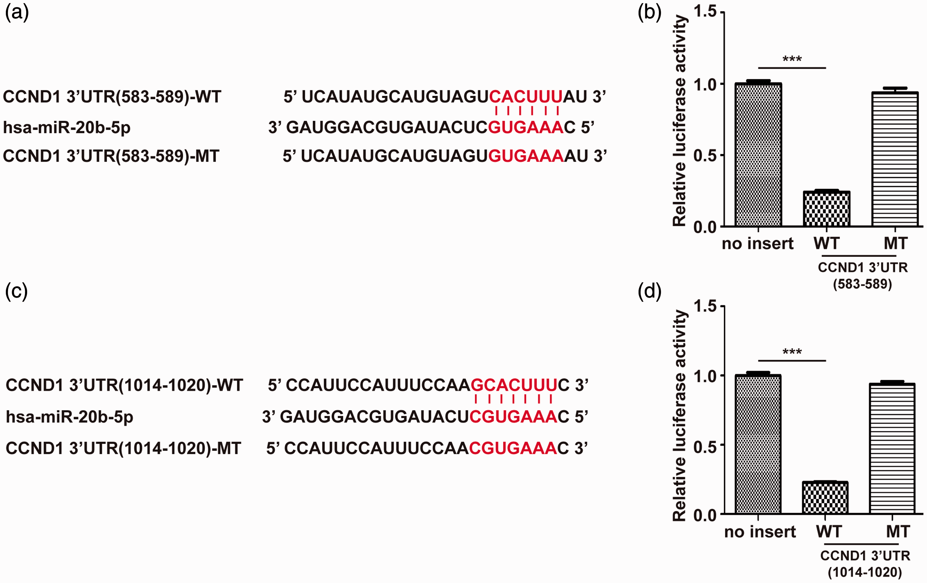
Targeting of cyclin D1 by miR-20b. (a) and (c) TargetScan predicted that two miR-20b binding sites were involved in the CCND1 3′-UTR, which are highly conserved among various species. (b) and (d) The luciferase reporter study indicated that miR-20b directly targeted the CCND1 3′-UTR. The SKOV3 cells were transfected with pRL-TK plasmid and control mimic or miR-20b mimic plus the parent luciferase expression vector, mutant or wild-type CCND1 3′-UTR plasmids. The reporter’s luciferase activity was normalized to the internal renilla luciferase activity. Results shown here were mean ± SD of triple experiments. ***P < 0.001. (A color version of this figure is available in the online journal.)
MiR-20b reduced cyclin D1 expression and induced cell cycle arrest in ovarian cancer cells
Based on the real-time qPCR results of miR-20b expression in three BRCA1 wild-type ovarian cancer cell lines (Figure 2(a)), and for consistency with our previous study, 11 SKOV3 and A2780 ovarian cancer cells were selected for experiments. Ovarian cancer cells were transfected with control mimic or miR-20b mimic. Real-time qPCR and Western blotting indicated that overexpression of miR-20b led to reduced expression of cyclin D1 mRNA and protein in the two cell lines. Conversely, cells treated with miR-20b inhibitor showed higher expression of CCND1 than cells transfected with the control inhibitor (Figure 2(b) and (c)). We further found that the miR-20b mimic induced a cell cycle arrest in G0/G1 phase, along with a reduction of S-phase cells. In contrast, the percentage of cells in G0/G1 phase was decreased after treatment with miR-20b inhibitor (Figure 3(a) and (b)). These phenomena were consistent with our previous study, in which we down-regulated or up-regulated CCND1. 11 These results suggested that miR-20b regulates the cell cycle through the regulation of CCND1. A previous study reported that olaparib-treated cells did not go through apoptosis. 17 Consistent with the previous study, the combination treatment of miR-20b mimic or inhibitor with olaparib had little effect on the number of sub-G1 cells, which suggested that the cells did not undergo apoptosis (data not shown).
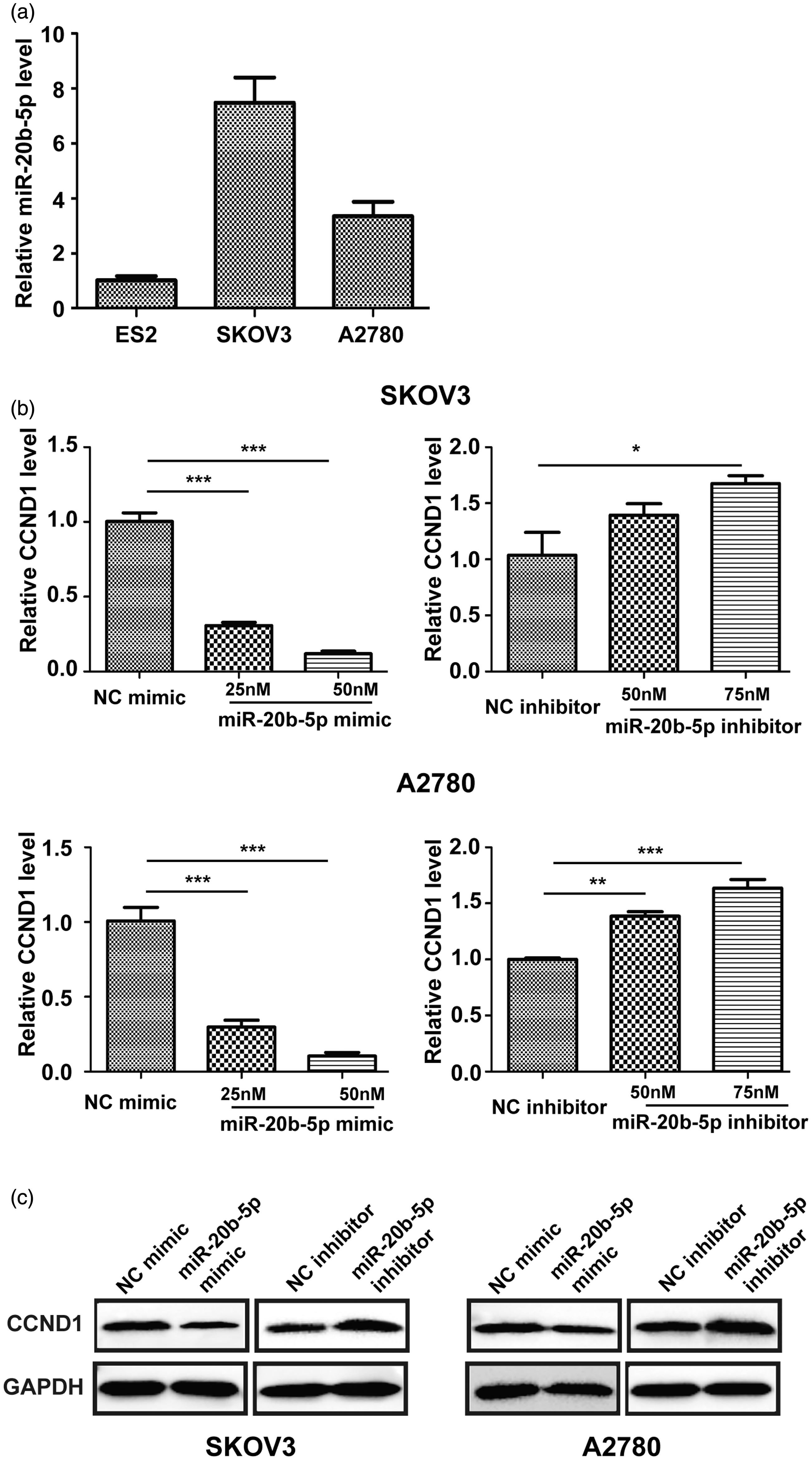
MiR-20b mimic or inhibitor treatment changed CCND1 expression. (a) Different expression level of miR-20b in three cell lines of ovarian cancer. (b) and (c) ovarian cancer A2780 and SKOV3 cells were treated with control mimic, miR-20b mimic, miR-20b inhibitor or the control inhibitor for 48 h; RT-qPCR and Western blot demonstrated the changes of CCND1 level. Results here were mean ± SD (repeated 3 times). * P< 0.05, ** P< 0.01, and *** P< 0.001.
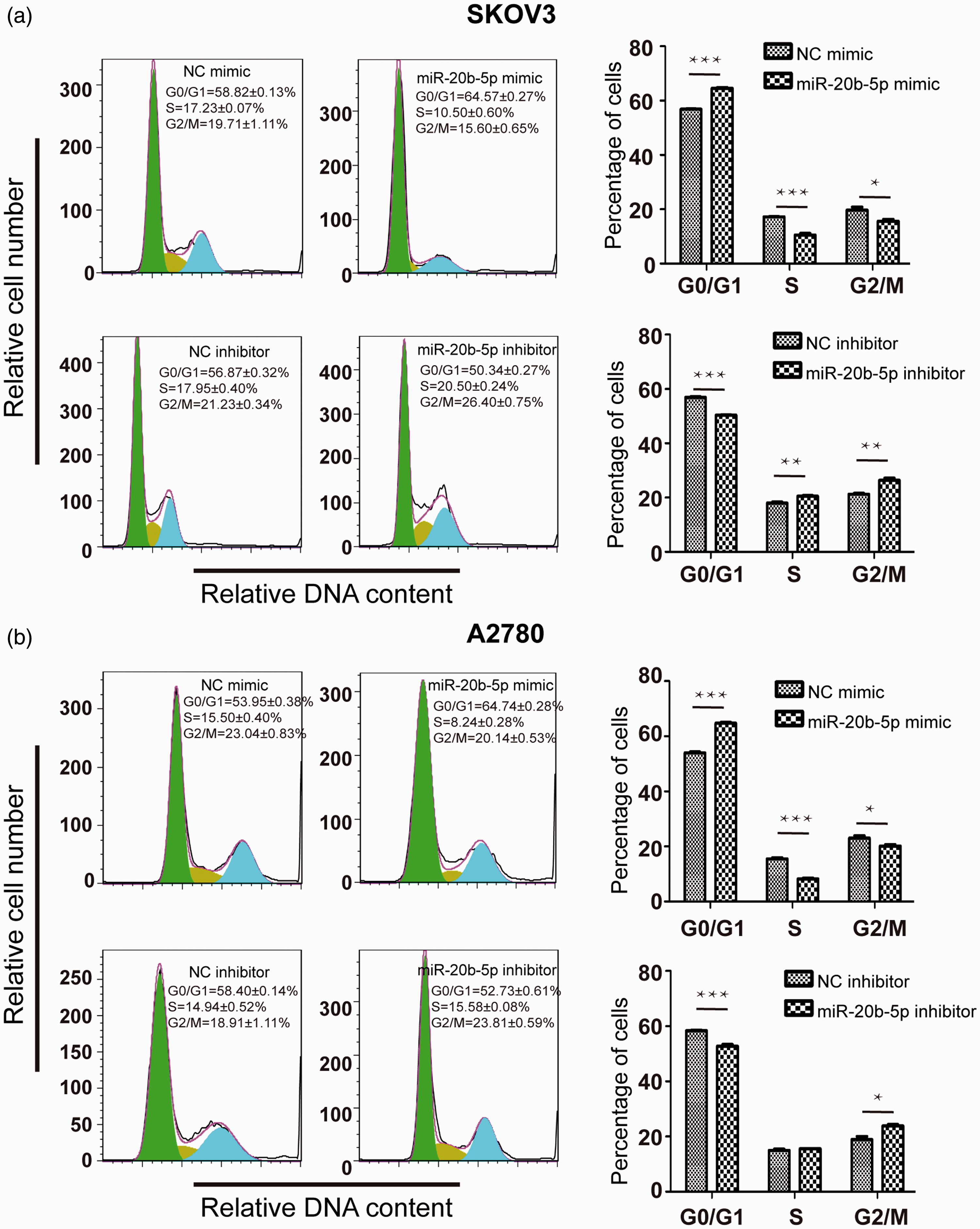
MiR-20b regulated cell cycle of the olaparib-treated cells. (a) Up-regulation or down-regulation of miR-20b by transfecting mimic or inhibitor in SKOV3 cells and 12 h later treated with olaparib at IC50 concentration (15.42 μM). Five days after treatment, cell cycle analysis was conducted by flow cytometry. (b) Up-regulation or down-regulation of miR-20b by transfecting mimic or inhibitor in A2780 cells and 12 h later incubated with olaparib at IC50 concentration (65.06 μM). Five days after treatment, cell cycle analysis was carried out by flow cytometry. Results here were mean ± SD (repeated three times). *P< 0.05, **P< 0.01, and ***P< 0.001. (A color version of this figure is available in the online journal.)
MiR-20b increased the sensitivity of ovarian cancer cells to olaparib in vitro
SKOV3 and A2780 ovarian cancer cells were treated with control mimic, miR-20b mimic, control inhibitor or miR-20b inhibitor. After 12 h, cells were incubated with olaparib at different concentrations (SKOV3 cells: 0, 4, 8, 16, 32 µM, and A2780 cells: 0, 16, 32, 64,128µM) for five days. We found that miR-20b mimic-treated SKOV3 and A2780 cells showed more pronounced response to olaparib than control mimic-treated cells. In contrast, the cells treated with miR-20b inhibitor showed resistance to olaparib compared with control cells in the two cell lines (Figure 4(a) and (b)). Upregulation of miR-20b markedly inhibited the growth ability of SKOV3 and A2780 cells, as indicated by decreased colony numbers (Supplementary Figure S2).
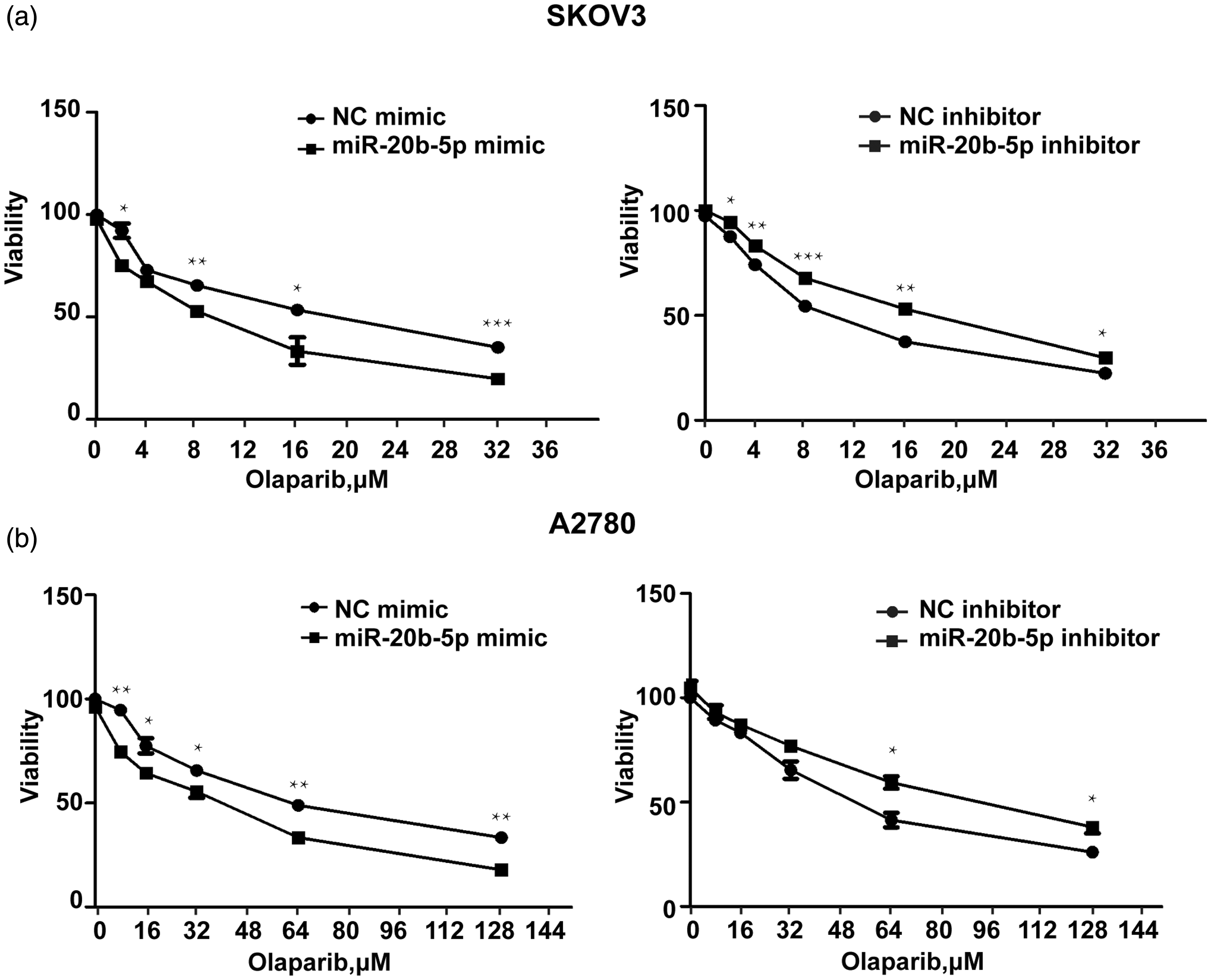
MiR-20b mimic or inhibitor treatment changed olaparib’s response in vitro. Ovarian cancer cells treated with control mimic, miR-20b mimic, miR-20b inhibitor, or the control inhibitor were put in the 37°C incubator for one night, then incubated with olaparib for five days at different concentrations. MTT assay analyzed the influence of miR-20b on cell viability of olaparib treated A2780 and SKOV3 cells. Results here were mean ± SD (repeated three times). * P< 0.05, ** P< 0.01, and *** P< 0.001.
MiR-20b increased the sensitivity of ovarian cancer cells to olaparib in vivo
To explore the impact of miR-20b on the sensitivity of ovarian cancer cells to olaparib in vivo, we studied the efficacy of a combination treatment comprising of miR-20b mimic and olaparib in mice carrying ovarian cancer peritoneal xenografts. An intraperitoneal carcinomatosis model was created in nude mice by intraperitoneal injection of SKOV3 cells. The treatment groups were as follows: (1) control; (2) control mimic; (3) miR-20b mimic; (4) control mimic + olaparib; (5) miR-20b mimic + olaparib. In comparison to mimic control-treated mice (tumor weight = 0.56 ± 0.069 g), the miR-20b mimic-treated mice (tumor weight = 0.33 ± 0.061 g) carried significantly less tumor burden (P <0.001). Although Olaparib + control mimic mice showed reduced tumor burden (tumor weight = 0.32 ± 0.029 g) compared with mimic control-treated mice, the combination of miR-20b mimic + olaparib led to significantly less tumor burden (tumor weight = 0.17 ± 0.024 g) compared with olaparib + control mimic mice (P < 0.01). The difference in tumor load between control mimic-treated mice and untreated mice (tumor weight = 0.56 ± 0.125 g) was not statistically significant (Figure 5(a) and (b)). These results showed that miR-20b may be a sensitizer of olaparib in ovarian cancer in vivo.
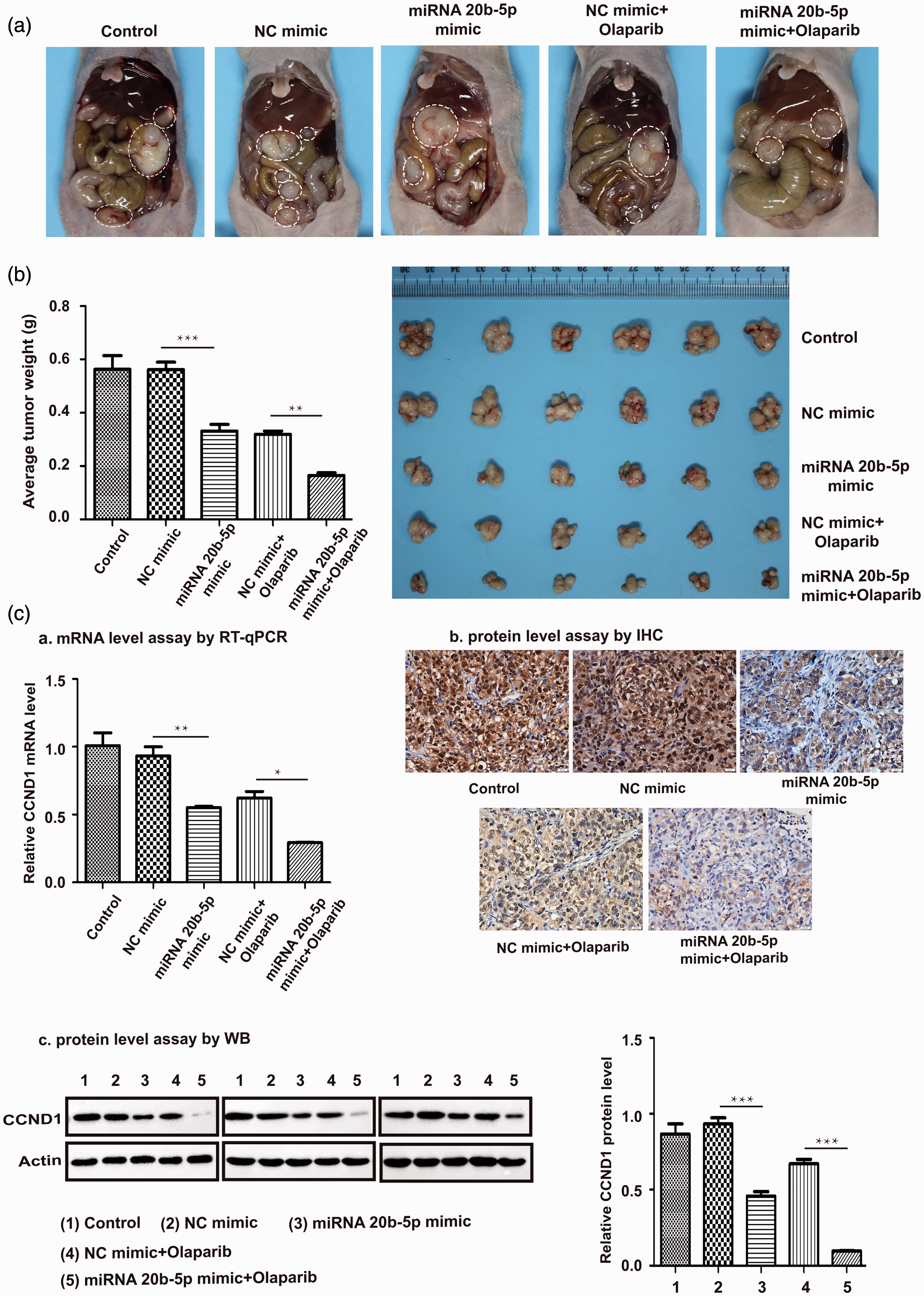
Combined treatment of miR-20b and olaparib against ovarian cancer in vivo. (a) and (b) Images of tumors inside the abdomen and tumor weight from five groups: 5% glucose solution (control), control mimic, miR-20b mimic, control mimic + olaparib and miR-20b mimic + olaparib group, respectively. (c) RT-qPCR analyzed CCND1 mRNA level in tumor tissue. Cyclin D1 protein level was shown by Western blot. Immunohistochemistry was done to verify CCND1 expression in tumor tissue, scale bar = 20 μm. Results here were mean ± SD (n=3). *P< 0.05, **P< 0.01, and ***P< 0.001. (A color version of this figure is available in the online journal.)
We next performed real-time qPCR, Western blot and immunohistochemistry studies to examine cyclin D1 expression in tumors. Consistent with the in vitro results, tumors in mice treated with miR-20b mimic showed significantly lower cyclin D1 mRNA and protein expression compared with control mimic-treated mice (P < 0.01). Cyclin D1 expression was significantly reduced by the combined treatment of miR-20b mimic + olaparib, compared with control mimic + olaparib (P < 0.05) (Figure 5(c)). γ-H2AX is a marker for DNA double-strand breaks, and RAD51 plays an important role in HR-mediated DNA repair.18–20 MiR-20b mimic-treated tumors showed reduced RAD51 expression (IHC score = 0.719 ± 0.035) and increased expression of γ-H2AX (IHC score = 1.96 ± 0.167) compared with tumors in the control mimic group (RAD51 IHC score =1.279 ± 0.246, P < 0.01; γ-H2AX IHC score = 0.920 ± 0.047, P < 0.05). RAD51 expression was significantly reduced in miR-20b mimic and olaparib co-treated tumors (IHC score = 0.307 ± 0.110) compared with tumors in the olaparib + control mimic group (IHC score = 0.837 ± 0.107) (P < 0.01). We further found that γ-H2AX expression was substantially increased in tumors from the miR-20b mimic + olaparib group (IHC score = 6.216 ± 1.201) compared with tumors in the olaparib + control mimic group (IHC score = 2.512 ± 0.286) (P < 0.001) (Figure 6(a) and (b)). These data suggested that miR-20b overexpression down-regulates CCND1 in ovarian cancer in vivo, thereby affecting DNA repair, which ultimately causes increased sensitivity to olaparib.
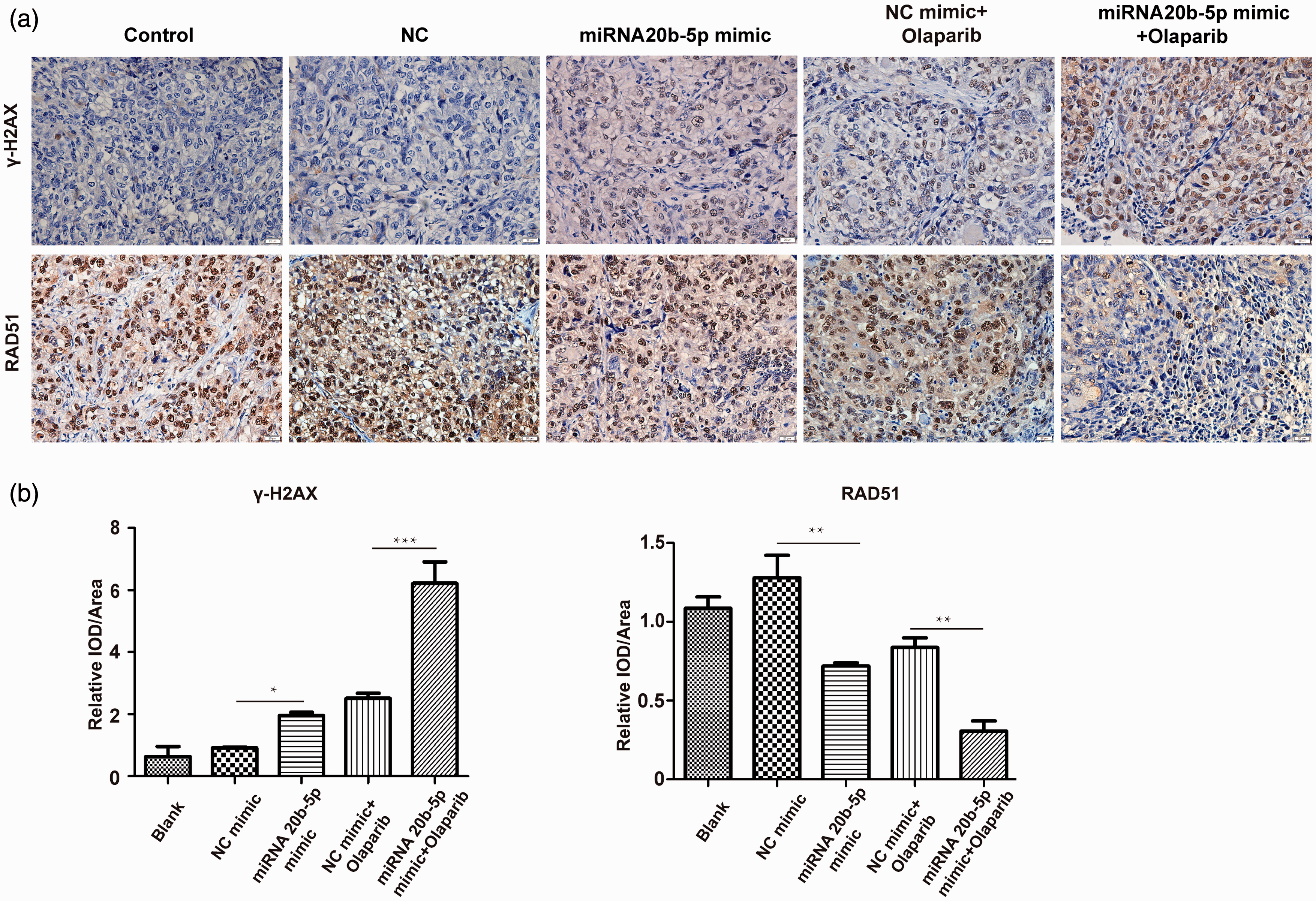
RAD51 and γ-H2AX level inside tumor tissue. (a) The typical sections from tumor tissue in various groups. (b) RAD51 and γ-H2AX expression were demonstrated by immunohistochemical scores (0–9). For instance, the scores were equivalent to the chroma of the signal(negative=0; light yellow=1; light brown=2; dark brown=3) ×percentage of positive cells (no signal=0; weak signal=0∼25%; intermediate signal=25∼50%; strong signal=above 50%). Scale bar: 20 μm. Results here were mean ± SD (n = 3). ***P< 0.001, **P< 0.01, and *P< 0.05. (A color version of this figure is available in the online journal.)
miR-20b expression and progression-free survival in ovarian cancer
We next investigated the relationship between miR-20b expression and survival of ovarian cancer patients by analyzing the TCGA dataset, which contains 367 high-grade serous ovarian cancer samples. 21 Increased miR-20b expression was significantly associated with longer progression-free survival in ovarian cancer (P = 0.027) (Figure 7).
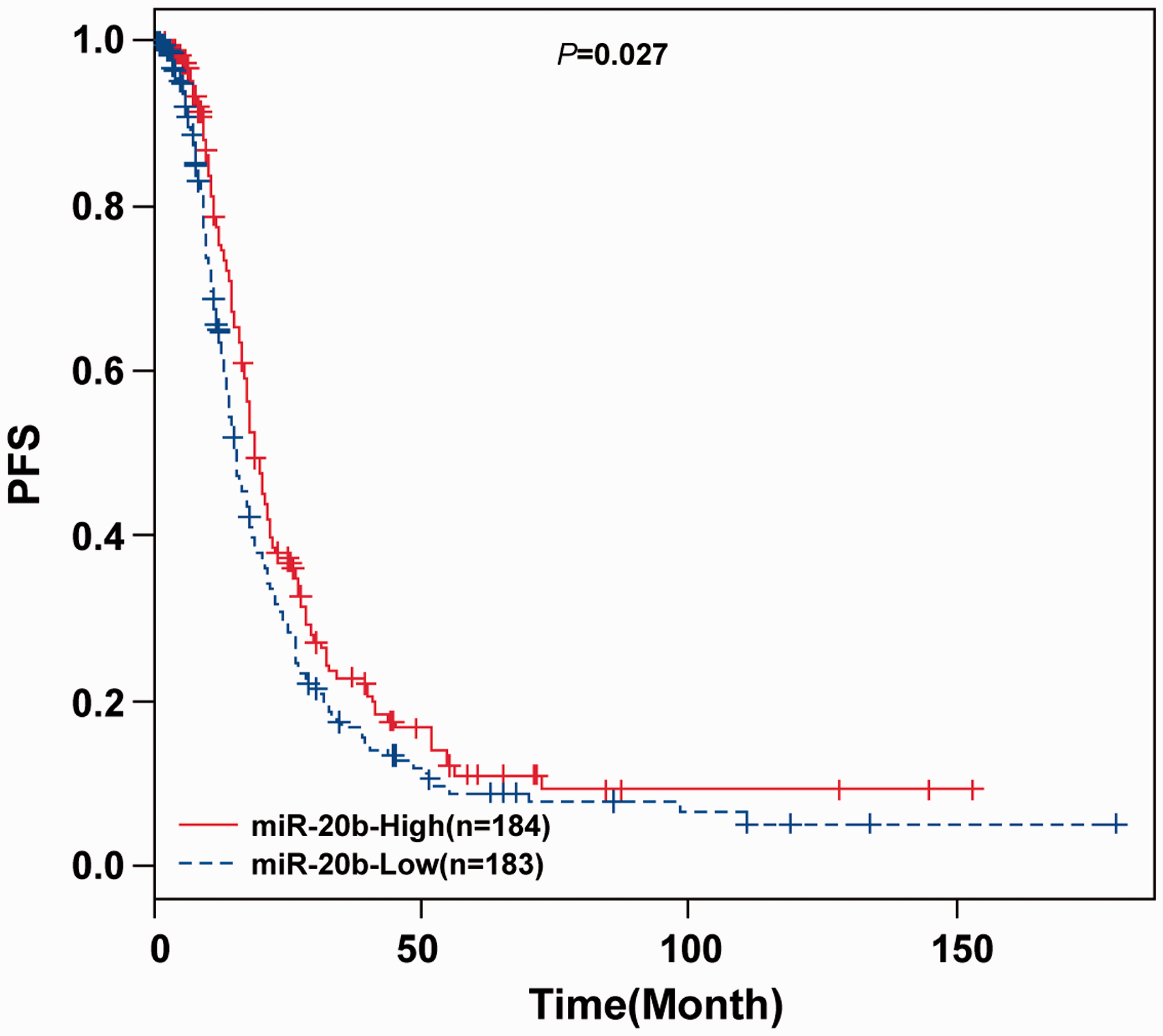
MiR-20b expression and ovarian cancer progression-free survival. Progression-free survival of 367 ovarian cancer patients in TCGA dataset with high miR-20b expression (red line) and low miR-20b expression (blue line) was analyzed by Kaplan-Meier survival curve. (A color version of this figure is available in the online journal.)
Discussion
Ovarian cancer carrying methylated or mutated BRCA1 or mutated BRCA2, account for 30% of all ovarian cancers, exhibit abnormal HR repair, are sensitive to PARP inhibitors, and show increased sensitivity to DNA-damaging anticancer agents. 22 Cyclin D1 is overexpressed in many cancers, 23 including ovarian cancer. Previous studies showed that cyclin D1 is a vital prognostic indicator for ovarian cancer. 24 Cyclin D1 plays a vital function in regulating the cell cycle transition from G1 phase to S phase. Another study showed that cyclin D1 plays a non-kinase-dependent role in DNA repair and affects HR repair through interacting with RAD51. 25 Our previous work demonstrated that downregulation of cyclin D1 expression augmented the response of BRCA1 wild-type ovarian cancer to olaparib by impairing DNA double-strand break repair. 11 However, the mechanisms underlying cyclin D1 overexpression in ovarian cancer have not been completely investigated. miRNAs are crucial for the expressions of many mRNAs. 26 Abnormal expression of miRNAs is often found in cancer. 27 Several miRNAs play an important role in regulating DNA repair and the susceptibility of cells to DNA-damaging drugs.26,28–30 The relationship between miRNAs and ovarian cancer has recently been investigated,13,31,32 and these studies have suggested that miRNAs may represent possible targets or strategies for prevention, early detection, and treatment of the disease. In this study, we performed a series of in vitro experiments and showed that miR-20b targets the CCND1 gene by binding to the 3′-UTR of the CCND1 mRNA in BRCA1 wild-type ovarian cancer cells. We found that overexpression of miR-20b increased G0/G1 cells and decreased S phase cells through the downregulation of cyclin D1. Conversely, cells in G0/G1 phase were significantly decreased after miR-20b inhibitor treatment, which was consistent with the results after overexpression of CCND1. miR-20b also inhibited the colony formation of ovarian cancer cells. We further demonstrated that miR-20b interfered with RAD51 accumulation, thereby affecting HR efficacy (as indicated by decreased γ-H2AX expression) and altered the sensitivity of ovarian cancer cells to olaparib. Finally, TCGA data demonstrated that high miR-20b expression was significantly associated with longer progression-free survival of ovarian cancer patients.
The miR-106a-363 cluster encodes miR-20b and belongs to a large family called the miR-17 family, which contains many highly similar miRNAs. 33 miR-20b is involved in the pathogenesis of many cancers and has been shown to inhibit or promote the proliferation, invasion, and metastasis of different types of tumors.34–40 These studies on miR-20b yielded controversial results, and the downstream target genes of miR-20b were not fully examined. Our current results suggest that miR-20b may function as a tumor suppressor miRNA in ovarian cancer. Other studies reported that miR-20b blocks bladder cancer EJ cells in G1 phase by targeting cyclin D1, 38 enhances the sensitivity of non-small cell lung cancer or breast cancer cells to cisplatin or taxol,34,39 and works as a tumor suppressor in papillary thyroid carcinoma. 40 These results were consistent with our findings.
The correlation between miR-20b and ovarian cancer has never been investigated before, let alone research on miR-20b and drug sensitivity in ovarian cancer. This report confirmed the potential of miR-20b combined with olaparib for targeted therapy of cyclin D1 in ovarian cancer without BRCA1 gene mutation. The combination therapy caused reduced tumor load and cell viability, induced cell cycle arrest, and blocked DNA repair in ovarian cancer cells. Furthermore, increased expression of miR-20b was related to the favorable progression-free survival of patients with ovarian cancer. To the best of our knowledge, this is the first report showing that miR-20b directly regulates CCND1 expression in ovarian cancer. Our data suggest the possibility for miRNAs as markers to predict drug sensitivity and the prognoses of patients in ovarian cancer. Our findings also indicate that PARPi application may be effective in treating BRCA wild-type tumors with the development of a miRNA-based drug delivery system.
Supplemental Material
sj-pdf-1-ebm-10.1177_1535370221994077 - Supplemental material for Enhancing the sensitivity of ovarian cancer cells to olaparib via microRNA-20b-mediated cyclin D1 targeting
Supplemental material, sj-pdf-1-ebm-10.1177_1535370221994077 for Enhancing the sensitivity of ovarian cancer cells to olaparib via microRNA-20b-mediated cyclin D1 targeting by Qian Zhong, Ying Xiong, Chen Ling, Yanping Qian, Xia Zhao and Hanshuo Yang in Experimental Biology and Medicine
Footnotes
Authors’ contributions
All the authors participated in the design, implement of the experiment and data analysis. QZ conceptualized the study, analyzed results and drafted the article. YX conducted the experiments. CL did statistical analysis. YPQ did literature search. XZ edited the article. HSY engaged in the experiment design, instructed students.
Acknowledgements
We thank Dr. Zhongyi Hu from Center for Research on Reproduction & Women’s Health, Perelman School of Medicine, University of Pennsylvania, who analyzed the TCGA dataset for us.
DECLARATION OF CONFLICTING INTERESTS
The author(s) declared no potential conflicts of interest with respect to the research, authorship, and/or publication of this article.
Funding
The author(s) disclosed receipt of the following financial support for the research, authorship, and/or publication of this article: This work was financially supported by the Science and Technology Support Program of Sichuan Province of China (2020YJ0295).
Supplemental material
Supplemental material for this article is available online.
References
Supplementary Material
Please find the following supplemental material available below.
For Open Access articles published under a Creative Commons License, all supplemental material carries the same license as the article it is associated with.
For non-Open Access articles published, all supplemental material carries a non-exclusive license, and permission requests for re-use of supplemental material or any part of supplemental material shall be sent directly to the copyright owner as specified in the copyright notice associated with the article.