



Abstract
We aimed to investigate the effects of melatonin administered before and during endotoxemia on the lung tissue of rats, cytokine, YKL-40, matrix metalloproteinase (MMP) and inhibitor levels, oxidative stress parameters, and energy balance. Sepsis was induced with lipopolysaccharide (LPS), the cell wall molecule of gram negative bacteria. Rats were divided into four groups, Control, LPS (Escherichia coli O127:B8, 20 mg/kg), melatonin (10 mg/kg), and melatonin+LPS (M+LPS). After injections, lung tissues samples were taken for experimental analyses. YKL-40, thiobarbituric acid reactive substances (TBARS), glutathione reductase (GR), glutathione peroxidase (GSH-Px), superoxide dismutase (SOD) enzymes levels were measured, high-energy components were analyzed; tumor necrosis factor-alpha (TNF-α), MMP-2, YKL-40, MMP-9, myeloperoxidase (MPO), tissue inhibitors of matrix metalloproteinase (TIMP)-1, and interleukin (IL)-10 immunoreactivities were investigated. In LPS group, YKL-40, creatine phosphate (both, p < 0.05), SOD, GR, adenosine mono-phophate (AMP), adenosine tri-phosphate (ATP) (for all, p < 0.01) were significantly decreased, while TBARS and adenosine di-phosphate (ADP) levels were increased (p < 0.01, p < 0.05; respectively) compared to other groups. MMP-2 and -9, TIMP-1, TNF-α, IL-10, and MPO immunoreactivity were investigated in LPS group. On the contrary, in M+LPS group, MMP-9, TIMP-1 immunoreactivities were not found and IL-10 and MMP-2 immunoreactivities were found with little involvement. In M+LPS group, YKL-40, GR, AMP, ATP, creatine phosphate (for all, p < 0.05), and SOD (p < 0.01) levels were significantly increased and TBARS levels were decreased (p < 0.05). In our study, we suggest that melatonin exerts a protective and curative effect by reducing the matrix metalloproteinase levels responsible for tissue damage balance, stimulating the release of antioxidant enzymes, regulating cytokines and energy balance during endotoxemia.
Impact Statement
Endotoxemia is a disease that has a high incidence and economic burden and its mechanism has not yet been clarified. It is claimed that the cytokine storm will play a role in causing death. Melatonin, one of the strongest antioxidants, has been emphasized in its role as an immunomodulator in inflammatory processes, regulating energy balance, and reducing the production of mediators responsible for tissue damage. We propose that the administration of melatonin before and during endotoxemia has an ameliorating effect, reducing the degree of activation of pro-inflammatory cytokines, enhancing anti-inflammatory cytokine levels, stimulating antioxidant enzyme production, regulating the energy balance, reducing free radical production and the levels of matrix metalloproteinase responsible for tissue damage.
Introduction
While endotoxemia, an increased inflammatory response to microorganisms caused by the host cell, is one of the oldest known and highest causes of mortality in the world, its mechanism is, as yet, not fully explained. 1
Bacterial lipopolysaccharide (LPS), which consists of lipid A, oligosaccharide core, and O-antigen, is involved in inflammatory reactions, and is used in experimental endotoxemia. LPS treatment mimics the clinical features of the initial phase of sepsis. Lipid A is the only site of LPS to affect the immune system, and it induces pro-inflammatory mediators, for example tumor necrosis factor-alpha (TNF-α). Additional effects include: the increase of free radical levels and rate of lipid peroxidation; the decrease of antioxidant enzymes superoxide dismutase (SOD), glutathione peroxidase (GSH-Px), and glutathione reductase (GR); and also mitochondrial dysfunction, disruption in energy and oxidative balance, and consequently tissue damage.2,3
The common conclusion obtained from numerous studies on the underlying causes is that of oxidative stress associated with disruption of oxidant/antioxidant balance, pro-inflammatory processes with increased cytokine release, defined as cytokine storm, leading to impaired energy balance, mitochondrial dysfunction, tissue damage with apoptosis, organ failure, and eventually the development of mortality.4,5 Lung is one of the first tissues to be affected in endotoxemia, and it has been reported that deterioration in lung tissue significantly affects homeostasis.6–8
Good endotoxemia prognosis is associated with its early detection. To achieve this, early stage markers are needed. YKL-40, is a 40 kDa glycoprotein molecule generated by inflammatory cells. In recent studies, it has been shown to be a candidate early marker of endotoxemia, and it has also been proposed to have a great influence on the prognosis of endotoxemia.9,10
Melatonin is a strong antioxidant and immunomodulatory molecule that is released endogenously from the pineal gland. Melatonin has pleitropic properties such as antioxidant, anti-apoptotic, and anti-inflammatory effects.11,12 The way in which melatonin achieves these effects is still a subject of research.
Matrix metalloproteinases (MMPs) are endoproteinases that have the ability to degrade extracellular matrix proteins, and their activities are suppressed by tissue inhibitors of matrix metalloproteinases (TIMPs). Studies have reported that stimulation of the fibrinogen/coagulation system in lung injury contributes to the inflammatory response in endotoxemia.13,14
Myeloperoxidase (MPO) is a lysosomal enzyme secreted from neutrophils in reply to oxidative stress. Studies have reported that MPO enzyme levels, which are abundant in leukocytes and generate reactive oxidant products, rise in oxidative stress caused by endotoxemia.15,16
In endotoxemia, the regulation of cellular energy balance, which is disrupted by oxidative stress, is essential for the continuity of organ functions.6,17
We proposed to investigate in our study the effects of a repeated dose of melatonin before and during LPS injection on the following: TNF-α, interleukin-10 (IL-10), GR, GSH-Px, SOD, thiobarbituric acid reactive substances (TBARS), adenosine mono-phosphate (AMP), YKL-40, adenosine di-phosphate (ADP), MMP-2, adenosine tri-phosphate (ATP), MMP-9, creatine, creatine phosphate (creatine p), TIMP-1, and MPO.
Materials and methods
Ethics and experimental groups
This study was approved by the Animal Experiments Local Ethics Committee of Istanbul University. Male Wistar albino rats weighing 200 to 250 g were used in the experiments. Rats were divided into four groups; control (n = 6), LPS (n = 8), melatonin (n = 8), and melatonin+LPS (M+LPS; n = 8).
Experimental procedures
An endotoxemia model was created with LPS (Escherichia coli O127: B8) application (10 mg/kg) and was in the form of LPS dissolved in 0.9% NaCl by intraperitoneal (i.p.) route.
The melatonin group received 10 mg/kg doses for three times at 2 h intervals by i.p. route (Sigma Aldrich, Product No: M5250). In the M+LPS group, a dose of melatonin was injected 30 min before the LPS application. Following LPS administration, doses of melatonin were applied twice, at 2 and 4 h (Figure 1).

Diagram of experimental procedure.
At the end of the sixth hour after the LPS application, all experimental groups were decapitated under the xylazine + Ketamine anesthesia by i.p. routes, and lung tissues were taken from all rats. The tissue samples were rapidly frozen by liquid nitrogen for analysis of creatine, creatine phosphate, high-energy compounds, YKL-40, GR, GSH-Px, and SOD. The remaining parts of the same tissue samples were separated in 10% formaldehyde for histological and immunohistochemical examinations.
Determination of energy metabolism
Lung tissue samples following homogenization were analyzed to evaluate adenine nucleotides, creatine, and creatine p, using a C18 column (5 µm, 250 mm × 4.6 mm, Nucleodur, USA) with isocratic elution using ion-paired reverse-phase chromatography in high-performance liquid chromatography (HPLC) at 214 nm 18 (Agilent 1100, USA). Adenine nucleotides, creatine, and creatine p were calculated from external standard curves.
Determination of lung tissue YKL-40
YKL-40 (Shangai Yehua Biological Technology, China, Lot: YHB20170315843), levels were measured in lung tissue homogenates by the enzyme-linked immunosorbent assay (ELISA) method using a biotin-based double antibody sandwich technique assay at 450 nm.
Determination of GR, GSH-Px, and SOD enzymes
GR (Shangai Yehua Biological Technology, China, Lot: YHB20171106220), GSH-Px (Shangai Yehua Biological Technology, China, Lot: YHB20171106219), and SOD (Shangai Yehua Biological Technology, China, Lot: YHB20171106218) levels were determined in tissue homogenates by the ELISA method using a biotin-based double antibody sandwich technique assay at 450 nm.
Lipid peroxidation procedure
TBARS levels of lung tissue homogenates were determined using the Bouge method, in a hot condition with thiobarbituric acid, as represented by the levels of lipid peroxidation end-products at 540 nm. 19
Immunohistochemical procedures
The incubation with primary antibodies was conducted at 37 °C for eight slides from each group as follows: TIMP-1, TNF-α, MMP-2, MMP-9, IL-10, MPO, and YKL-40. The slides were incubated with the secondary antibody (859043 Histostain-Plus Kit, Invitrogen, USA) at 37 °C for 30 min, washed with phosphate buffered saline (PBS) buffer, and then 3-amino-9-ethylcarbazole (AEC; 2007, AEC reagent set, Invitrogen) staining was applied. Histological sections were counterstained with Mayer’s hematoxylin (MHS16, Sigma-Aldrich, USA). All the sections were examined by two blinded observers. The specialist photographed the sections using an Olympus C-5050 digital camera. Counting of cells was done with the ImageJ program (ImageJ is an image analysis package produced at the National Institutes of Health and the Laboratory for Optical and Computational Instrumentation). For the area of interest, one score was assigned according to the percentage of positive cells: < 5% of the cells: 1 point; 6–35% of the cells: 2 points; 36–70% of the cells: 3 points; and > 71% of the cells: 4 points. Another score was assigned according to the intensity of staining, with negative staining equal to 0 point; no involvement, 1 point; mild involvement, 2 points; moderate involvement and intense involvement 3 points. For each sample, the histological score (H-score) was determined by adding the percentages of cells in the lung tissue that were stained at each intensity and multiplying the weighted intensity of the stain. (H-score = ΣPi(i + 1), where i is the staining intensity, Pi is the percentage of stained cells for each intensity varying from 0 to 100%, and 1 is a correction for optical density.)
Statistical analysis
All analyses were performed using The Statistical Package for the Social Sciences (SPPS) 21.0 Statistical Software (SPSS, Inc., Chicago, IL, USA). All parametric data were evaluated by one-way analysis of variance (ANOVA) and Tukey test, and non-parametric data were evaluated by Kruskal–Wallis analysis of variance and the Mann–Whitney U test. Data are presented as the mean ± Standard error of the mean (SEM). Statistical analyses were considered to be significant when p < 0.05.
Results
Findings of energy metabolism
In our study, one-way between-subjects ANOVA was conducted to compare the results of lung tissue HPLC analysis for AMP, ATP, creatine, and creatine p levels. There were significant differences in levels of AMP, ADP, ATP, creatine, and creatine p levels (F (3.16) = 12.098, p = 0.0043, for AMP; F (3.15) = 9.085, p = 0.0374, for ADP; F (3.19) = 17.589, p = 0.0078, for ATP; F (3.14) = 2.458, p = 0.0503, for creatine; F (3.16) = 4.8273, p = 0.0129, for creatine p).
Post hoc comparisons using the Tukey honestly significant difference (HSD) test indicated significant decreases of AMP, ATP, and creatine p levels in the LPS group compared to the control, melatonin and M+LPS groups; also there were significant increases of ADP levels in LPS group compared to the control, melatonin and M+LPS groups. However, creatine levels did not show significant differences in any of the groups (Figure 2).
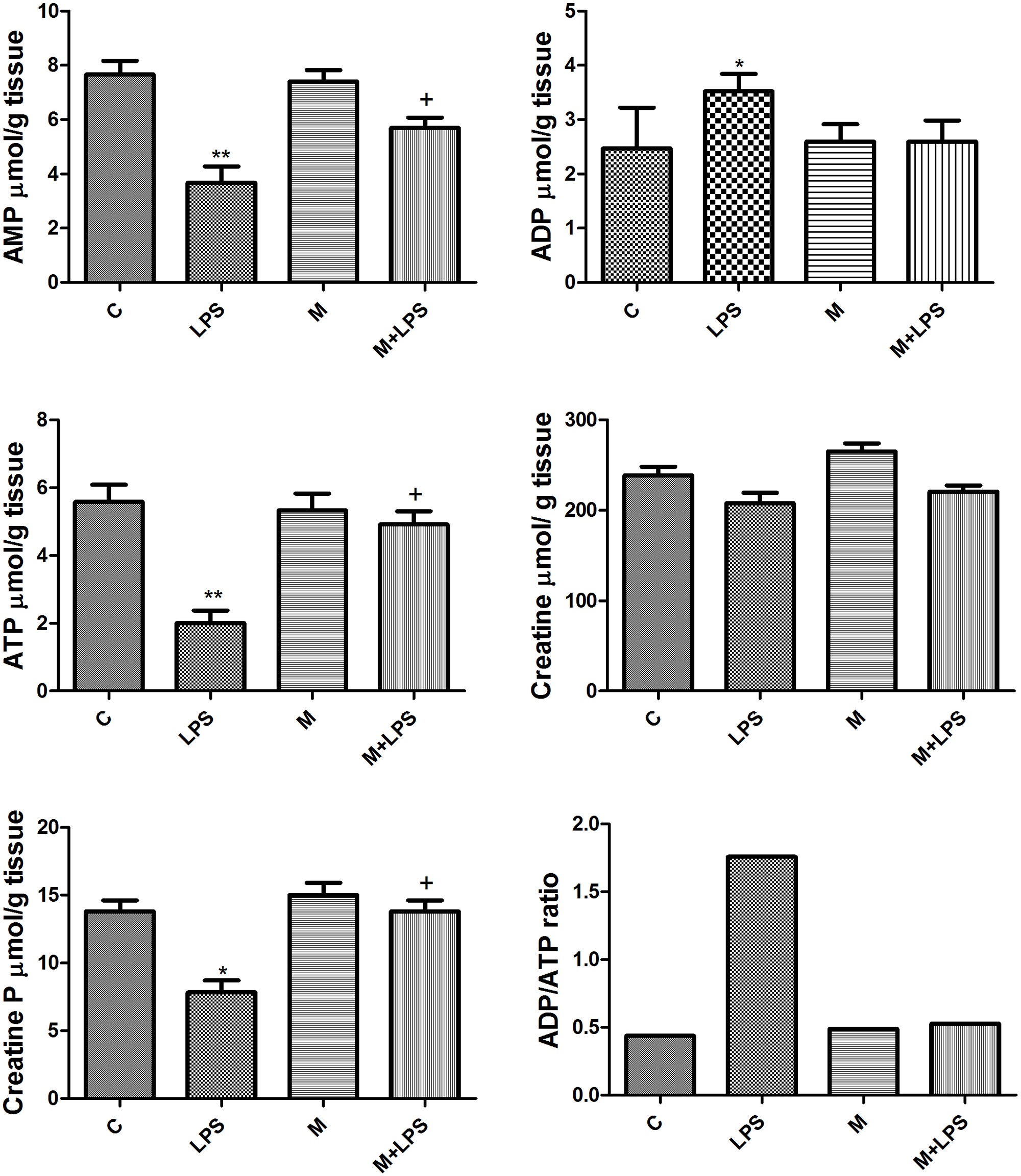
AMP, ADP, ATP, creatine, creatine p, ADP/ATP ratio levels in lung tissue were measured with HPLC in all experimental groups; control (n = 6), LPS (n = 8; 10 mg/kg i.p.), melatonin (n = 8; 10 mg/kg × 3 doses i.p.), melatonin+LPS (n = 8; LPS group treated with melatonin).
Findings of ELISA analysis of lung tissue YKL-40 determinations
In our study, one-way between-subjects ANOVA was conducted to compare the results of lung tissue YKL-40 levels (F (3.14) = 6.243, p = 0.0374). Post hoc comparisons using the Tukey HSD test showed a significant decrease in the LPS group compared to the control and melatonin groups. In the M+LPS group, YKL-40 levels were increased compared to the LPS group. No significant difference was observed between the control, melatonin and M+LPS groups (Figure 3).
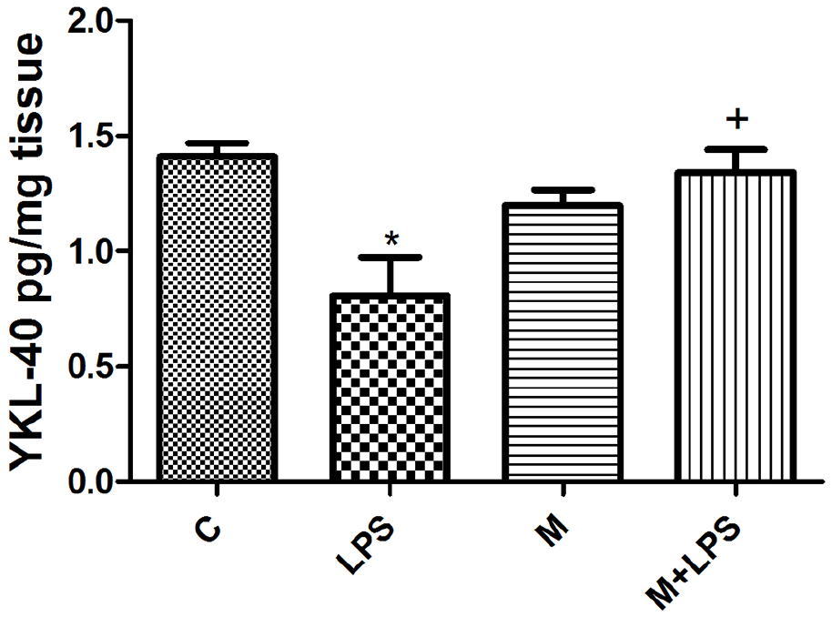
YKL-40 levels in lung tissue of all experimental groups; control (n = 6), LPS (n = 8; 10 mg/kg i.p.), melatonin (n = 8; 10 mg/kg × 3 doses i.p.), melatonin+LPS (n = 8; LPS group treated with melatonin).
Findings of ELISA analysis of lung tissue antioxidant enzyme chain (GR, GSH-Px, and SOD) determinations
In our study, one-way between-subjects ANOVA was conducted to compare the findings of ELISA analysis of lung tissue GR, GSH-Px, and SOD enzymes levels (F (3.15) = 7.027, p = 0.0034, for GR; F (3.14) = 3.024, p = 0.078, for GSH-Px; F (3.21) = 19.030, p = 0.001, for SOD).
Post hoc comparisons using the Tukey HSD test showed a significant decrease of GR levels in the LPS group compared to the control, melatonin and M+LPS groups, and a significant decrease of SOD levels in the LPS group compared to the control and melatonin and M+LPS groups. No significant difference was observed between the control, melatonin and M+LPS groups in GR or SOD enzymes levels. Meanwhile, GSH-Px levels did not show significant difference in any of the groups (Figure 4(A) to (C)).
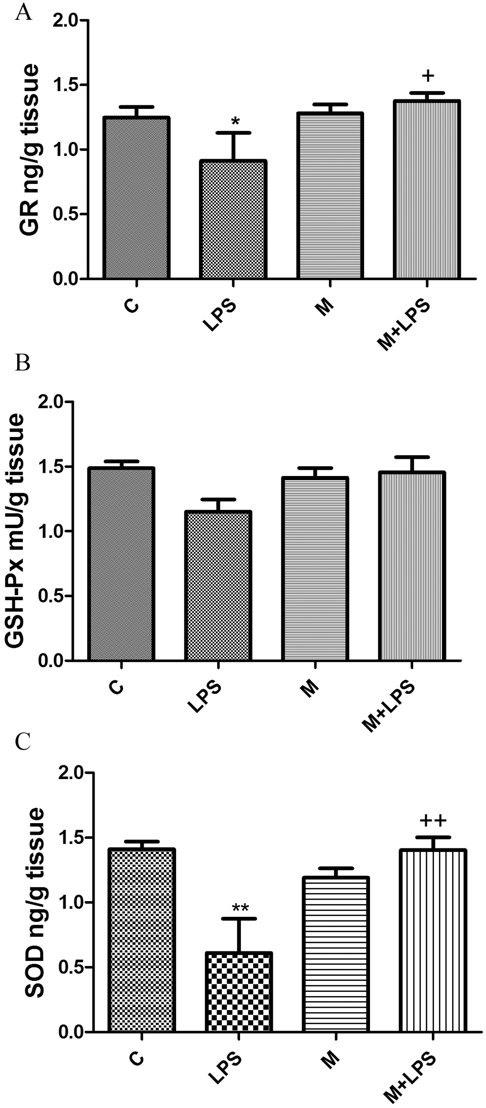
Antioxidant enzymes chain (GR, GSH-Px, SOD) levels in lung tissue of all experimental groups; control (n = 6), LPS (n = 8; 10 mg/kg i.p.), melatonin (n = 8; 10 mg/kg × 3 doses i.p.), melatonin+LPS (n = 8; LPS group treated with melatonin).
Findings of lipid peroxidation
In our study, one-way between-subjects ANOVA was conducted to compare the results of lung tissue TBARS levels (F (3.21) = 24.74, p = 0.0048). Post hoc comparisons using the Tukey HSD test indicated a significant decrease of TBARS levels in the LPS group compared to the control, melatonin and M+LPS groups. No significant difference was observed between the control, melatonin and M+LPS groups (Figure 5).

Lipid peroxidation (TBARS) in lung tissue of all experimental groups; control (n = 6), LPS (n = 8; 10 mg/kg i.p.), melatonin (n = 8; 10 mg/kg × 3 doses i.p.), melatonin+LPS (n = 8; LPS group treated with melatonin).
Findings of immunohistochemical staining
In the control and melatonin groups, there were no immunoreactivities with MMP-9, TNF-α, MMP-2, TIMP-1, IL-10, YKL-40, and MPO antibodies. In the LPS group, there was intense involvement with TNF-α and MMP-9 antibodies, moderate involvement with MMP-2 and MPO antibodies, and mild involvement with IL-10 and TIMP-1 antibodies. There was no involvement with YKL-40 antibodies in the LPS group. In the M+LPS group, there was mild involvement with TNF-α and MMP-2 antibodies and moderate involvement with IL-10. There were no immunoreactivities with MMP-9, TIMP-1, YKL-40, and MPO antibodies in the M+LPS group. H-scores of sections of the lung tissues immunoreactivity from all groups are shown in Figures 6 and 7. (F = 5.463; Figures 6 and 7).
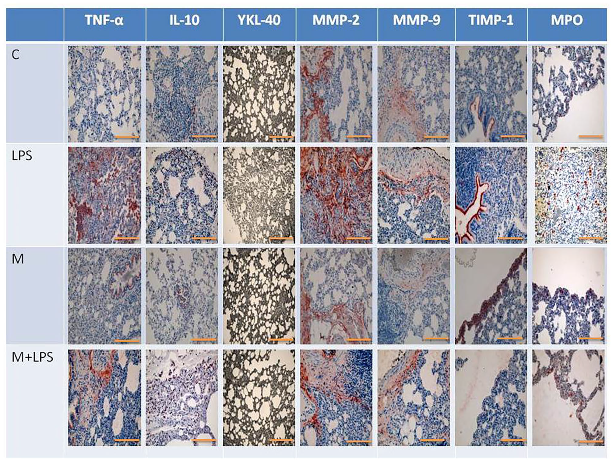
Sections of the lung tissue immunoreactivity from all groups; control (n = 6), LPS (n = 8; 10 mg/kg i.p.), melatonin (n = 8; 10 mg/kg × 3 doses i.p.), melatonin+LPS (n = 8; LPS group treated with melatonin) stained with TNF-α, IL-10, YKL-40, MMP-2, MMP-9, TIMP-1, and MPO staining. For each specimen, one score was assigned according to the percentage of positive cells: < 5% of the cells: 1 point; 6–35% of the cells: 2 points; 36–70% of the cells: 3 points; and > 71% of the cells: 4 points. Another score was assigned according to the intensity of staining, with negative staining equal to 0 point; no involvement, 1 point; mild involvement, 2 points; moderate involvement and intense involvement 3 points. (A color version of this figure is available in the online journal.)

H-scores of sections of the lung tissue immunoreactivity from all groups. “a” indicates p < 0.05 versus all experimental groups; “b” indicates p < 0.05 versus control group (F = 5.463).
Findings of histological investigation
Normal alveoli structure in lung tissue was observed in the control and melatonin-treated groups, whereas damaged alveoli, septum thickening, and immune cell infiltration were evident in the lung tissue of LPS-infected groups. It was observed that the damage caused by LPS decreased in the M+LPS group (Figure 8; (a) Control, (b) Melatonin, (c) LPS, (d) M + LPS group).
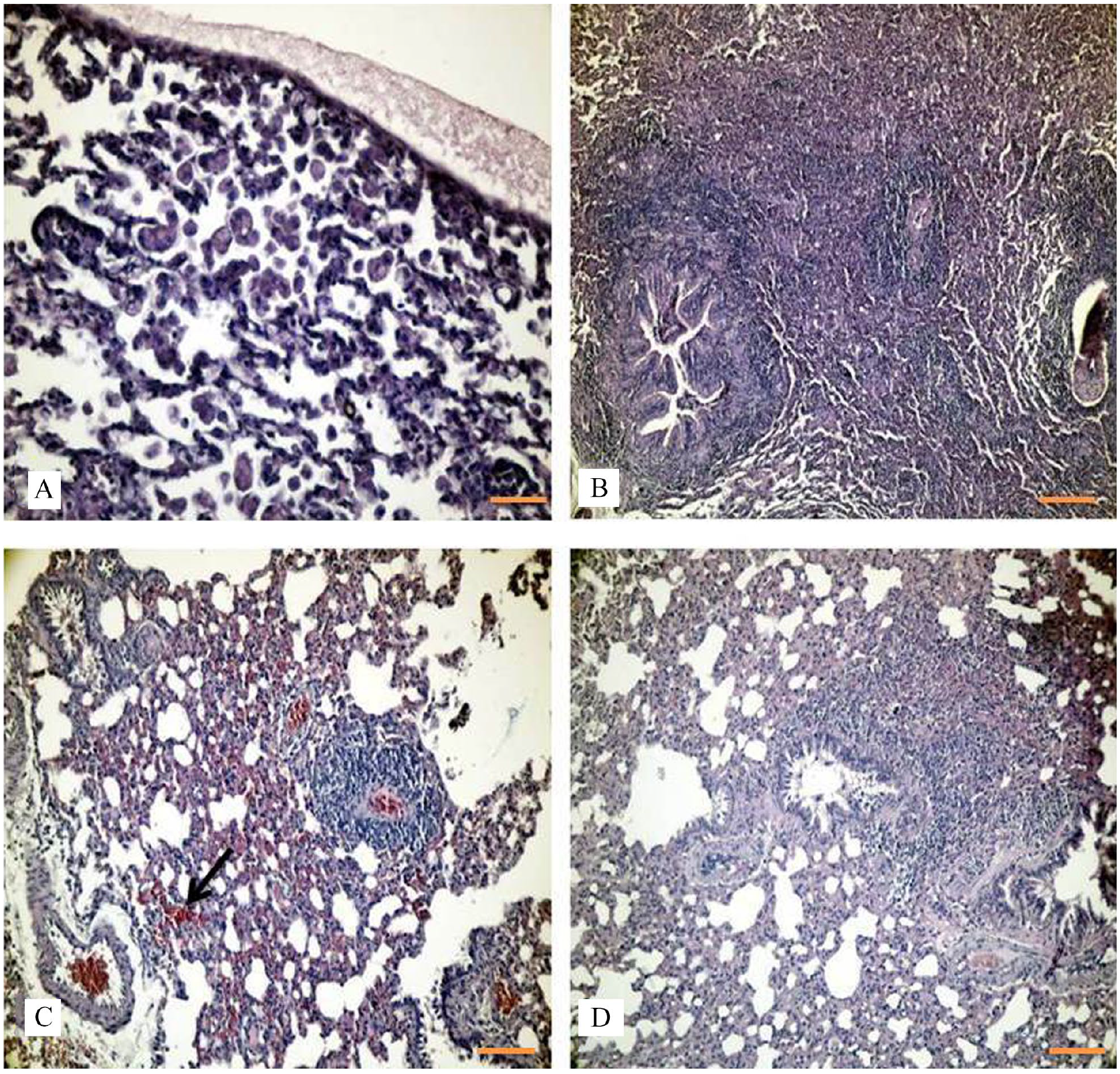
Sections of the lung tissue from all groups stained with H&E. (a) Control, (b) melatonin, (c) LPS, and (d) M+LPS group, 50 µm scale bar. (A color version of this figure is available in the online journal.)
Discussion
We demonstrated in this study the effects of melatonin, which we applied in the three time dosages before and during LPS injection, on antioxidant enzyme levels, lipid peroxidation, high-component energetics, YKL-40, MMPs and their inhibitor, MPO and cytokine levels.
In the literature, it has been stated that the dose required for the antioxidant and anti-inflammatory effects of melatonin should be much higher than that given for the sleep–wake cycle regulation. In parallel with these studies, 10 mg/kg was chosen as the ideal therapeutic dose, and it was applied as three doses at different stages to be used as a preventive and therapeutic agent.12,15,20,21
Increased oxidative stress, impaired energy balance, mitochondrial dysfunction, disruption in the antioxidant enzyme chain and increased inflammatory cytokine release were observed in rats with endotoxemia induced with LPS injection.6,15,16
Melatonin, in addition to its circadian rhythm regulator and antioxidant roles, has been reported in many studies to be an immunomodulating agent. Studies have reported that exogenous melatonin administration has immunostimulating, anti-inflammatory, and antioxidant properties.22,23 However, it has also been suggested that it can function as both a pro- and anti-inflammatory regulator. Melatonin can stimulate pro-inflammatory cytokines, nitric oxide (NO), and other mediators under different conditions. However, it has also been reported that it stimulates anti-inflammatory mediator production such as IL-10 and Sirtuin-1, and suppresses the pro-inflammatory process by inhibiting nuclear factor kappa β (NF-κβ).24,25
In our study, we observed that TNF-α involvement increased excessively in rats with endotoxemia, and melatonin administration stimulated IL-10 release in endotoxemic rats. Similar to our study, it has been reported in other studies that melatonin regulates the immune response by stimulating the release of IL-10 in endotoxemia.12,26–28
Melatonin reduced TNF-α and superoxide activation in the aorta and liver of LPS-induced endotoxemic rats. 21 Melatonin injected to rats (10–60 mg/kg) before and/or after LPS depressed lipid peroxidation and elevated the NO level caused by LPS in the liver and lungs in a dose-dependent manner. Melatonin blocked systemic alterations due to LPS treatment. 29
The complex pathogenesis of endotoxemia can prolong the time of diagnosis and poor prognosis. YKL-40 is a candidate early biomarker molecule for endotoxemia. It has been reported to be released from activated neutrophils and macrophages. 30 However, discordant results have been reported in studies of YKL-40 in serum and bronchoalveolar fluid in clinical practice.31,32
In our study, lung tissue YKL-40 levels were found to be significantly lower in the LPS group. Melatonin administration also increased YKL-40 to levels similar to that of the control group. It has also been reported to cause fibrous tissue changes by inducing MMP-9 release from stimulated alveolar macrophages in lung injury.33,34
MMPs are important in initiating the inflammatory response of leukocytes in endotoxemia. It has been reported that there is a relationship between the increase of MMPs and cytokine levels and tissue/organ damage due to infiltration of monocytes. 14
Elevated MMP-9 levels have been reported after endotoxin injection in healthy volunteers. It was found that enhanced MMP-9 levels are associated with lung tissue degeneration.35,36
It has been reported that TIMPs are important in the control of MMP tissue activity. 37 It is suggested that increased levels of MMP, released in response to the increased inflammatory process, cause insufficient blood supply of tissues and organs by disrupting blood flow. 38
In our study, involvement of MMP-2 and TIMP-1, and more intense involvement of MMP-9, were found in the lung tissue of rats in the LPS group compared to the other groups.
There are parallels between the study of Ganguly et al. and our results; endotoxemia-induced MMP-9 and TIMP-1 involvement were not observed in the melatonin-administered endotoxemic group, whereas MMP-2 involvement was observed mildly.39,40
Melatonin (5 mg/kg) decreased cerebral MMP-9 activity in a meningitis model induced by LPS. In addition, pretreatment or co-treatment with melatonin significantly blocked LPS-induced MMP-9 activation. 41
Studies have reported that endotoxemia disrupts the oxidant/antioxidant balance, causes mitochondrial damage and irregularities in energy balance.4,11 Application of LPS has been shown to result in higher production of superoxide and lipid peroxides, especially malondialdehyde (MDA), 4-hydroxylalkenal, and TBARS.
Gitto et al. 42 showed that melatonin decreases oxidative stress and excessive free radical production in newborns with sepsis.
In our study, it was observed that TBARS levels increased significantly in the LPS group compared to the control group. On the contrary, compared to the LPS group, a decrease of TBARS was observed in the M+LPS group.11,23,24,43
The levels of GR, GSH-Px, and SOD enzymes shows that antioxidant activity was significantly decreased in the endotoxemic group compared to the other groups. We observed that melatonin administration raised the decreased antioxidant enzyme levels in endotoxemic rats.12,22,23 Li Volti et al. 44 reported that melatonin showed antioxidant activity in sepsis conditions. It has been shown that melatonin ameliorates the oxidant damage caused by LPS in sepsis.45,46
Studies have reported enhanced levels of MPO in endotoxemia, and its role in the leukocyte infiltration observed in lung tissue with endotoxemia.15,16,47 In parallel with these studies, a moderate level of MPO immunoreactivity was shown in the lung tissue of LPS-treated rats. However, increased leukocyte infiltration was found in the lung tissue in the LPS group in histological examinations.
It has been reported that melatonin administration showed a significant decrease in MPO levels in rats with endotoxemia.15,47 We observed that melatonin administration prevents the increase in MPO due to endotoxemia. In histological examinations, we observed that alveolar deterioration, thickening of the septum, and increased leukocyte infiltration observed in the lung tissue of endotoxemic rats improved with melatonin administration.
Mitochondrial damage due to sepsis causes pathophysiology of organ dysfunction. Li et al. 48 found that following LPS administration, the congestion and infiltration of inflammatory cells was observed, which appeared to be predominantly neutrophils, and in parallel with our study, it was reported that ATP depletion, depletion of intracellular antioxidant systems, hypoxia and mitochondrial respiratory chain disorders, caused by endotoxemia, results in excessive production of free radicals, energy disturbances, and multiple organ dysfunction shock. It was observed that the damage caused by LPS decreased in the M+LPS group.
Li et al. 49 found that melatonin could protect against organ injury during sepsis. Melatonin significantly attenuated lung inflammation as shown by the reduction in lung injury score during sepsis.
The production of ATP is essential for the signal cascade of the regulation of immune cell responses. ATP content does not activate adhesion and migration of leucocytes, but ADP is involved in this major role toward cytokine production and migration in affected tissues. 50
As it depends on the oxygen consumption by the mitochondrial electron transport chain, the conversion rate of ADP to ATP can be considered as an indicator of mitochondrial respiration rate. The organism uses creatine group molecules to maintain energy production with degraded ATP. Disruption of cellular respiration may lead to circulatory disorders, impaired perfusion, and secondary hypoxia, together with multi-organ failure.4,11,51,52
In our study, we observed that ATP, AMP, and creatine p levels significantly decreased in rats treated with LPS, but ADP levels significantly increased compared to the other groups.
Studies have reported that Melatonin prevents mitochondrial dysfunction by increasing ATP production and complex IV activity. Melatonin has also been reported to normalize mitochondrial ATP production in endotoxemic mice.17,51,52 Melatonin treatment (60 mg/kg, i.p.) importantly prevented LPS-induced blocking of complexes I and IV of the electron transport chain and decreased mitochondrial nitric oxide synthase (NOS) activity and NO production, thereby preventing LPS toxicity. 53
In similar studies, it has been reported that the deterioration of ATP production correlates with the increase of ADP stores. This is also an indicator of impaired cellular respiration. However, we observed that ADP and ATP levels improved energy levels in endotoxemic rats treated with melatonin.11,51,52
It was observed that the ratio of ADP/ATP increased by approximately four times in the LPS group compared to the other experimental groups, and that the melatonin application approached the control levels. Similarly, an increase in the ratio of ADP/ATP in muscle tissue was reported in septic patients in a human study. 54
We found that there was no significant difference in creatine levels. Studies have reported that creatine levels are required in addition to ATP in case of activity requiring high metabolism. 52
We hypothesize that the administration of melatonin before and during endotoxemia has an effect of improvement through reducing the level of pro-inflammatory cytokines, activating anti-inflammatory cytokines, stimulating the release of antioxidant enzymes, regulating the energy balance, and reducing free radical production and the levels of MMP responsible for tissue damage.
In our study, we examined the effects of acute sepsis induced by LPS with the mechanisms of inflammation and oxidative stress in terms of lung tissue damage and serum protein levels. In addition to the acute sepsis model, creating the chronic model, gene expressions effective on these mechanisms and the inability to examine the relevant pathways are the limitations of our study. In our future studies, we aim to investigate the chronic sepsis model, to investigate effective biomarkers for sepsis and inflammatory pathways, and to search for molecules that are effective both at the cellular level and in inflammatory diseases.
Footnotes
Authors’ Contributions
GA, ST, HY, and EO designed, conducted, and analyzed the experiments and prepared all the figures. SM, VO, AA, and NBC provided technical support. GA, ST, HY, and EO wrote the article.
Declaration of conflicting interests
The author(s) declared no potential conflicts of interest with respect to the research, authorship, and/or publication of this article.
Funding
The author(s) disclosed receipt of the following financial support for the research, authorship, and/or publication of this article: This study was supported by Istanbul University Research Project (Project Number: TSA-2017-25047).
