



Abstract
Zebrafish patient-derived xenograft (zPDX) models have shown great potential in predicting the short-term treatment response in various types of tumor cases. However, few studies have used zPDX models for drug screening in non–small cell lung cancer (NSCLC). We aimed to compare the treatment responses of patients with NSCLC with those of the corresponding zPDX models. Tumor cells were obtained from pleural fluid or biopsy procedures from patients with NSCLC and injected into the perivitelline space of zebrafish larvae. Then, the same antineoplastic drugs administered to the corresponding patient were tested in the successfully constructed zPDX model, for 3 days. Responses to treatment were compared. A total of 21 patients with advanced NSCLC were enrolled in our study, and 13 corresponding zPDX models were successfully established. Based on the clinical medication of enrolled patients, we provided a corresponding drug treatment to these zebrafish embryos, including epidermal growth factor receptor tyrosine kinase inhibitors (EGFR TKIs), pemetrexed/platinum (AP), or docetaxel/platinum (DP) administration. The chemosensitivity consistency rate between the clinical responses and those obtained from zPDXs was 76.9% (10/13). There was a high correlation between patient responses and the corresponding zPDX drug responses. Thus, zPDX can accurately and quickly reproduce patient responses to treatment with EGFR TKIs, AP, and DP and has a considerable potential to serve as a biological platform for predicting treatment effect on patients with NSCLC.
Impact Statement
Zebrafish patient-derived xenograft (zPDX) models are an increasingly popular and powerful animal model applied in the field of cancer biology. A growing body of evidence suggests that the zPDX model can serve as an effective pre-clinical model to guide personalized treatment of patients with cancer. Here, we performed a retrospective study comparing the response of patients with non–small cell lung cancer (NSCLC) to anticancer therapy with that of the corresponding zPDX models. Our study targeted the commonly used clinical treatment regimen for lung cancer; primary lung cancer cells were collected from patients using targeted drugs, chemotherapy drugs, and combination chemotherapy drugs, which can more comprehensively demonstrate the application value of the zPDX model in drug screening in lung cancer. Our results suggest that zPDX models have considerable potential to serve as a biological platform for personalized NSCLC treatment.
Introduction
Lung cancer is the leading cause of mortality worldwide, with a considerable impact on the disease burden worldwide. 1 Clinicians determine the appropriate drug regimens for patients according to the stage and type of tumor, which is supported by large-scale clinical trials. However, treatments selected by this “one-size-fits-all” approach have proven efficient for only a subset of patients because of high individual variations. 2 With the emergence of targeted drugs in the past 10 years, molecular genetic testing, including the detection of epidermal growth factor receptor (EGFR), serine/threonine protein kinase b-Raf (BRAF), and mesenchymal-epithelial transition factor mutations, as well as the analysis of anaplastic lymphoma kinase (ALK), ROS1 proto-oncogene receptor tyrosine kinase (ROS1), rearranged during transfection (RET), and neurotrophic tyrosine kinase alterations, is paramount for developing personalized treatment, which has significantly improved the prognosis of patients with cancer. 3 Unfortunately, some patients still fail to receive effective treatment even if they harbor targetable genetic alterations.4,5
Increasing evidence suggests that patient-derived xenograft (PDX) models constructed from implantation of patient tumor fragments directly into immunodeficient mice can closely replicate parent tumor biology, mimic disease response, accurately predict the clinical curative effect, and address the need for personalizing therapeutics.6 –10 However, there are some important limitations rendering the use of these models unviable in the clinic. First, mouse-PDX models require a sufficient volume of tumor tissues for transplantation, which is difficult to obtain from patients for whom there is no plan for surgery. In addition, a previous study using such models suggested an unsatisfactory engraftment rate, particularly for breast cancer. 8 Finally, the extensive time for engraftment and tumor passages, generally 4–6 months, is a major obstacle to providing direct advice for clinical treatment in an actionable time frame.
In recent years, the great potential of the application of zebrafish to compensate for the deficiency of mice models with the advantages of ease, speed, and relative cost-effectiveness has emerged.11,12 Evidence indicates that the heterogeneity of drug responses could be preserved in zebrafish patient-derived xenograft (zPDX) models constructed with patient-derived cell lines and that drug response is consistent in PDX model and patients.13,14 Several clinical studies have conducted proof-of-concept experiments to predict short-term treatment responses in patients with colorectal cancer, gastric cancer, multiple myeloma, and pancreatic ductal adenocarcinoma by constructing corresponding zPDX models with 75–100% accuracy.13,15 –18 Here, we performed a retrospective study comparing the response of patients with non–small cell lung cancer (NSCLC) to anticancer therapy with that of the corresponding zPDX models.
Materials and methods
Reagents
The experiment involved the following drugs: osimertinib (AstraZeneca, London, UK), docetaxel (Hengrui, Jiangsu, China), pemetrexed (Simcere, Jiangsu, China), and cisplatin (Qilu Pharmaceutical, Shandong, China). Fetal bovine serum (FBS), Hanks’ balanced salt solution (HBSS), penicillin and streptomycin, Liberase, and Dulbecco’s modified eagle medium (DMEM) were purchased from Gibco (Waltham, MA, USA).
Primary tissue dissociation and cell culture
Between September 2020 and February 2021, we obtained 21 NSCLC samples from the Department of Respiratory and Critical Care Medicine, Jinling Hospital, Medical School of Nanjing University. The study was conducted in accordance with the Declaration of Helsinki and was approved by the Institutional Ethics Committee (approval number: DBNJ20228). Written informed consent was obtained from the participants. The obtained samples included malignant pleural effusion and cancer tissue obtained by pneumocentesis. As this was a retrospective study, all patients underwent anticancer therapy prior to sample collection. Primary single cells from the pleural effusion were isolated through centrifugation, HBSS flushing, and filtration collection. Tissue samples were processed by HBSS flushing, Liberase digestion (400 µg/mL, 37°C, 75 min), culture medium (10% FBS, 90% DMEM) termination digestion, and filtration collection.
If for some reason the fluorescence labeling and xenograft injection was not administered on the day of sample collection (such as in cases wherein sufficient zebrafish embryos were not available at that time), the primary cells were cultured in vitro for a specific time period. These tumor cells were grown in RPMI1640 medium with 10% FBS and 1% penicillin–streptomycin.
Cell labeling and xenograft
Primary cells were fluorescently labeled with CellTracker™ CM-Dil (Invitrogen, CA, USA) according to the instructions. Labeled cells were washed in HBSS three times and re-suspended in 5–20 μL of HBSS for injection.
Approximately 800 labeled cells were injected into the perivitelline space of zebrafish larvae, at 48 hours post fertilization (hpf), obtained from Xinjia Medical Technology Co., Ltd. (Nanjing, China). After injection, larvae were placed at 34°C in six-well cell culture plates containing embryonic medium (15.0 mM NaCl, 0.5 mM KCl, 1.0 mM MgSO4, 0.15 mM KH2PO4, 0.05 mM Na2HPO4, 1.0 mM CaCl2, and 0.7 mM NaHCO3 in distilled water) until the end of the experiments. At 1 day post injection (dpi), successfully injected xenografts with similar tumor sizes were selected and randomly assigned to the control or treatment group (<30 embryos per group).
Drug administration by soaking
Toxicity assay was first performed in zebrafish embryos without tumor cell injection to determine the maximum tolerated dose (MTD) of the testing drugs, which is defined as the highest dose that is tolerated by the embryos without showing toxic signs, such as death or deformity (curvature of body, edema). Specifically, 72-hpf zebrafish embryos were exposed to embryonic medium supplemented with various concentrations of drugs for 3 days, replaced daily. According to the number of zebrafish embryos with no toxic reactions at the end of treatment, the MTD of the drug in the study was determined as osimertinib (1 µM), AP (10 mM pemetrexed/2 µg/mL cisplatin), and DP (10 µM docetaxel/2 µg/mL cisplatin).
At 1 dpi, successfully injected xenografts received drug administration by soaking based on the determined MTD concentration for 72 hours.
Imaging in vivo and quantitative analysis
At 4 dpi, the zebrafish larvae were mounted using 1.2% low-melting gel for the imaging experiments. The red fluorescence area representing tumor size was observed in vivo and photographed using a stereo microscope (MVX10, Olympus, Japan). The spatial resolution of the images was 1600 × 1200. We quantified the tumor area with CM-DiI positive signals and performed image processing using ImageJ. Finally, the tumor size in each drug treatment group was divided by the tumor size in the control group to obtain the relative tumor size at 4 dpi.
Statistical analysis
We used GraphPad Prism 8.0 for statistical analysis. The statistical data were analyzed using unpaired Student’s t-tests. The level of significance was set at P < 0.05.
Results
Exploring the MTD of drugs
For targeted and chemotherapy drugs commonly used in patients with lung cancer, we first explored the MTDs of these drugs in zebrafish embryos. The drugs included osimertinib, docetaxel, pemetrexed, and cisplatin. We set up four to six concentration gradients for each drug and set the reference concentration based on our previous experience and published research.19 –22 Although there is no clear conversion formula, the maximum plasma concentration of medication for patients has also been used as a reference to set the reference concentration.13,17 However, the final MTD concentration is still to be determined by setting the concentration gradient and conducting toxicity tests in zebrafish embryos. The patient clinical dose, corresponding maximum plasma concentration,23 –26 and testing MTD concentration in our study are listed in Supplementary Table 1. For each concentration gradient group, we used 15 zebrafish embryos; 72-hpf zebrafish embryos without tumor cell injection, as the experimental subjects, were exposed to the corresponding embryonic medium, soaked with the corresponding drugs. The number of embryos that survived without any toxic reaction was recorded for three consecutive days after soaking. This survival rate was used as a reference to determine the MTD in our study. If the appropriate concentration could not be determined in the first round, another round was conducted until the concentration of the drug to be used was determined. The toxicity curves for these drugs are presented in Figure 1.

Toxicity curves for osimertinib, pemetrexed, docetaxel, and cisplatin. Zebrafish embryos at 72 hours post fertilization were exposed to embryonic medium supplemented with varying concentrations of osimertinib, pemetrexed, docetaxel, and cisplatin for three consecutive days. The percentage of viable embryos without toxic reaction were plotted versus the drug dose.
On day 3 of soaking the drug osimertinib with concentrations of 2, 5, and 10 µM, all the embryos died. When the drug concentration was reduced to 1.5 µM, the embryos still had a high mortality rate. Therefore, we chose 1 µM as the final soaking concentration because the survival rate of the embryos was 100% using this dose. Similarly, the MTDs of pemetrexed, docetaxel, and cisplatin were confirmed as 10 mM, 10 µM, and 2 µg/mL, respectively.
Clinical information and medication sensitivity of included patients
In this study, we initially enrolled 21 patients with advanced NSCLC, including 20 cases of lung adenocarcinoma and one case of squamous cell carcinoma. We constructed zPDX models with cancer cells from malignant pleural fluid or cancer tissue obtained by pneumocentesis. The zPDX model is regarded as successfully established if the xenograft grow successfully in zebrafish embryos. Finally, 13 cases were successfully established, and a further eight cases failed. The reasons for the failure included the following: poor condition of tumor cells when cultured in vitro; insufficient number of tumor cells; the state of zebrafish embryos was not good after tumor cell transplantation or drug immersion; and the mortality rate was high. The specific reasons for each model failure are summarized in Supplementary Table 2.
The clinicopathological information of the 13 patients enrolled is presented in Table 1, including their age, gender, pathological type, clinical stage, genetic mutation information, surgical history, and clinical medication. All patients underwent treatment, and their serum tumor markers and imaging examination reports before and after drug treatment are shown in Table 2. Patients’ imaging findings after drug treatment were assessed by a clinician based on the Response Evaluation Criteria in Solid Tumors (RECIST) guidelines (version 1.1), 27 and the following four outcomes were determined: complete response (CR), partial response (PR), stable disease (SD), and progressive disease (PD). Patients with imaging findings for outcomes of CR, PR, or SD or patients with significantly decreased serum tumor marker levels were considered “Responders” (R) or sensitive to treatment, whereas patients with imaging findings of PD or a significant increase in serum tumor marker levels were considered “Non-Responders” (NR) or resistant to treatment 28 (Table 2).
The clinicopathological features of patients with lung cancer enrolled in our study.

TNM stage: tumor-node-metastasis stage; Genetic mutation: this part mainly showed the information on epidermal growth factor receptor mutations; No mention: no genetic test results were provided in this patient’s clinical data; surgical history: whether to perform lung cancer therapy surgery.
Clinical basis for sensitivity to clinical drug treatment.
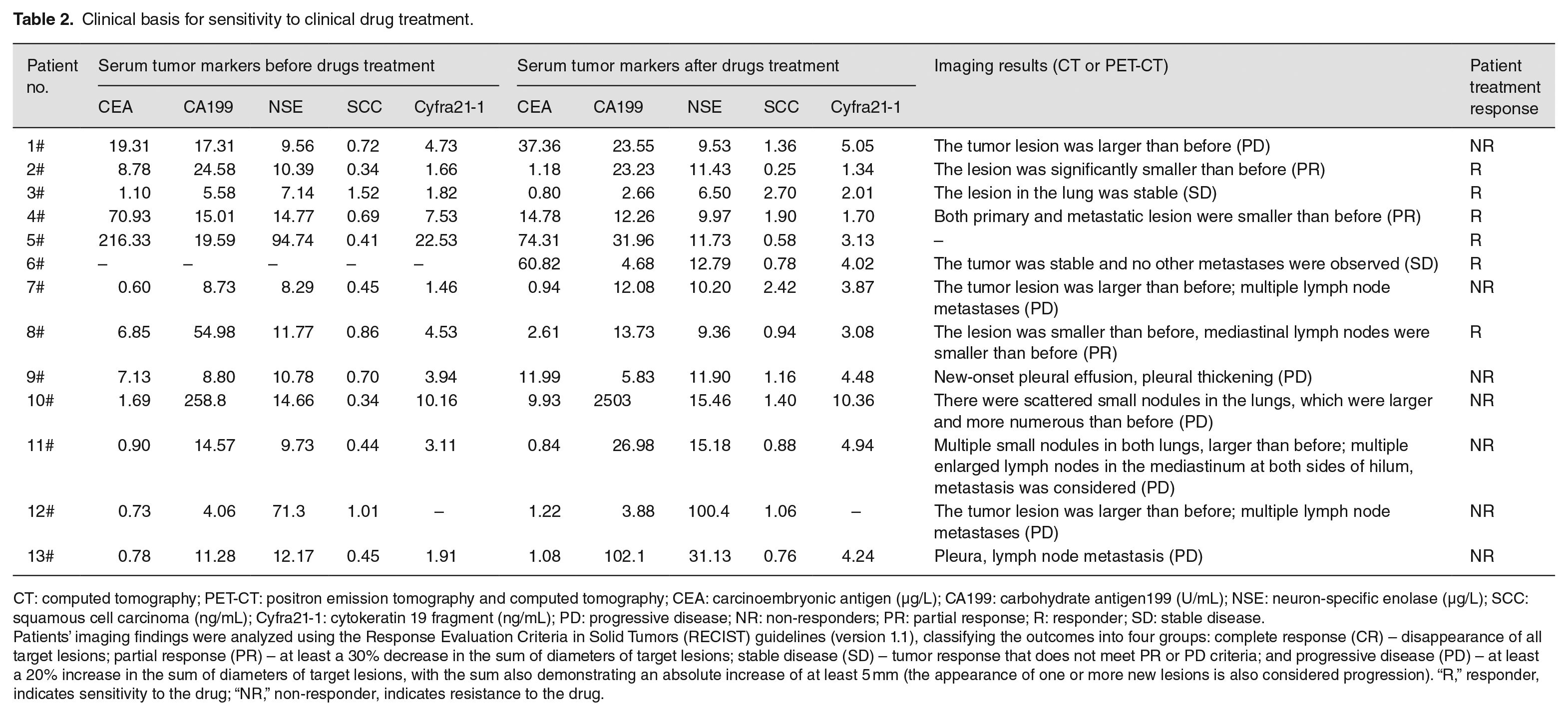
CT: computed tomography; PET-CT: positron emission tomography and computed tomography; CEA: carcinoembryonic antigen (µg/L); CA199: carbohydrate antigen199 (U/mL); NSE: neuron-specific enolase (µg/L); SCC: squamous cell carcinoma (ng/mL); Cyfra21-1: cytokeratin 19 fragment (ng/mL); PD: progressive disease; NR: non-responders; PR: partial response; R: responder; SD: stable disease.
Patients’ imaging findings were analyzed using the Response Evaluation Criteria in Solid Tumors (RECIST) guidelines (version 1.1), classifying the outcomes into four groups: complete response (CR) – disappearance of all target lesions; partial response (PR) – at least a 30% decrease in the sum of diameters of target lesions; stable disease (SD) – tumor response that does not meet PR or PD criteria; and progressive disease (PD) – at least a 20% increase in the sum of diameters of target lesions, with the sum also demonstrating an absolute increase of at least 5 mm (the appearance of one or more new lesions is also considered progression). “R,” responder, indicates sensitivity to the drug; “NR,” non-responder, indicates resistance to the drug.
Consistency analysis of zPDX model and clinical drug sensitivity of patients with lung cancer
Primary tumor cells from patients were collected, centrifuged, and stained and then transplanted into zebrafish embryos to establish a xenograft model. The same class of drugs was used in zebrafish embryos as that administered to the patients in clinical practice. We used the MTD of the drug to soak the zebrafish embryos, which was obtained from previous screening. Drug susceptibility was evaluated using the quantifications of changes in relative tumor size in zebrafish embryos after drug administration, which was determined by tumor size in each drug treatment group compared with the blank control group. Finally, drug response from the zPDX model was compared with that of the actual effect in clinical settings. Specific results for each sample are presented in Table 3 and Figure 2. In addition, the brightfield image for these 13 corresponding zPDX models is shown in Supplementary Figure 1.
The results of zPDX model established in our study.

zPDX: zebrafish patient-derived xenograft; EGFR TKI: epidermal growth factor receptor tyrosine kinase inhibitors (1 µM osimertinib); “R”: responder, indicating that the treatment reduced the CM-DiI-positive area representing tumor area significantly; DP: docetaxel/platinum (10 µM docetaxel/2 µg/mL cisplatin); “NR”: non-responder, indicating that the treatment did not reduce the tumor area significantly; AP: pemetrexed/platinum (10 mM pemetrexed/2 µg/mL cisplatin).
P < 0.05; **P < 0.01; ***P < 0.001.

The images of tumor size in zPDX model. Thirteen corresponding zPDX models were successfully established. Based on the treatment of the enrolled patients, we provided a corresponding drug treatment for these zebrafish embryos, including epidermal growth factor receptor tyrosine kinase inhibitors (EGFR TKIs), pemetrexed/platinum (AP), or docetaxel/platinum (DP) administration. The red fluorescence area representing the tumor size was observed in vivo at 4 days post injection by a fluorescence microscopy. The number of xenografts analyzed is indicated in the corresponding images. Tumor area was reduced significantly in the 1#–6#, 8#, 12# (TKI), and 13# (TKI) experimental groups compared with the blank control group (NC). Scale bar, 500 μm.
In the corresponding zPDX model, six models were treated with monotherapy (EGFR-TKI), five with two drugs (DP/AP), and two with three drugs (TKI combined with DP or AP). Subsequently, we compared the consistency between zPDX and patient outcomes. As summarized in Table 4, drug sensitivity was consistent in 10/13 of the cases (5/6 in the monotherapy group, 5/5 in the two-drug group, and 0/2 in the three-drug group, respectively). We reviewed three cases with inconsistent results and tried to determine the possible reasons. Only case 1# in the monotherapy group presented inconsistent drug sensitivity. This patient was treated with osimertinib and gefitinib. However, the disease still progressed, as seen by the elevated serum tumor indicators, and the imaging findings showed that the tumor lesion was larger than pre-treatment. Although the effect of osimertinib on reducing tumor area was observed in zebrafish embryos, this effect was not very significant (P = 0.0432). We considered that increasing the number of embryos tested could improve the accuracy of the results. In addition, Patients 12# and 13# were treated with three drugs in clinical practice but showed drug resistance to targeted and chemotherapeutic drugs. Consistently, the corresponding zPDX model showed that the primary tumor cells were resistant to DP and AP, respectively. However, in the corresponding xenograft model, the therapeutic effect of TKI was inconsistent with that observed in patients. We postulated that the ability to test drug sensitivity in the zPDX model may decrease with the increase in drug types. Overall, our study suggested that the reproduction of positive and negative clinical models by zPDX models was relatively accurate.
Confusion matrix of the correlation between the response of drug treatments in patients and in zPDX model.
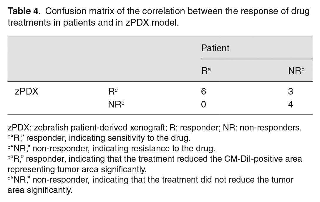
zPDX: zebrafish patient-derived xenograft; R: responder; NR: non-responders.
“R,” responder, indicating sensitivity to the drug.
“NR,” non-responder, indicating resistance to the drug.
“R,” responder, indicating that the treatment reduced the CM-DiI-positive area representing tumor area significantly.
“NR,” non-responder, indicating that the treatment did not reduce the tumor area significantly.
Discussion
Here, we constructed a zPDX model of NSCLC, which had many advantages compared with the mouse-PDX model, including the ability to use fresh patient tumor tissues, the small number of cancer cells obtained through nonsurgical procedures, and the short latency of drug effects that could be shown within 3 days after administration. In this study, a consistency analysis was conducted between the therapeutic effect of the drugs used in clinical patients and the corresponding effects in zebrafish xenograft models. Interestingly, we found that the consistency rate was 76.9% (10/13).
The zPDX model has great application potential for predicting drug efficacy and guiding the personalized treatment of patients. 29 Attempts have been made to use the zebrafish model for drug screening and drug efficacy observation for various tumor types. However, there are few studies on lung cancer drug screening. Fan et al. 21 explored the inhibitory effect of dosimertinib and gefitinib in a zebrafish brain metastasis model. Li et al. 20 used the zPDX model to study the inhibitory effect of osimertinib on NSCLC with EGFR mutation and T790M resistance mutation. However, all these studies used passaging cell lines as the experimental material, and the results still need to be confirmed using clinical specimens. In the largest scale NSCLC patient-derived zPDX model study to date, Ali et al. 14 enrolled a total of 43 patient cases and found that the zPDX model (constructed by transplanting tissue fragments from mouse-PDX models) can accurately reproduce the response to paclitaxel or erlotinib in the corresponding mouse-PDX model and clinical patients themselves, and predict lymph node metastasis with a 91% sensitivity. On this basis, our study targeted the commonly used clinical treatment regimen for lung cancer; primary lung cancer cells were collected from patients using targeted drugs, chemotherapy drugs, and combination chemotherapy drugs, which further contributed to the application value of the zPDX model in the drug screening of lung cancer.
This study had some limitations. First, we collected the primary tumor cells from patients who received targeted or chemotherapy drugs, and then the corresponding drugs were applied in the zebrafish transplantation model. The drugs used for patients and their corresponding zPDX models were of the same class, but not necessarily the same drugs. Specifically, in the monotherapy (EGFR-TKI) group, we used osimertinib as the symbol of EGFR-TKI in zPDX models. However, these patients may have been treated with other targeted drugs such as gefitinib or erlotinib. Similarly, we used docetaxel/cisplatin or pemetrexed/cisplatin as the symbol of DP or AP, although some patients were also treated with nedaplatin. Although the drugs are of the same class, the accuracy of the results may be affected by the differences in the specific drugs. Second, we collected 21 samples and successfully established 13 xenograft models, with a success rate of 61.9%. In the future, this success rate can be increased by improving sampling standardization and the operating proficiency of technicians. In addition, we should consider enrolling a similar number of sensitive and resistant cases for each treatment mode to reduce the result bias caused by sample selection in the future. Finally, the zebrafish xenograft model itself has some limitations. Although zebrafish share high homology with mammalian organ systems and physiology, orthotopic engraftment is impossible because zebrafish lack some mammal-specific tissues, such as lungs and mammary glands. 30 Furthermore, there are only few studies on the equivalent conversion between drug administration in zebrafish embryos by soaking and clinical dose. The selection of optimal dose and whether different degrees of absorption in fish due to pharmacokinetic differences of the drug itself can cause differences of the results remain to be verified. 31 Therefore, the combined use of mouse and zebrafish PDX models to complement each other is encouraged to conduct rapid screening and advanced research in the future. A separate xenograft model could be established for different stages of treatment response in the same patient, considering that drug therapy is a long-term process in patients with advanced lung cancer, but drug sensitivity may change with the treatment cycle, and that the zebrafish zPDX model validation is for a short-term immediate response. Furthermore, a large-scale prospective study is required to more accurately test the effectiveness of zebrafish as an early screening for drug sensitivity.
Conclusions
To sum up, our retrospective analysis suggests that the reproduction of patients’ responses to EGFR-TKI, AP (pemetrexed/platinum), and DP (docetaxel/platinum) by zPDX models is relatively accurate and rapid and has considerable potential to serve as a biological platform for personalized NSCLC treatment.
Supplemental Material
sj-pdf-1-ebm-10.1177_15353702221142612 – Supplemental material for Zebrafish patient-derived xenografts accurately and quickly reproduce treatment outcomes in non–small cell lung cancer patients
Supplemental material, sj-pdf-1-ebm-10.1177_15353702221142612 for Zebrafish patient-derived xenografts accurately and quickly reproduce treatment outcomes in non–small cell lung cancer patients by Xin Hua, Xiaodi Wu, Ke Xu, Ping Zhan, Hongbing Liu, Fang Zhang, Tangfeng Lv and Yong Song in Experimental Biology and Medicine
Footnotes
Authors’ Contributions
XH and XDW designed the study and were major contributors in writing the manuscript. KX completed the figures and tables. XH and PZ were responsible for the experimental operation. HBL completed the data statistics. FZ, TFL, and YS provided the experimental designs and analyzed the experimental results. All authors participated in the review of the final draft of the manuscript.
Declaration of Conflicting Interests
The author(s) declared no potential conflicts of interest with respect to the research, authorship, and/or publication of this article.
Funding
The author(s) disclosed receipt of the following financial support for the research, authorship, and/or publication of this article: The authors disclose receipt of the following financial support for the research: this work was supported by the Natural Science Foundation of Jiangsu province [grant numbers BE2019719, BE2019718].

Supplemental Material
Supplemental material for this article is available online.
References
Supplementary Material
Please find the following supplemental material available below.
For Open Access articles published under a Creative Commons License, all supplemental material carries the same license as the article it is associated with.
For non-Open Access articles published, all supplemental material carries a non-exclusive license, and permission requests for re-use of supplemental material or any part of supplemental material shall be sent directly to the copyright owner as specified in the copyright notice associated with the article.
